Contents
- What is granuloma annulare
What is granuloma annulare
Granuloma annulare is a common skin condition in which there are smooth discolored plaques. They are usually thickened and ring-shaped or annular in shape. Granuloma annulare is more correctly known as necrobiotic papulosis.
No one knows exactly what causes granuloma annulare. But it may be triggered by minor skin injuries and certain medications. Some types of granuloma annulare affect adults, and others typically affect children.
Granuloma annulare can occur on any site of your body and is occasionally quite widespread. Granuloma annulare only affects the skin. Granuloma annulare affects the skin of children, teenagers or young adults (or older adults, less commonly).
Granuloma annulare may cause no symptoms, but affected areas are often tender when knocked. The plaques tend to slowly change shape, size and position.
Granuloma annulare is harmless, does not affect general health, is not infectious or contagious, doesn’t run in families and is not due to allergies. Some types of granuloma annulare have been linked with diabetes, but this is very uncommon.
There are several types of granuloma annulare. The most common is called localized granuloma annulare. This is characterized by skin-colored, pink or purple patches which usually appear on only one or two sites of the body. The patches can appear anywhere, but typically develop on bony areas such as the back of the hands, the feet, elbows or knees. Involvement of the face is very uncommon. Usually the rings start as small in size and then slowly grow to 2.5-5 cm (1 or 2 inches) in diameter. As they enlarge they become flatter and sometimes more purple in color, and then gradually fade.
Occasionally, people with granuloma annulare can develop a more widespread rash called disseminated (generalized) granuloma annulare, but this occurs in less than 1 in 10 cases.
Granuloma annulare treatment
Unfortunately there is no really effective treatment. Decisions are usually made based on the subtype of granuloma annulare.
As localized granuloma annulare is limited to only a few sites and tends to improve spontaneously with time, treatment is usually unnecessary. Symptomatic or obviously visible patches may improve using steroid creams or ointments. While steroid injections into the rings may be helpful, this is not always the case and so this treatment is not recommended as there is some risk of thinning the skin. Cryotherapy, which refers to the treatment of skin lesions by freezing them, may be an option for treating very small patches, but can leave a permanent scar.
Evidence for treatments that work in disseminated (generalized) granuloma annulare is limited with no ideal treatment available. Ultraviolet light treatment and powerful drugs such as steroid tablets, antibiotics, antimalarial drugs, isotretinoin, ciclosporin and dapsone have been reported to help in individual cases but, for the vast majority of patients, the symptoms of granuloma annulare do not justify the use of these drugs which all have significant side-effects.
Can granuloma annulare be cured?
No, but in over half of cases it will clear by itself within two years. However, this cannot be predicted accurately on an individual basis.
If you’re bothered by how your skin looks, your doctor can prescribe medications that will speed the disappearance of the lesions.
If your child has bumps or a ring-shaped lesion on the skin for more than several weeks, see his or her doctor or a dermatologist who can tell whether the bumps are due to granuloma annulare or conditions such as ringworm, insect bites, or even Lyme disease.
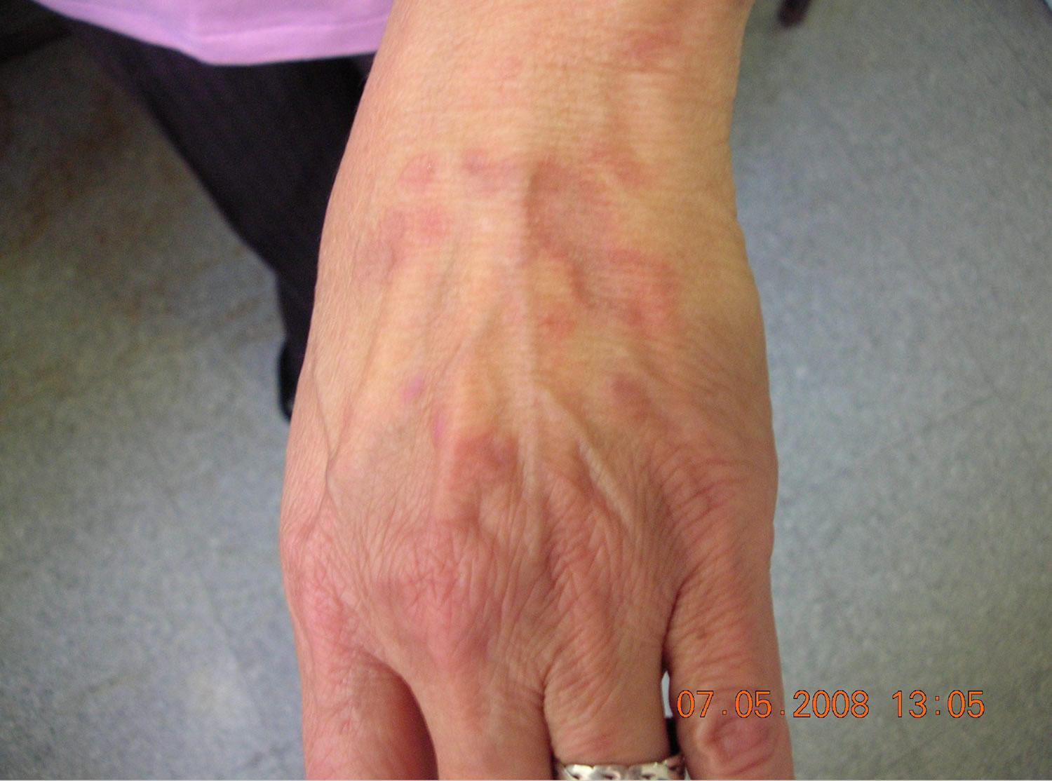
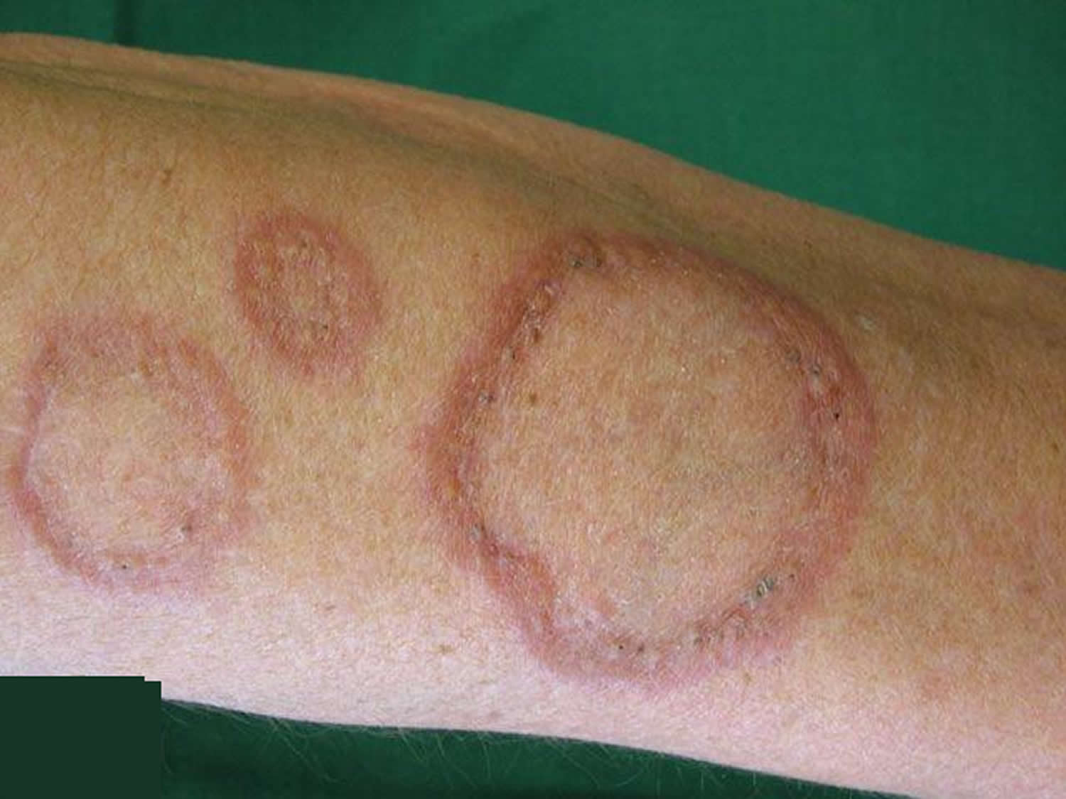
Figure 1. Granuloma annulare rash
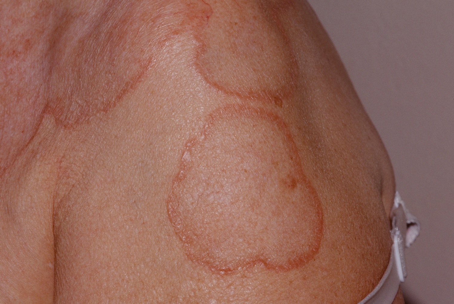
Figure 2. Granuloma annulare
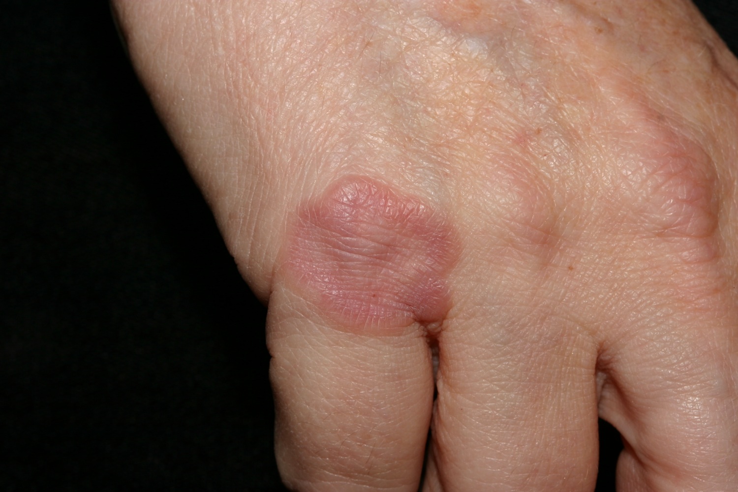
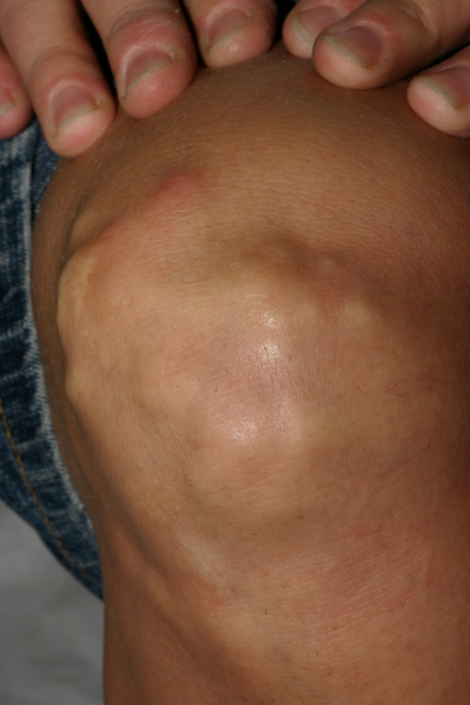
Localized granuloma annulare
The localized form is the most common type of granuloma annulare in children. One or more skin colored bumps occur in rings (circular or semicircular shape) , with a diameter up to 2 inches (5 centimeters) in the skin over joints, particularly the knuckles. The center of each ring is often a little depressed. Localized granuloma annulare usually affects the fingers or the backs of both hands, but is also common on top of the foot or ankle, and over one or both elbows.
Figure 3. Localized granuloma annulare
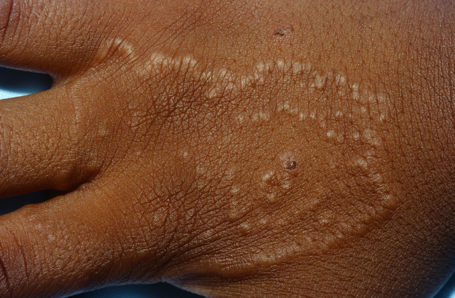
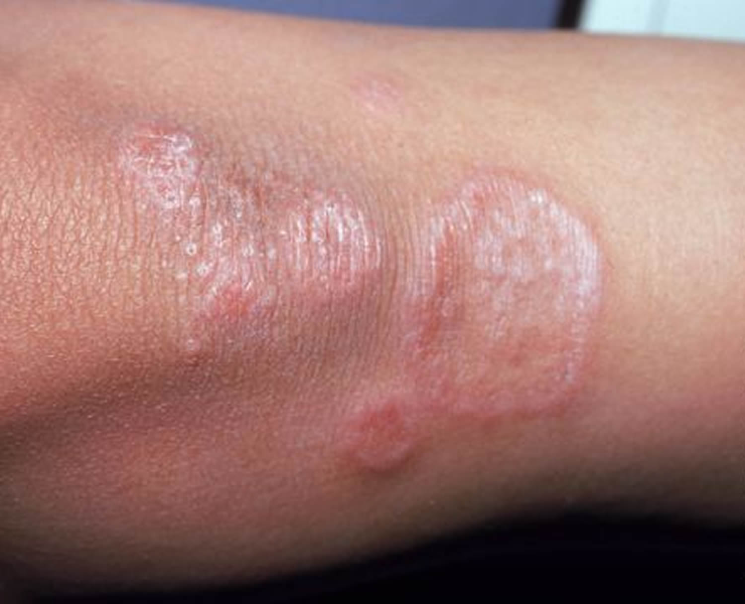
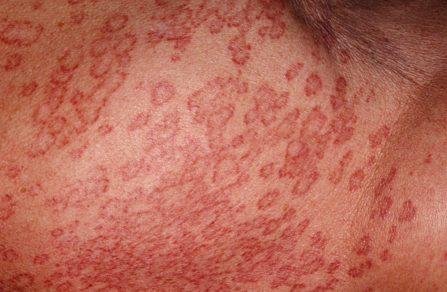
Disseminated granuloma annulare
Generalised granuloma annulare usually presents in adults, as widespread skin-coloured, pinkish or slightly mauve-colored patches. The disseminated type is composed of small papules, usually arranged symmetrically in rings 10 cm or more in diameter. They are often found around the skin folds of the trunk (armpits, groin). Up to 15 percent of the people who have granuloma annulare have lesions over a large portion of their bodies. This type is more likely to be itchy and to affect adults.
Figure 4. Disseminated granuloma annulare
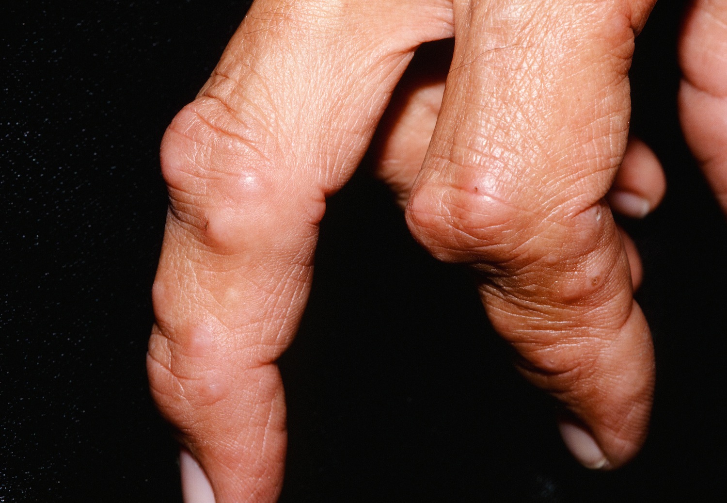
Subcutaneous granuloma annulare
Subcutaneous granuloma annulare most often occurs in children and presents as rubbery lumps. It produces firm, usually painless, lumps under the skin instead of a rash. The lumps are usually less than 1.4 inches (3.5 centimeters) in diameter and appear on scalp margins, fingertips and shins. Subcutaneous granuloma annulare is also called pseudo-rheumatoid nodules because the subcutaneous lesions look rather like rheumatoid nodules. However, they arise in people that do not suffer from rheumatoid arthritis.
Figure 5. Subcutaneous granuloma annulare
Perforating granuloma annulare
Perforating granuloma annulare describes plaques in which damaged collagen is eliminated through the epidermis. Perforating granuloma annulare is usually localized to the hands but plaques may occasionally arise on any body site, especially within scars. Dermatoscopy helps to confirm the presence of perforations in small papules arising within otherwise typical plaques of granuloma annulare. Perforating lesions are frequently itchy or tender.
Figure 6. Perforating granuloma annulare
Atypical granuloma annulare
Atypical granuloma annulare describes:
- Granuloma annulare in unusual sites, such as face, palms and ears
- Granuloma annulare with photosensitive distribution
- Unusually severe or symptomatic granuloma annulare
Interstitial granulomatous dermatitis
Interstitial granulomatous dermatitis is a pathological finding noted in some patients with extensive granuloma annulare or other disorders with similar clinical presentation.
Granuloma annulare prognosis
Localized granuloma annulare tends to clear up within a few months or years, although it may recur. Disseminated (generalized) and atypical variants are more persistent, sometimes lasting decades.
Granuloma annulare causes
Granuloma annulare is a delayed hypersensitivity reaction to some component of the dermis. Inflammation is mediated by tumor necrosis factor alpha (TNFα). The reason that this occurs is unknown.
Localized granuloma annulare is sometimes associated with autoimmune thyroiditis but it does not clear up with thyroid replacement. Extensive granuloma annulare is sometimes associated with diabetes mellitus, hyperlipidaemia, and rarely with lymphoma, HIV infection and solid tumors.
In some people, granuloma annulare may be triggered by:
- Animal or insect bites
- Infections, including hepatitis
- Tuberculin skin tests
- Vaccinations
- Sun exposure
- Other minor injury to the skin
Granuloma annulare is not contagious.
Risk factors for granuloma annulare
Granuloma annulare is occasionally associated with diabetes or thyroid disease, most often when lesions are numerous or widespread.
Granuloma annulare symptoms
In most cases granuloma annulare causes no symptoms, or it may be associated with a mild itch. If knocked the skin can be tender.
Granuloma annulare occurs most frequently over the joints or in areas that have had mild injury. The most common locations for granuloma annulare include:
- Backs of the hands and tops of the fingers
- Tops of the feet
- Around the elbows
- Around the knees
The lesions of granuloma annulare are usually found in the same areas on both sides of the body (symmetrically).
Granuloma annulare appears as small (1–3 mm), skin-colored or pink bumps. These bumps, which are smooth rather than scaly, may occur singly or in groups. Each bump may expand in size, leaving a shallow dent in the center, which may be lighter or darker than the normal skin. Granuloma annulare may also appear as several small bumps that merge to form a ring, 1–5 cm in diameter. Healed lesions of granuloma annulare do not leave scars.
Rarely, granuloma annulare may be widespread, called disseminated (generalized) granuloma annulare. Disseminated granuloma annulare tends to appear in adults over 30 years old. The condition may consist of hundreds to thousands of 1–2 mm bumps that appear on the arms, legs, and upper trunk. These skin-colored or pink bumps may be quite itchy.
Granuloma annulare diagnosis
Most often granuloma annulare is recognized clinically because of its characteristic appearance. But sometimes the diagnosis is not obvious, and other conditions may be considered. Skin biopsy usually shows necrobiotic degeneration of dermal collagen surrounded by an inflammatory reaction. It is not a true granuloma.
If the diagnosis of granuloma annulare is not obvious, the doctor may want to perform a skin biopsy. The procedure involves:
- Numbing the skin with an injectable anesthetic.
- Sampling a small piece of skin by using a flexible razor blade, a scalpel, or a tiny cookie cutter (called a “punch biopsy”). If a punch biopsy is taken, a stitch (suture) or two may be placed and will need to be removed 6–14 days later.
- Having the skin sample examined under the microscope by a specially trained physician (dermatopathologist).
Once the diagnosis of granuloma annulare is confirmed, you and your physician may decide to not treat it. Up to 70% of cases of granuloma annulare go away by themselves (spontaneous resolution) within 2 years, even without treatment. However, up to 40% of people may experience a return of the lesions (recurrence), usually at the same site(s) of the original rash.
Granuloma annulare treatment
In most cases of granuloma annulare, no treatment is required because the patches disappear by themselves in a few months, leaving no trace. However, sometimes they persist for years. Treatment is not always successful.
Local therapy
Options to consider include:
- Topical corticosteroid ointment under occlusion
- Intralesional steroid injections
- Destruction by cryotherapy or laser ablation
- Imiquimod cream
- Topical calcineurin inhibitors (tacrolimus and pimecrolimus)
Systemic therapy
Systemic therapy may be considered in widespread granuloma annulare. The following treatments have been reported to help at least some cases of disseminated granuloma annulare. None of these can be relied upon to clear it, and there are some potential adverse effects.
- Systemic steroids
- Isotretinoin
- Methotrexate
- Potassium iodide
- Dapsone
- Hydroxychloroquine
- Pentoxifylline
- Allopurinol (note: allopurinol has also be cited as cause of disseminated granuloma annulare)
- Combination of antibiotics once monthly: rifampicin, oflaxacin, minocycline
- Photochemotherapy (PUVA)
- UVA1 phototherapy
- Photodynamic therapy
- Ciclosporin
- Biologics particularly TNFα inhibitors such as adalimumab and infliximab
If few in number, triamcinolone 5 mg/cc may be injected into the border. Usually one injection is sufficient for any single lesion. If many lesions are present, and IL steroid is impractical or not desired, a potent topical steroid may be tried.
Any sort of trauma may improve granuloma annulare. All of the following have been reported to improve granuloma annulare:
- Intralesional saline,
- Biopsy,
- Incision of lesions,
- Scarification (i.e., the use of a 19-gauge needle drawn across the lesion until capillary bleeding occurs),
- Repetitive pricking and cryotherapy.
Tumor necrosis factor (TNF)-α inhibitors have been reported to improve granuloma annulare. Adalimumab seems to be the most promising with anecdotal reports of rapid clinical response, generally within 2 to 6 weeks, with sustained remission after therapy discontinuation 1. Caution must be exercised here as biologic agents have been reported to trigger granuloma annulare.
Other treatments that have been employed for disseminated granuloma annulare include hydroxychloroquine, methotrexate 2, low dose dapsone, isotretinoin 0.5 mg/kg/day, SSKI, chlorambucil and CSA. In one review, all 6 patients treated with chloroquine improved, 5 of whom had failed previous treatment with hydroxychloroquine 3. Narrow band UVB, and PUVA (both standard and bath0 have been reported useful. Phototherapy (5/week) with UVA-1 (340-400 nm) alone has been shown effective. Caution must be exercised here however as some cases of granuloma annulare may be photo induced.
Six patients (5 with disseminated granuloma annulare) were treated with once monthly single doses of rifampicin 600 mg, ofloxacin 400 mg, and minocycline hydrochloride 100 mg until the skin cleared. All patients cleared after 4-8 pulses 4. If ofloxacin is not available, ciprofloxacin 500 mg has been substituted.
Treatment in Children
Low dose chloroquine 1.5-3.0 mg/kg/day or hydroxychloroquine 1-6 mg/kg/day cleared 6 children in one study 5.
Self care
There is not a great deal you can do to influence the course of granuloma annulare. There is no current evidence on whether diet is an influence. Skin camouflage products may help to conceal the affected patches.
If the lesions of granuloma annulare are itchy, an over-the-counter cortisone cream may be helpful.


