Contents
- What causes armpit rash
- Infections causing armpit rash (intertrigo)
- Thrush: Candida albicans
- Erythrasma: Corynebacterium minutissimum
- Tinea: Trichophyton rubrum + Tinea interdigitale
- Impetigo: Staphylococcus aureus and Streptococcus pyogenes
- Boils: Staphylococcus aureus
- Folliculitis: Staphylococcus aureus
- Common inflammatory skin conditions causing intertrigo
- Infections causing armpit rash (intertrigo)
What causes armpit rash
Armpit rash is also called intertrigo, it’s a rash in the flexures or body folds, such as behind the ears, in the folds of the neck, under the arms (armpits), under a protruding abdomen, in the groin, between the buttocks, in the finger webs or toe spaces. Although intertrigo may affect one skin fold, it is common for it to involve multiple sites.
However, it is difficult to say exactly what is causing the armpit rash and itching you are experiencing without seeing the rash in person. However, there are a few possibilities of what commonly cause an itchy rash in an individual’s armpits.
Intertrigo is due to genetic and environmental factors.
- Flexural skin has relatively high surface temperature
- Moisture from insensible water loss and sweating cannot evaporate due to occlusion.
- Friction from movement of adjacent skin results in chafing.
The microorganisms that are normally resident on flexural skin, the microbiome, include corynebacteria, other bacteria and yeasts. These multiply in warm moist environments and may cause disease.
Doctors can classify intertrigo into infectious and inflammatory origin but there is often overlap.
- Infections tend to be unilateral and asymmetrical.
- Inflammatory disorders tend to be symmetrical affecting armpits, groins, under the breasts and the abdominal folds, except atopic dermatitis, which more often arises on the neck, and in elbow and knee creases.
Infections causing armpit rash (intertrigo)
Thrush: Candida albicans
Candida infection (candidiasis) can cause or aggravate intertrigo, an inflammatory rash arising in body folds.
- Rapid development
- Itchy moist peeling red and white skin
- Small superficial papules and pustules
What is candida ?
Candida is the name for a group of yeasts (a type of fungus) that commonly infect the skin. The name ‘candida’ refers to the white colour of the organisms in culture. Candidal infection is known as ‘candidiasis’, ‘candidosis’ or ‘moniliasis’ (monilia is also a genus of ascomycete fungi).
The most common Candida (C) species to result in candidiasis is Candida albicans. Other species are:
- Candida tropicalis
- Candida parapsilosis
- Candida glabrata
- Candida guilliermondii
Who gets candida ?
Candida depends on a living host for survival. It is a normal inhabitant of the human digestive tract from early infancy, where it lives without causing any disease most of the time. However, if the host’s defences are lowered, the organism can cause infection of the mucosa (the lining of the mouth, anus and genitals), the skin, and rarely, deep-seated infection.
Figure 1. Candida (thrush) armpit rash (Candidal intertrigo)
Predisposing factors for candida infection
- Infancy or old age
- Warm climate
- Occlusion eg, plastic pants (babies), nylon pantyhose (women), dental plates
- Broad spectrum antibiotic treatment
- High-oestrogen contraceptive pill or pregnancy
- Diabetes mellitus, Cushing syndrome and other endocrine conditions
- Iron deficiency
- General debility eg, from cancer or malnutrition
- Underlying skin disease eg, psoriasis, lichen planus
- Immunodeficiency eg, low levels of immunoglobulins, infection with human immunodeficiency virus (HIV)
- Chemotherapy or immunosuppressive medications including systemic steroids.
Treatment of candida infection
- Keep the affected skin clean and dry. Wash daily. Take care to dry between the toes and in the skin folds; use a hair dryer if necessary. Use your own towel.
- Correct predisposing factors where possible. For example, treat diabetes, lose weight, etc.
Topical antifungal medications
Topical antifungal medications are creams, solutions, lotions, powders, gels, sprays and lacquers applied to the skin surface to treat fungal infections. They can often cure localised infections, although recurrence is common. Many suitable creams can be obtained over the counter without a doctor’s prescription.
Many antifungal medications are suitable for both dermatophyte and yeast infections. Others are more specific to one or the other type of fungus. The medications are listed below, with their trade names in parentheses.
Those unsuitable for dermatophyte fungal infections are marked with an asterix (*).
Preparations for general fungal skin infections
Topical antifungal creams can be used to treat:
- Dermatophyte infections such as tinea corporis, tinea cruris, tinea faciei, tinea manuum, tinea pedis.
- As an adjunct to oral therapy for tinea capitis and tinea barbae.
- Yeast infections such as candida intertrigo, pityriasis versicolor.
- Mould skin infections such as tinea nigra and nail plate infections.
The creams are applied to the affected area twice daily for two to four weeks, including a margin of several centimetres of normal skin. Continue for one or two weeks after the last visible rash has cleared. Repeated treatment is often necessary.
- Whitfield’s ointment (benzoic acid)
- Undecylenic alkanolamide
- Ciclopirox olamine (Batrafen® cream, powder, solution)
- Polyenes * – Nystatin (Nilstat® cream, ointment; Mycostatin® cream, ointment, paste)
- Imidazoles
- Bifonazole (Canesten® Once Daily Bifonazole Cream)
- Clotrimazole (Canesten® cream, solution; Clocreme® cream; Clomazol®, Fungizid® spray)
- Econazole (Ecreme® cream; Pevaryl® cream, powder, foaming solution)
- Ketoconazole (Nizoral® cream, Daktagold® cream)
- Miconazole (Daktarin® cream, lotion, dusting powder, spray powder; Micreme® cream, Resolve® solution, tinea cream, jock itch cream, thrush cream, powder; Tinasolve® Alpha cream)
- Tioconazole
- Allylamine (higher cure rates and more rapid responses than older topical antifungals for dermatophyte infections)
- Terbinafine (Lamisil® cream, gel, spray)
- Thiocarbamates
- Tolciclate
- Tolnaftate
In other countries, additional antifungal agents include the azoles, bifonazole, tioconzaole, sulconazole, efinaconazole and luliconazole; naftifine; and a benzoxaborole, tavaborole.
Combination products
Topical antifungals may be sold with an oral antifungal, e.g. Canesten® combination pack (fluconazole capsule and clotrimazole cream duo).
Antifungal creams are sometimes combined with:
- Hydrocortisone or other topical steroid (e.g. Resolve® Plus cream)
- Antibacterial agents
- Both topical steroid and antibacterial agent
Oral antifungal medications may be required for a fungal infection if:
- It is extensive or severe.
- It resists topical antifungal therapy.
- It affects hair-bearing areas (tinea capitis and tinea barbae).
Oral antifungal medications
Oral antifungal medications may be required for a fungal infection if:
- It is extensive or severe.
- It resists topical antifungal therapy.
- It affects hair-bearing areas (tinea capitis and tinea barbae).
The choice of oral antifungal medication, its dose and the duration of treatment depends on:
- The type of fungus i.e. candida, dermatophyte (tinea), malassezia, or mould.
- The site affected i.e. skin, mucosa, nails.
- Other co-existing diseases.
- Interactions with other medications.
Medications for both candida and dermatophyte infections (azoles)
The trade names of the medications are given in parentheses.
- Itraconazole (Sporanox® capsules).
- Ketoconazole (Nizoral® tablets).
- Fluconazole (Diflucan® capsules).
Voriconazole IVfend® tablets) and posaconazole are reserved for serious invasive Candida and mould infections.
Some species of candida are resistant to azoles, and azole resistance is increasing especially in immunosuppressed patients who are prescribed long courses.
Medications only suitable for candida infections
- Nystatin (Mycostatin®, Nilstat®), a polyene. This is not absorbed into the blood stream through the gut.
- Flucytosine (Alcobon® infusion), usually used with amphotericin. It can cause bone marrow depression.
- Amphotericin B (Ambisome® injection; Fungizone® infusion), a polyene. This is not absorbed into the blood stream through the gut.
- Caspofungin, anidulafungin and micafungin (echinocandins)
Medications only suitable for dermatophyte infections
- Griseofulvin, derived from Penicillium
- Terbinafine (Lamisil®, Terbafin® tablets), an allylamine.
Home remedies
Fungal spores can survive long periods. To reduce the chance of reinfection:
- Do not share towels, sheets or personal clothing.
- Carefully clean the shower or bath using bleach.
- Avoid walking bare foot where others may tread – wear jandals, sandals or aquasocks at the public pools and sports changing rooms.
- Avoid long periods wearing the same clothing, or wearing occlusive clothing such as wet weather gear and nylon pantyhose.
- Wear open-toed sandals when possible. Avoid long periods in occlusive footwear such as gum boots or tramping boots.
- Use antifungal foot powder e.g. ciclopirox (Batrafen®), econazole (Pevaryl®), miconazole (Daktarin®, Fungo®), tolciclate (Tolmicen®), tolnaftate (Tinaderm®), undecylenic acid (Mycota®). Sprinkle it in your shoes.
- In the case of zoophilic fungal infections, infected animals should be identified and treated.
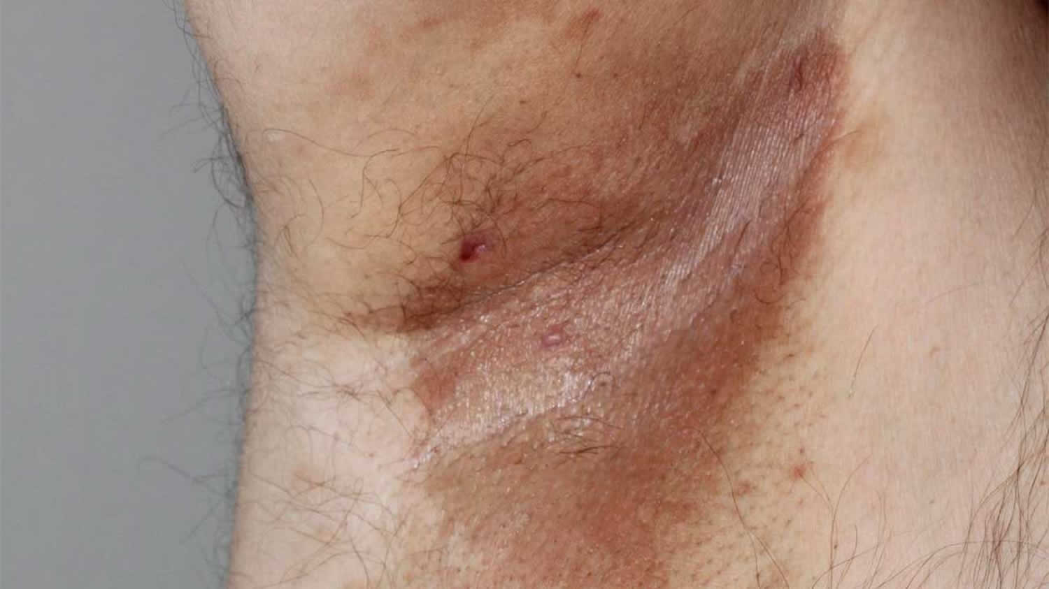
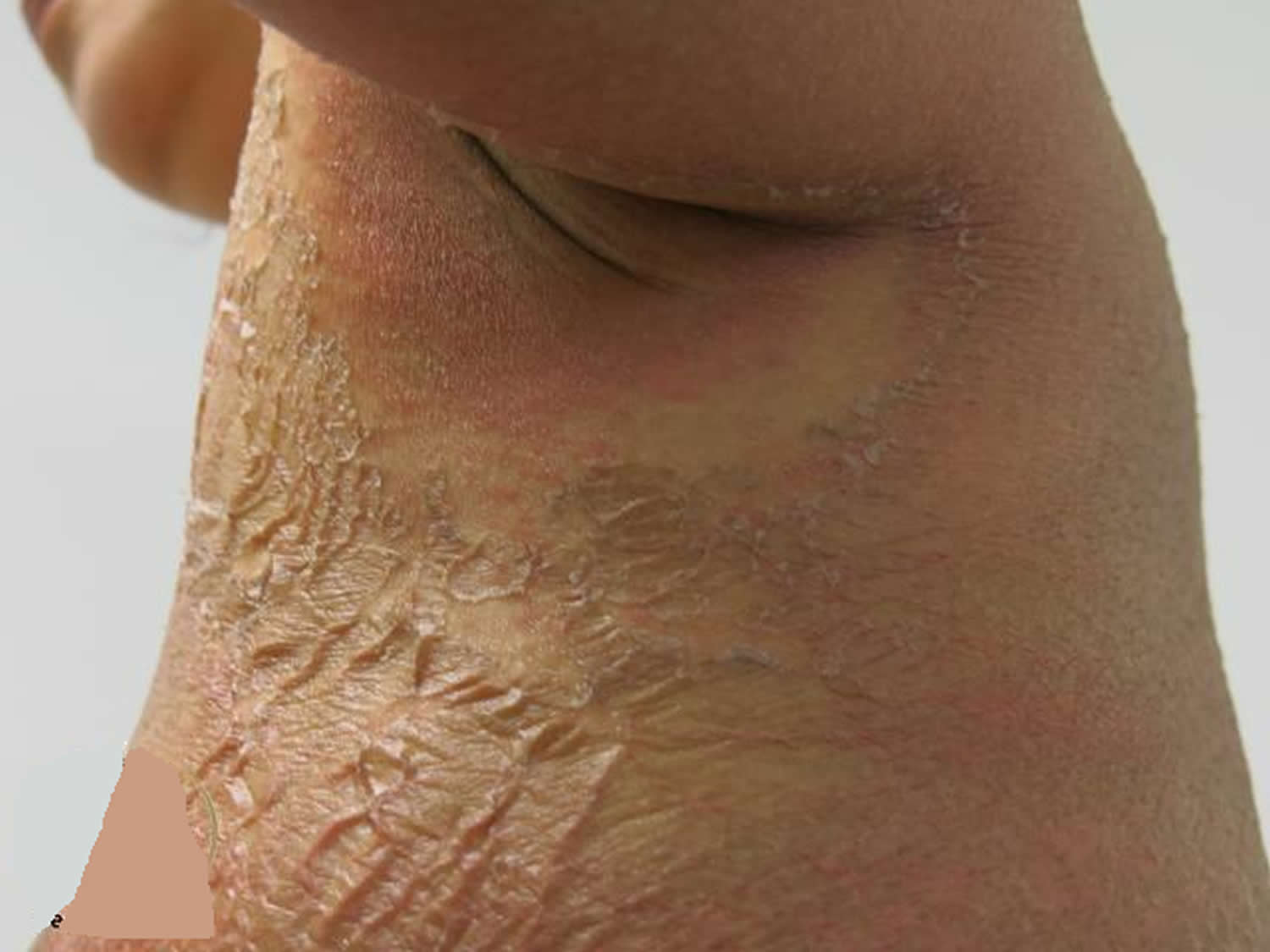
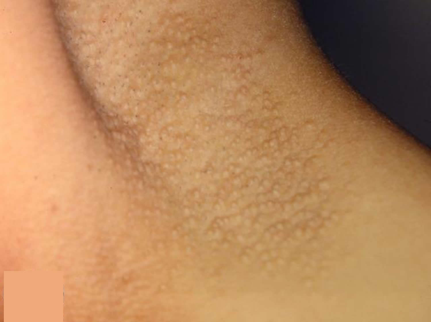
Erythrasma: Corynebacterium minutissimum
Erythrasma is a common skin condition affecting the skin folds such as the armpits, in the groin and between the toes. It causes dark inner armpits.
The bacteria responsible for erythrasma are gram-positive, non-spore-forming, aerobic or facultative bacilli called Corynebacterium minutissimum.
- Erythrasma may coexist with or be confused with other causes of intertrigo (rashes in the skin folds) including fungal infections such as tinea or Candida albicans (thrush).
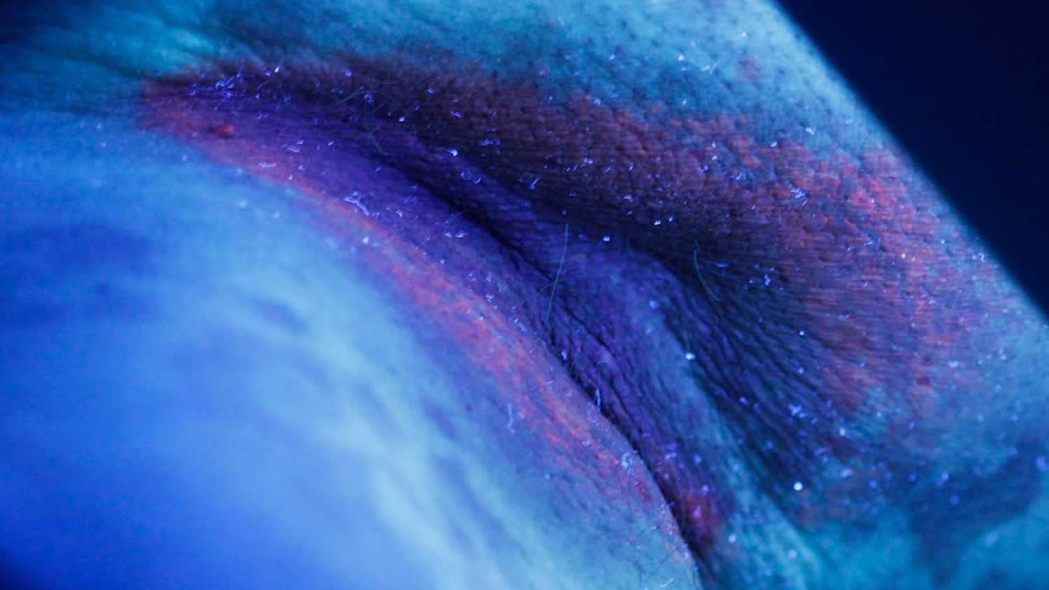
Although erythrasma may be confused with a fungal infection, doctors can easily diagnose erythrasma because skin infected with Corynebacterium glows coral-red under an ultraviolet light (Wood’s lamp).
- Persistent brown patches
- Minimal scale
- Asymptomatic
Who gets erythrasma ?
Erythrasma affects males and females, but it is thought to be more common in the groin of males and between the toes of females.
It is reported to be more prevalent in the following circumstances:
- Warm climate
- Excessive sweating
- Skin of color
- Diabetes
- Obesity
- Poor hygiene
- Advanced age
- Other immunocompromised states
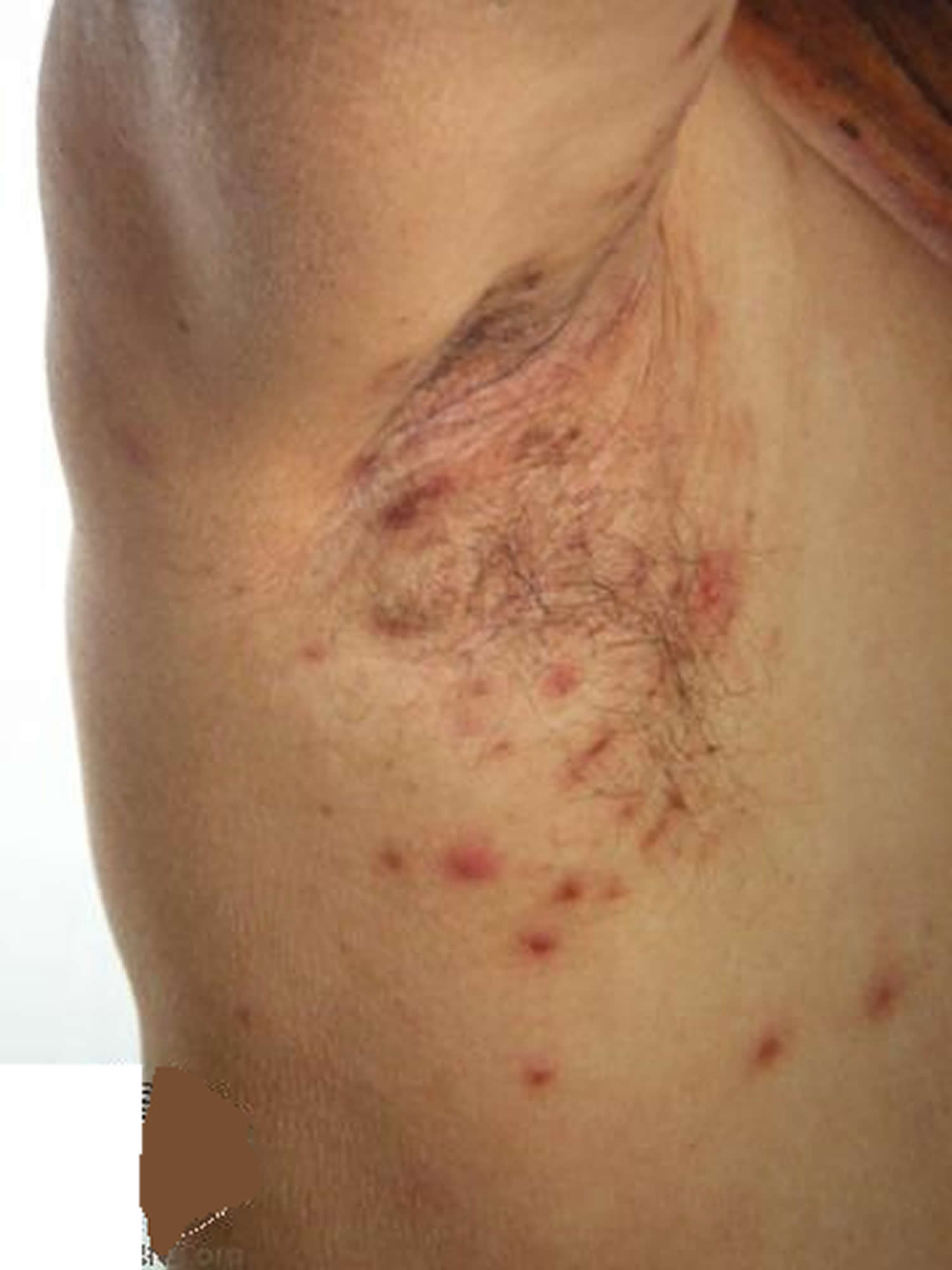
What are the clinical features of erythrasma ?
Erythrasma presents as well-defined pink or brown patches with fine scaling and superficial fissures. Mild itching may be present.
Common sites for erythrasma are armpits, groin and between the toes. Intergluteal fold, submammary, and periumbilical skin may also be affected. Widespread infections are associated with diabetes mellitus.
Erythrasma is classified into 3 types according to location.
- Interdigital erythrasma: between 3rd, 4th and 5th toe web spaces
- Intertriginous erythrasma: in armpits, groin, under the breasts and umbilicus
- Generalised/disciform erythrasma: on the trunk
Figure 2. Erythrasma armpit
How is the diagnosis of erythrasma made ?
Erythrasma has a typical clinical appearance. Diagnosis may be supported by the following investigations.
- Wood’s lamp skin examination: long wavelength ultraviolet radiation causes erythrasma to fluoresce a coral-pink colour due to coproporphyrin III released by the bacteria.
- Swab or skin scrapings: microscopy may reveal gram-positive filamentous rods. Methylene blue also stains C minutissimum.
Figure 3. Armpit erythrasma under Wood’s lamp
What are the complications of erythrasma ?
Erythrasma is usually self limiting. It can be complicated by contact dermatitis, lichenification, postinflammatory pigmentation, and coinfection with other bacteria, yeasts, and dermatophytes.
Serious complications are very rare. Corynebacteria have been reported to causes abscess, cellulitis, cutaneous granuloma, endocarditis, pyelonephritis, endophthalmitis, arteriovenous fistula infection and meningitis.
Treatments for Erythrasma
Once the diagnosis of erythrasma is established, the doctor may try one of the following treatments:
- Topical antibiotic lotions such as erythromycin or clindamycin
- Whitfield’s ointment (a mixture of benzoic acid and salicylic acid)
- Aluminum chloride solution to inhibit sweating and moisture
- Oral antibiotics such as erythromycin or clarithromycin.
Treatment is a single dose of clarithromycin 1 gram orally. One to two treatments (80 J/cm2) of broadband red light (635 nm) have been successful in a small case series. Topical erythromycin or clindamycin is also effective. Recurrence is common 1.
Home remedies for prevention of armpit erythrasma
Recurrence of erythrasma is common. Antibacterial soap can be used to prevent recurrence. Treatment can be repeated if necessary.
If you are overweight, it may help to lose weight and often change your body position.
Other things you can do are:
- Separate skin folds with dry towels.
- Blow a fan on moist areas.
- Wear loose clothing and moisture wicking fabrics.
- Keep skin cool and dry.
- Do not wear tight shoes or clothing. Wear a bra that has good support.
- Wear clothes made with absorbent fabrics, such as cotton. Avoid nylon or other synthetic (manmade) fibers.
- After exercising, shower and dry off completely. Use a hair dryer with a cool setting to dry areas that can trap wetness, such as under your arms or breasts.
- Gently cleanse the affected areas daily with mild soap substitutes.
Antibacterial soap
Antibacterial soaps for household use generally contain the active ingredient triclosan at concentrations between 0.1% and 0.45% weight/volume. Triclosan has varying effectiveness across bacterial and fungal species, and is less effective against viruses. A related compound, triclocarban, is used in antibacterial bar soaps.
Laboratory-based studies have shown triclosan at high concentration (1.0% weight/volume or higher) can reduce bacterial counts on the hands compared with plain soap.
However, community-based studies have shown that triclosan, at usual household concentrations of 0.1%-0.45% weight/volume, is generally no more effective than plain soap in reducing bacterial levels on the hands or in reducing infectious illnesses. Interestingly, even in studies conducted in areas with high rates of infectious illnesses, such as squatter settlements in Pakistan, antibacterial soaps offered little benefit compared with plain soap.
As most community infections are viral, the lack of a clear benefit for antibacterial household soap is not unexpected. However, even when examining the incidence of bacterial infections, such as impetigo, triclosan provided no benefit over plain soap.
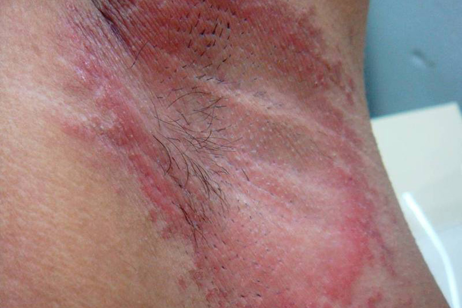
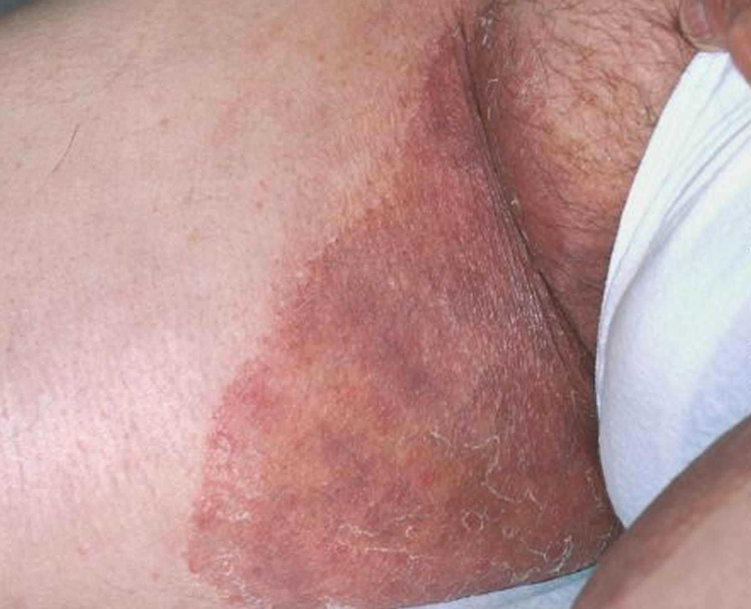
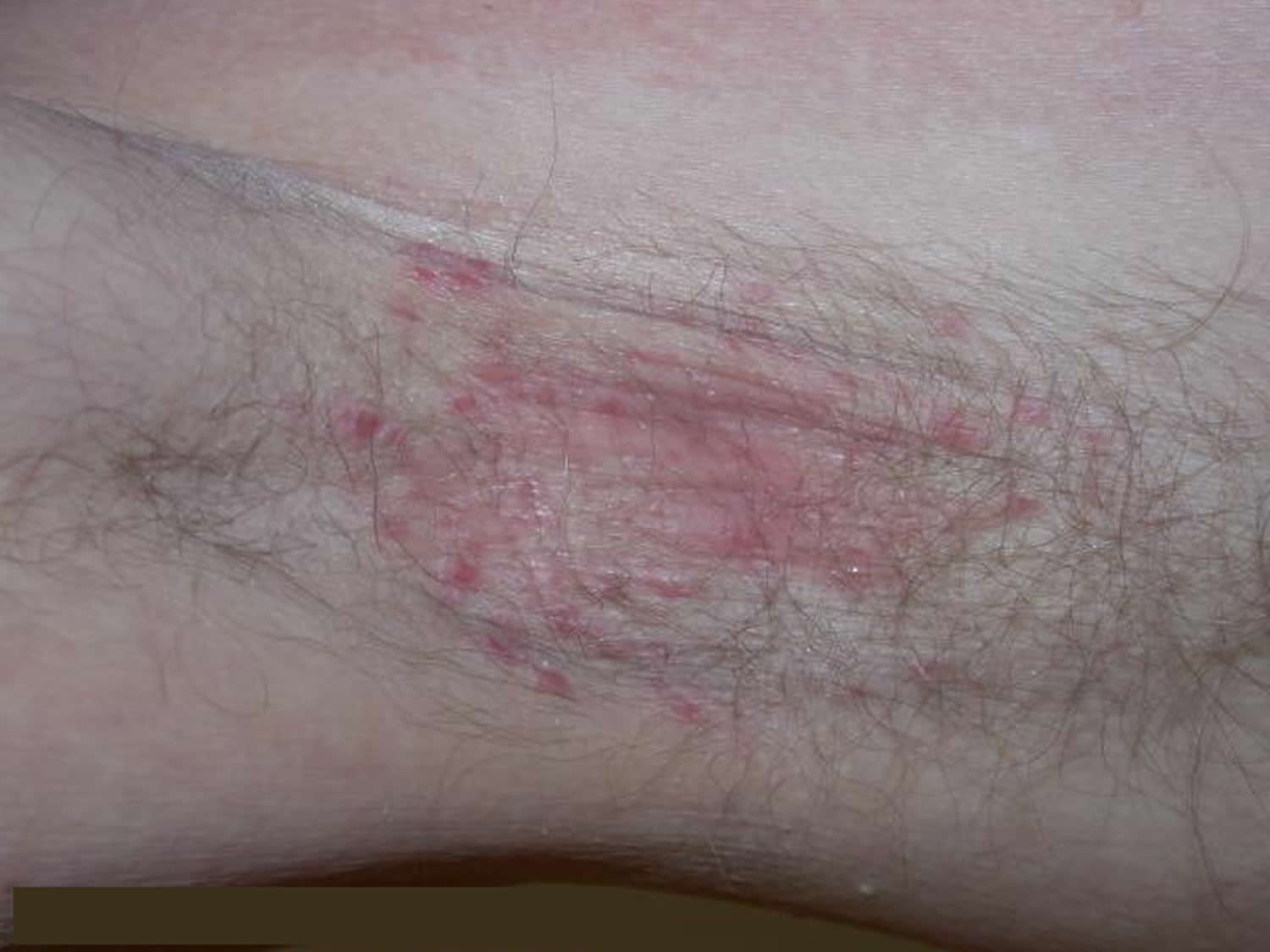
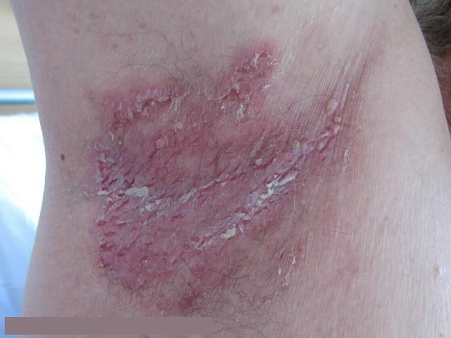
Tinea: Trichophyton rubrum + Tinea interdigitale
“Tinea” refers to a skin infection with a dermatophyte (ringworm) fungus. Types of tinea include ringworm, athlete’s foot and jock itch. These infections are usually not serious, but they can be uncomfortable. You can get them by touching an infected person, from damp surfaces such as shower floors, or even from a pet.
- Tinea cruris (groin) and athletes foot (between toes)
- Slowly spreads over weeks to months
- Irregular annular plaques
- Peeling, scaling
The appearance is similar to ringworm (tinea corporis). The rash has a scaly raised red border that spreads down the inner thighs from the groin or scrotum. Tinea cruris may form ring-like patterns on the buttocks. It is not often seen on the penis or vulva or around the anus. Tinea cruris can be very itchy.
In different parts of the world, different species cause tinea cruris. For example, Trichophyton rubrum and Epidermophyton floccosum are the most common causes. Infection often comes from the feet (tinea pedis) or nails (tinea unguium) originally, spread by scratching or the use of an infected towel.
Figure 4. Tinea cruris
Tinea cruris can be confused with other forms of intertrigo such as:
- Candida
- Erythrasma
- Psoriasis
- Seborrhoeic dermatitis
Tinea cruris quite often recurs after apparently successful treatment. To reduce the chance of reinfection:
- Treat the feet if tinea pedis is present.
- Dry the groin carefully after bathing using a separate towel.
- Do not share towels, sheets or personal clothing.
- Avoid wearing occlusive or synthetic clothing.
- If you are overweight, try to lose weight to reduce chafing and sweating.
Diagnosis of tinea cruris
The diagnosis of tinea cruris is confirmed by microscopy and culture of skin scrapings.
Treatment of tinea cruris
Tinea cruris is usually treated with topical antifungal agents (see above under treatment for candida infection). Sometimes hydrocortisone is added, for faster relief of itch. Topical steroids should not be used on their own. If the treatment is unsuccessful, oral antifungal medicines may be considered, including terbinafine and itraconazole.
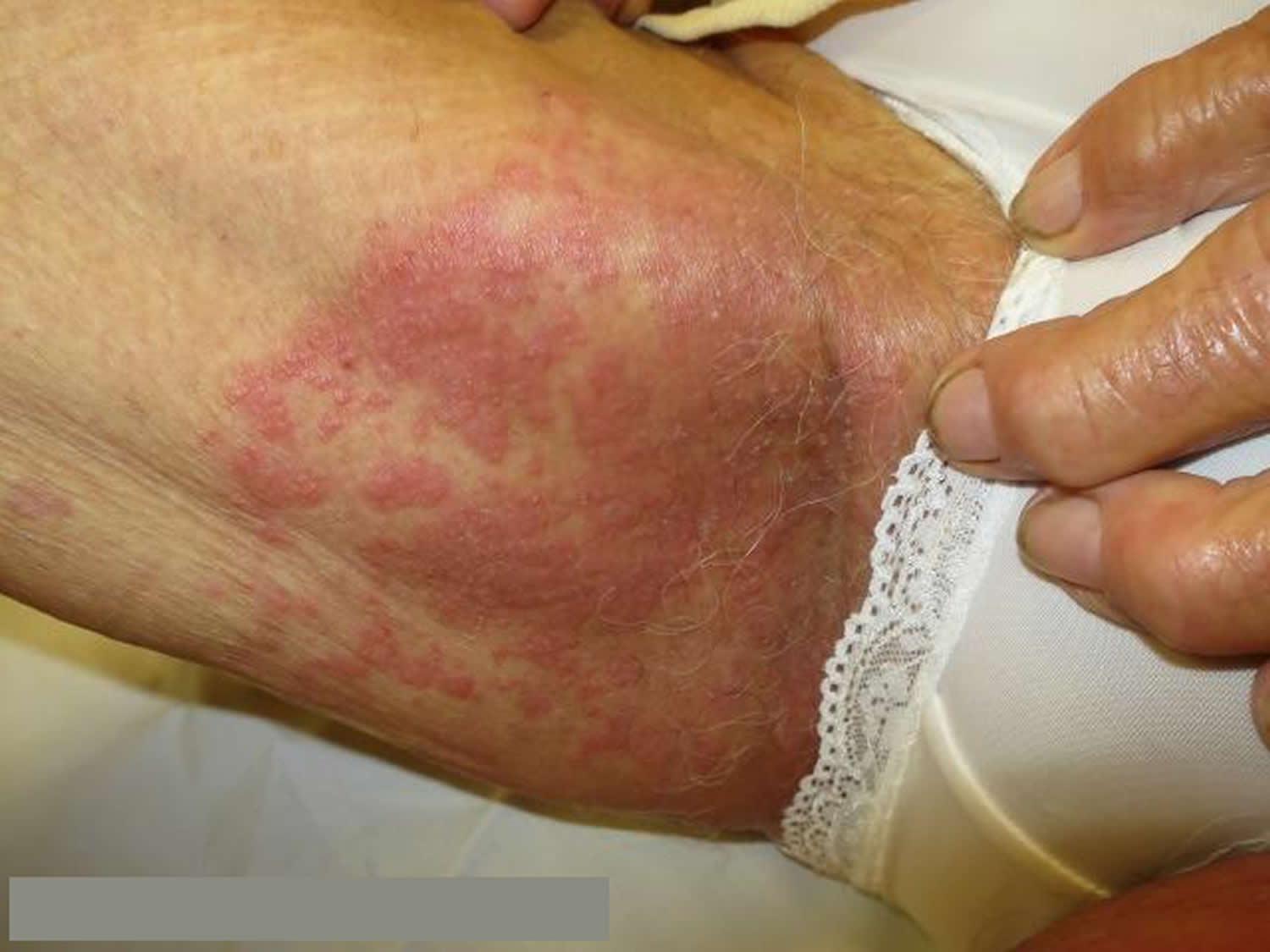
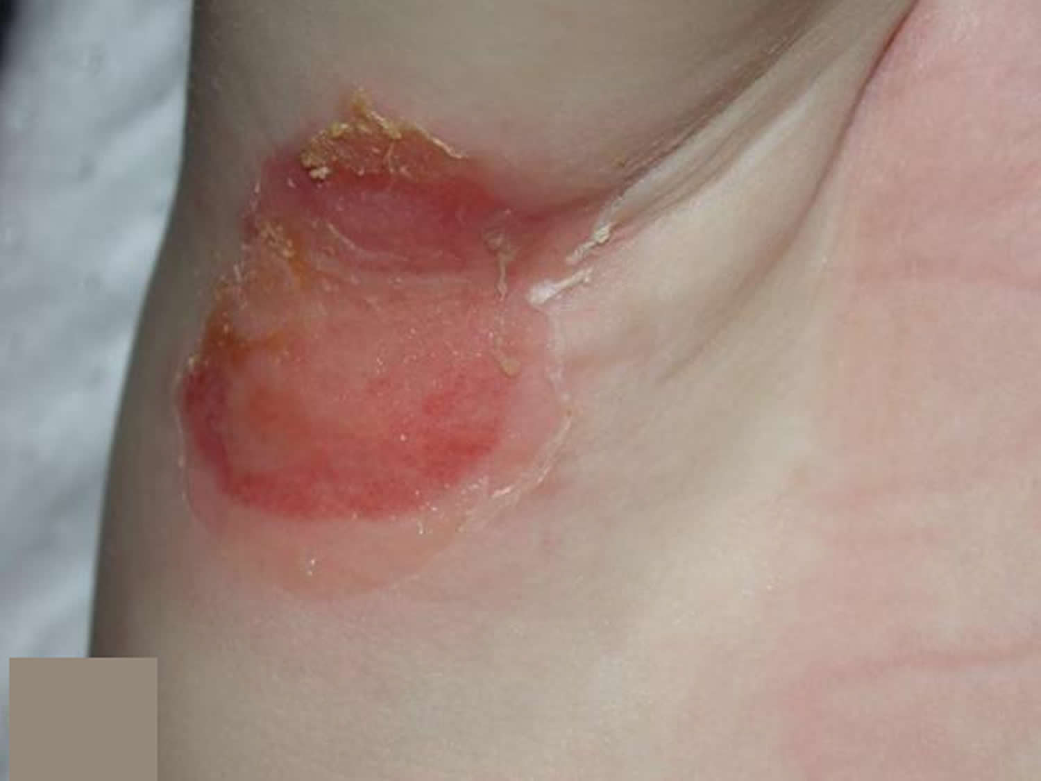
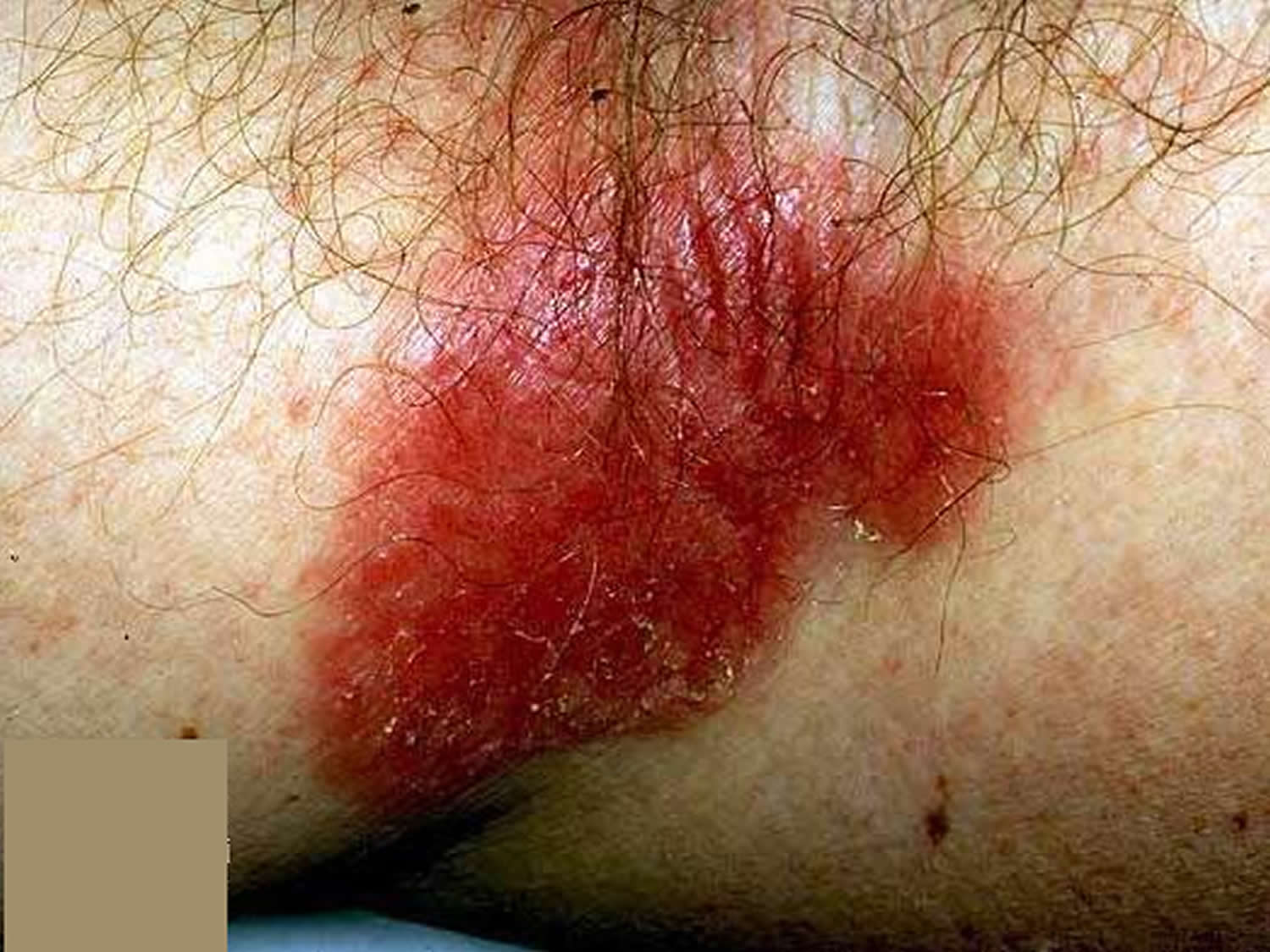
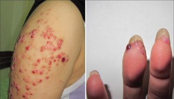
Impetigo: Staphylococcus aureus and Streptococcus pyogenes
Impetigo is a common acute superficial bacterial skin infection (pyoderma). It is characterised by pustules and honey-coloured crusted erosions (“school sores”).
Impetigo is most often caused by Staphylococcus aureus. Non-bullous impetigo can also be caused by group A beta-haemolytic streptococcus (Streptococcus pyogenes).
- Rapid development
- Moist blisters and crusts on red base
- Contagious, so other family members may also be affected
Figure 5. Armpit Impetigo
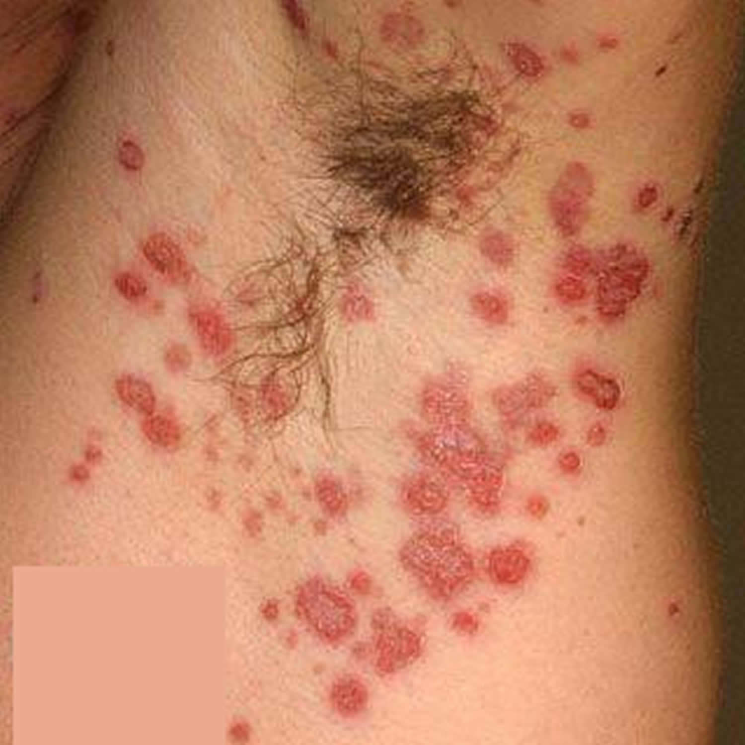
Figure 6. Armpit Impetigo
Types of impetigo
- Nonbullous impetigo: In nonbullous impetigo, staphylococci and/or streptococci invade a site of minor trauma where exposed proteins allow the bacteria to adhere.
- Bullous impetigo: Bullous impetigo is due to staphylococcal exfoliative toxins (exfoliatin A–D), which target desmoglein 1 (a desmosomal adhesion glycoprotein), and cleave off the superficial epidermis through the granular layer. No trauma is required, as the bacteria can infect intact skin.
- Ecthyma: Ecthyma is usually due to Streptococcus pyogenes, but co-infection with Staphylococcus aureus may occur.
Who gets impetigo ?
Impetigo is most common in children (especially boys), but may also affect adults if they have low immunity to the bacteria. It is prevalent worldwide. Peak onset is during summer and it is more prevalent in developing countries.
The following factors predispose to impetigo.
- Atopic eczema
- Scabies
- Skin trauma: chickenpox, insect bite, abrasion, laceration, thermal burn, dermatitis, surgical wound.
What are the clinical features of impetigo
Primary impetigo mainly affects exposed areas such as the face and hands, but may also affect trunk, perineum and other body sites. It presents with single or multiple, irregular crops of irritable superficial plaques. These extend as they heal, forming annular or arcuate lesions.
Although many children are otherwise well, lymphadenopathy, mild fever and malaise may occur.
Nonbullous impetigo
Nonbullous impetigo starts as a pink macule that evolves into a vesicle or pustule and then into crusted erosions. Untreated impetigo usually resolves within 2 to 4 weeks without scarring.
Ecthyma
Ecthyma starts as nonbullous impetigo but develops into a punched-out necrotic ulcer. This heals slowly, leaving a scar.
Bullous impetigo
Bullous impetigo presents with small vesicles that evolve into flaccid transparent bullae. It heals without scarring.
Complications from impetigo
Impetigo may lead to:
- Spread of the infection to other parts of the body (common)
- Kidney inflammation or failure (rare)
- Cellulitis. This potentially serious infection affects the tissues underlying your skin and eventually may spread to your lymph nodes and bloodstream. Untreated cellulitis can quickly become life-threatening.
- Permanent skin damage and scarring (very rare)
Soft tissue infection
The bacteria causing impetigo can become invasive, leading to cellulitis and lymphangiitis; subsequent bacteraemia might result in osteomyelitis, septic arthritis or pneumonia.
Staphylococcal scalded skin syndrome
In infants under 6 years of age or adults with renal insufficiency, localised bullous impetigo due to certain staphylococcal serotypes can lead to a sick child with generalised staphylococcal scalded skin syndrome (SSSS). Superficial crusting then tender cutaneous denudation on face, in flexures, and elsewhere is due to circulating exfoliatin/epidermolysin, rather than direct skin infection. It does not scar.
Toxic shock syndrome
Toxic shock syndrome is rare and rarely preceded by impetigo. It causes fever, diffuse erythematous then desquamating rash, hypotension and involvement of other organs.
Post-streptococcal glomerulonephritis
Group A streptococcal infection may rarely lead to acute post-streptococcal glomerulonephritis 3–6 weeks after the skin infection. It is associated with anti-DNase B and antistreptolysin O (ASO) antibodies.
Rheumatic fever
Group A streptococcal skin infections have rarely been linked to cases of rheumatic fever and rheumatic heart disease. It is thought that this occurs because strains of group A streptococci usually found on the skin have moved to the throat (the more usual site for rheumatic fever-associated infection).
How is impetigo diagnosed ?
Impetigo is usually diagnosed clinically but can be confirmed by bacterial swabs sent for microscopy (gram positive cocci are observed), culture and sensitivity.
Blood count may reveal neutrophil leucocytosis when impetigo is widespread.
Skin biopsy is rarely necessary. Histological features are characteristic.
Non-bullous impetigo
- Gram-positive cocci
- Intraepidermal neutrophilic pustules,
- Dense inflammatory infiltrate in upper dermis
Bullous impetigo
- Split through granular layer of epidermis without inflammation or bacteria
- Acantholytic cells
- Minimal inflammatory infiltrate in upper dermis
- Resembles pemphigus foliaceus
Ecthyma
- Full thickness skin ulceration
- Gram stain shows cocci within dermis
What is the treatment for impetigo ?
The following steps are used to treat impetigo.
- Cleanse the wound; use moist soaks to gently remove crusts
- Apply antiseptic (povidone iodine, hydrogen peroxide cream, chlorhexidine, superoxidised solution and others) or antibiotic ointment (fusidic acid, mupirocin or retapamulin) as prescribed
- Cover the affected areas.
- If impetigo is extensive, oral antibiotics are recommended, often flucloxacillin.
To prevent recurrence:
- Treat carrier sites: apply antibiotic ointment to nostrils
- Wash daily with antibacterial soap or soak in a bleach bath
- Identify and treat the source of re-infection, ie another infected person or carrier
To reduce the chance of passing the infection to another person:
- Avoid close contact with others
- Children must stay away from school until crusts have dried out
- Use separate towels and flannels
- Change and launder clothes and linen daily
Boils: Staphylococcus aureus
Boils (also called furuncles) are a deep bacterial infection of hair follicles.
Boils present as one or more tender red spots, lumps or pustules. Careful inspection reveals that the boil is centred on a hair follicle. A boil is a deep form of bacterial folliculitis; superficial folliculitis is sometimes present at the same time. Staphylococcus aureus can be cultured from the skin lesions.
If there are multiple heads, the lesion is called a “carbuncle”. Large boils form abscesses, defined as an accumulation of pus within a cavity. Cellulitis may also occur, i.e. infection of the surrounding tissues, and this may cause fever and illness.
Figure 7. Armpit Boil
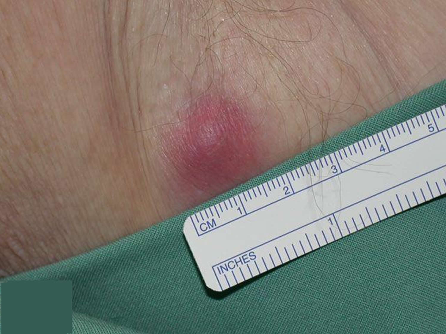
- Rapid development
- Very painful follicular papules and nodules
- Central pustule or abscess
Why do boils occur ?
Most people with boils are otherwise healthy and have good personal hygiene. They do however carry Staphylococcus aureus on the surface of their skins (staphylococcal carrier state). Why this occurs is usually not known, but it is estimated that 10–20% of the population are staphylococcal carriers.
Staphylococcus aureus is most commonly carried in the nostrils, armpits, between the legs and in the cleft between the buttocks. It may be transferred to other sites from the nostrils via the finger nails.
Tiny nicks or grazes or something rubbing against the skin can innoculate the bacteria into the wall of a hair follicle which is a weak point in the skin’s defences. Once innoculated, the bacteria cause a boil which goes on to run its usual course of about 10 days.
Although most people with boils are otherwise healthy, boils are sometimes related to immune deficiency, anemia, diabetes, smoking or iron deficiency.
What is the treatment for boils ?
Medical treatment of boils
Treatment of boils depends on their severity. Your doctor may give you specific advice and medical treatment, some are listed below:
- Antiseptic or antibacterial soap in your daily bath or shower for a week then twice weekly for several weeks. The cleanser may cause a little dryness.
- Use a hand sanitiser regularly to reduce the chance of reinfecting yourself or others with contaminated hands.
- Antiseptic or antibiotic ointment or gel to apply to the inside of the nostrils.
- Wipe the entire skin surface daily for a week with 70% isopropyl alcohol in water (this will make the skin dry).
- Apply a topical antiseptic such as povidone iodine or chlorhexidine cream to the boils and cover with a square of gauze.
- Your doctor may prescribe an oral antibiotic (usually the penicillin antibiotic flucloxacillin), sometimes for several weeks.
- Other members of the family with boils should also follow a skin cleansing regime. Your doctor may also advise the family to apply topical antibiotic to their nostrils in case they are Staphylococcus aureus carriers as well.
- If the boils fail to clear up, a swab should be taken for microbiological culture, in case of methicillin (meticillin) resistant staphylococci.
- Sometimes, special antibiotics may be prescribed on the recommendation of a specialist, including fusidic acid, clindamycin, rifampicin and cephalosporins.
General measures to prevent boils
- Consult your doctor about your general health.
- If you are overweight, try to reduce your weight; take regular exercise.
- Follow a balanced healthy diet with meat, plenty of fruit and vegetables.
- Avoid smoking.
- Wash your whole body once a day with soap or cleanser and water. Wash your hands several times daily or use antiseptic hand rubs.
- Don’t share your flannel or towel with other family members.
- Maintain a clean handkerchief and don’t pick your nose!
- Change your underclothes and night attire regularly.
- Consider modifying leisure activities that cause sweating and friction from clothing, such as squash and jogging.
- If you are iron deficient, a course of iron tablets may help reduce infection.
- 1000 mg of vitamin C each day has also been advocated to improve deficient neutrophil function.
Folliculitis: Staphylococcus aureus
Folliculitis can be due to infection, occlusion (blockage), irritation and various skin diseases.
To determine if folliculitis is due to an infection, swabs should be taken from the pustules for cytology and culture in the laboratory.
Folliculitis due to infection
Bacteria
Bacterial folliculitis is commonly due to Staphylococcus aureus. If the infection involves the deep part of the follicle, it results in a painful boil. Recommended treatment includes careful hygiene, antiseptic cleanser or cream, antibiotic ointment, and/or oral antibiotics.
Spa pool folliculitis is due to infection with Pseudomonas aeruginosa, which thrives in inadequately chlorinated warm water. Gram negative folliculitis is a pustular facial eruption also due to infection with Pseudomonas aeruginosa or other similar organisms. When it appears, it usually follows tetracycline treatment of acne, but is quite rare.
Yeasts
The most common yeast to cause a folliculitis is Pityrosporum ovale, also known as Malassezia. Malassezia folliculitis (Pityrosporum folliculitis) is an itchy acne-like condition usually affecting the upper trunk of a young adult. Treatment includes avoiding moisturisers, stopping any antibiotics and topical antifungal or oral antifungal medication for several weeks.
Candida albicans can also provoke a folliculitis in skin folds (intertrigo) or in the beard area. It is treated with topical or oral antifungal agents.
Fungi
Ringworm of the scalp (tinea capitis) usually results in scaling and hair loss, but sometimes results in folliculitis. Cat ringworm (Microsporum canis) is the commonest organism causing scalp fungal infection. Other fungi such as Trichophyton tonsurans are increasingly reported. Treatment is with oral antifungal agents for several months.
Viral infections
Folliculitis may caused by herpes simplex virus. This tends to be tender, and resolves without treatment in around 10 days. Severe recurrent attacks may be treated with aciclovir and other antiviral agents.
Herpes zoster (the cause of shingles) may also present as folliculitis with painful pustules and crusted spots within a dermatome (an area of skin supplied by a single nerve). It is treated with hihg-dose aciclovir.
Molluscum contagiosum, common in young children, may also cause follicular umbilicated papules, usually clustered in and around a body fold. Molluscum may provoke dermatitis.
Parasitic infection
Folliculitis on the face or scalp of older or immunosuppressed adults may be due to colonisation by hair follicle mites (demodex). This is known as demodicosis.
The human infestation, scabies, often provokes folliculitis, as well as non-follicular papules, vesicles and pustules.
Folliculitis due to irritation from regrowing hairs
Folliculitis may arise as hairs regrow after shaving, waxing, electrolysis or plucking. Swabs taken from the pustules are sterile ie there is no growth of bacteria or other organisms. In the beard area irritant folliculitis is known as pseudofolliculitis barbae.
Irritant folliculitis is also common on the lower legs of women (shaving rash). It is frequently very itchy. Treatment is by stopping hair removal, and not beginning again for about three months after the folliculitis has settled. To prevent reoccurring irritant folliculitis, use a gentle hair removal method, such as a lady’s electric razor. Avoid soap and apply plenty of shaving gel, if using a blade shaver.
Folliculitis due to contact reactions
Occlusion
Paraffin-based ointments, moisturisers, and adhesive plasters may all result in a sterile folliculitis. If a moisturiser is needed, choose an oil-free product, as it is less likely to cause occlusion.
Chemicals
Coal tar, cutting oils and other chemicals may cause an irritant folliculitis. Avoid contact with the causative product.
Topical steroids
Overuse of topical steroids may produce a folliculitis. Perioral dermatitis is a facial folliculitis provoked by moisturisers and topical steroids. Perioral dermatitis is treated with tetracycline antibiotics for six weeks or so.
Folliculitis due to immunosuppression
Eosinophilic folliculitis is a specific type of folliculitis that may arise in some immune suppressed individuals such as those infected by human immunodeficiency virus (HIV) or those who have cancer.
Folliculitis due to drugs
Folliculitis may be due to drugs, particularly corticosteroids (steroid acne), androgens (male hormones), ACTH, lithium, isoniazid (INH), phenytoin and B-complex vitamins. Protein kinase inhibitors (epidermal growth factor receptor inhibitors) and targeted therapy for metastatic melanoma (vemurafenib, dabrafenib) nearly always result in folliculitis.
Folliculitis due to inflammatory skin diseases
Certain uncommon inflammatory skin diseases may cause permanent hair loss and scarring because of deep seated sterile folliculitis. These include:
- Lichen planus
- Discoid lupus erythematosus
- Folliculitis decalvans
- Folliculitis keloidalis
Treatment depends on the underlying condition and its severity. A skin biopsy is often necessary to establish the diagnosis.
Acne variants
Acne and acne-like or acneform disorders are also forms of folliculitis. These include:
- Acne vulgaris
- Nodulocystic acne
- Rosacea
- Scalp folliculitis
- Chloracne
The follicular occlusion syndrome refers to:
- Hidradenitis suppurativa (acne inversa)
- Acne conglobata (a severe form of nodulocystic acne)
- Dissecting cellulitis (perifolliculitis capitis abscedens et suffodiens)
- Pilonidal sinus.
Treatment of the acne variants may include topical therapy as well as long courses of tetracycline antibiotics, isotretinoin (vitamin-A derivative) and in women, antiandrogenic therapy.
Figure 8. Armpit Folliculitis
- Acute or chronic
- Superficial tender red papules
- Pustules centred on hair follicles
- May be provoked by shaving, waxing, epilating
Common inflammatory skin conditions causing intertrigo
Flexural psoriasis
- Well-defined smooth or shiny red patches
- Very persistent
- Common in submmamary and groin creases
- Symmetrical involvement
- May fissure (crack) in the crease
- Red patches on other sites are scaly
Figure 9. Armpit psoriasis
Seborrheic dermatitis
Seborrheic or seborrhoeic dermatitis is a common, chronic or relapsing form of eczema/dermatitis that mainly affects the sebaceous, gland-rich regions of the scalp, face, and trunk .
There are infantile and adult forms of seborrhoeic dermatitis. It is sometimes associated with psoriasis (sebopsoriasis). Seborrhoeic dermatitis is also known as seborrhoeic eczema.
Dandruff (also called ‘pityriasis capitis’) is an uninflamed form of seborrhoeic dermatitis. Dandruff presents as bran-like scaly patches scattered within hair-bearing areas of the scalp.
What causes seborrhoeic dermatitis ?
The cause of seborrhoeic dermatitis is not completely understood. It is associated with proliferation of various species of the skin commensal Malassezia, in its yeast (non-pathogenic) form. Its metabolites (such as the fatty acids oleic acid, malssezin, and indole-3-carbaldehyde) may cause an inflammatory reaction. Differences in skin barrier lipid content and function may account for individual presentations.
Who gets seborrhoeic dermatitis ?
Infantile seborrhoeic dermatitis affects babies under the age of 3 months and usually resolves by 6–12 months of age.
Adult seborrhoeic dermatitis tends to begin in late adolescence. Prevalence is greatest in young adults and in the elderly. It is more common in males than in females.
The following factors are sometimes associated with severe adult seborrhoeic dermatitis:
- Oily skin (seborrhoea)
- Familial tendency to seborrhoeic dermatitis or a family history of psoriasis
- Immunosuppression: organ transplant recipient, human immunodeficiency virus (HIV) infection and patients with lymphoma
- Neurological and psychiatric diseases: Parkinson disease, tardive dyskinesia, depression, epilepsy, facial nerve palsy, spinal cord injury and congenital disorders such as Down syndrome
- Treatment for psoriasis with psoralen and ultraviolet A (PUVA) therapy
- Lack of sleep, and stressful events.
What are the clinical features of seborrhoeic dermatitis ?
Infantile seborrhoeic dermatitis
Infantile seborrhoeic dermatitis causes cradle cap (diffuse, greasy scaling on scalp). The rash may spread to affect armpit and groin folds (a type of napkin dermatitis).
- There are salmon-pink patches that may flake or peel.
- It is not especially itchy, so the baby often appears undisturbed by the rash, even when generalised.
Adult seborrhoeic dermatitis
Seborrhoeic dermatitis affects scalp, face (creases around the nose, behind ears, within eyebrows) and upper trunk.
Typical features include:
- Winter flares, improving in summer following sun exposure
- Minimal itch most of the time
- Combination oily and dry mid-facial skin
- Ill-defined localised scaly patches or diffuse scale in the scalp
- Blepharitis: scaly red eyelid margins
- Salmon-pink, thin, scaly, and ill-defined plaques in skin folds on both sides of the face
- Petal or ring-shaped flaky patches on hair-line and on anterior chest
- Rash in armpits, under the breasts, in the groin folds and genital creases
- Superficial folliculitis (inflamed hair follicles) on cheeks and upper trunk
Extensive seborrhoeic dermatitis affecting scalp, neck and trunk is sometimes called pityriasiform seborrhoeide.
Figure 10. Armpit Seborrhoeic dermatitis
- Ill-defined salmon-pink thin patches
- Common in axilla and groin creases
- Fluctuates in severity
- May be asymmetrical
- Often unnoticed
- Red patches on face and scalp tend to be flaky
How is seborrhoeic dermatitis diagnosed ?
Seborrhoeic dermatitis is diagnosed by its clinical appearance and behaviour. As malassezia are a normal component of skin flora, their presence on microscopy of skin scrapings is not diagnostic.
Skin biopsy may be helpful but is rarely indicated. Histological findings specific to seborrhoeic dermatitis are superficial perivascular and perifollicular inflammatory infiltrates, psoriasiform hyperplasia, and parakeratosis around follicular openings.
What is the treatment for seborrhoeic dermatitis ?
Treatment of seborrhoeic dermatitis often involves several of the following options.
- Keratolytics can be used to remove scale when necessary, eg salicylic acid, lactic acid, urea, propylene glycol
- Topical antifungal agents are applied to reduce malassezia eg ketoconazole, or ciclopirox shampoo or and/or cream. Note, some strains of malassezia are resistant to azole antifungals. Try zinc pyrithione or selenium sulphide
- Mild topical corticosteroids are prescribed for 1–3 weeks to reduce the inflammation of an acute flare
- Topical calcineurin inhibitors (pimecrolimus cream, tacrolimus ointment) are indicated if topical corticosteroids are often needed, as they have fewer adverse effects on facial skin.
In resistant cases in adults, oral itraconazole, tetracycline antibiotics or phototherapy may be recommended. Low dose oral isotretinoin has also been shown to be effective for severe or moderate seborrhoeic dermatitis.
Scalp treatment
- Medicated shampoos containing ketoconazole, ciclopirox, selenium sulfide, zinc pyrithione, coal tar, and salicylic acid, used twice weekly for at least a month and if necessary, indefinitely.
- Steroid scalp applications reduce itching, and should be applied daily for a few days every so often.
- Calcineurin inhibitors such as tacrolimus can be used as steroid alternatives.
- Coal tar cream can be applied to scaling areas and removed several hours later by shampooing.
- Combination therapy is often advisable.
Face, ears, chest and back
- Cleanse the affected skin thoroughly once or twice each day using a non-soap cleanser.
- Apply ketoconazole or ciclopirox cream once daily for 2 to 4 weeks, repeated as necessary.
- Hydrocortisone cream can also be used, applied up to twice daily for 1 or 2 weeks. Occasionally a more potent topical steroid may be prescribed.
- Topical calcineurin inhibitors such as pimecrolimus cream or tacrolimus ointment may be used instead of topical steroids.
- A variety of herbal remedies are commonly used, but their efficacy is uncertain.
Management in infants
Regular washing of the scalp with baby shampoo or aqueous cream is followed by gentle brushing to clear the scales.
- White petrolatum may be useful.
- Topical antifungals are often prescribed, depending on the extent of the rash.
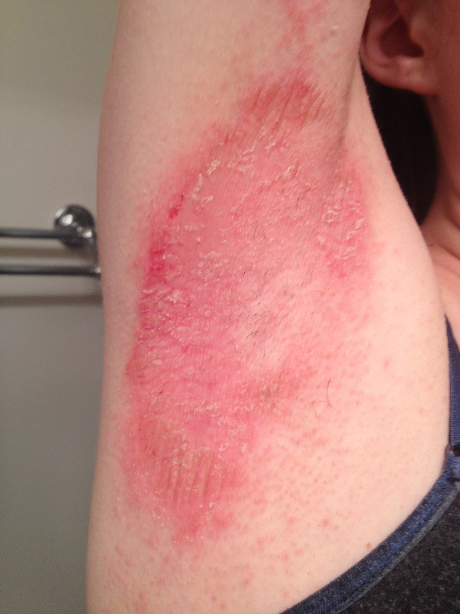
Atopic dermatitis
Atopic dermatitis is a chronic, itchy skin condition that is very common in children but may occur at any age. It is also known as eczema and atopic eczema, and was formerly known as Besnier prurigo. It is the most common form of dermatitis.
Atopic dermatitis usually occurs in people who have an ‘atopic tendency’. This means they may develop any or all of three closely linked conditions; atopic dermatitis, asthma and hay fever (allergic rhinitis). Often these conditions run within families with a parent, child or sibling also affected. A family history of asthma, eczema or hay fever is particularly useful in diagnosing atopic dermatitis in infants.
Atopic dermatitis arises because of a complex interaction of genetic and environmental factors. These include defects in skin barrier function making the skin more susceptible to irritation by soap and other contact irritants, the weather, temperature and non-specific triggers.
See other article on Dermatitis for more details here : Dermatitis
- First occurs in infancy
- Common in elbow and knee creases
- Characterised by flares
- Very itchy
- Acute eczema is red, blistered, swollen
- Chronic eczema is dry, thickened, lined (lichenified)
Figure 11. Armpit atopic dermatitis
Contact allergic dermatitis
Acute or relapsing
Allergen may be:
- Fragrance, preservative or medicament in deodorant, wet-wipe or other product
- Component of underwear (rubber in elastic, nickel in bra wire)
See other article on Dermatitis for more details here : Dermatitis
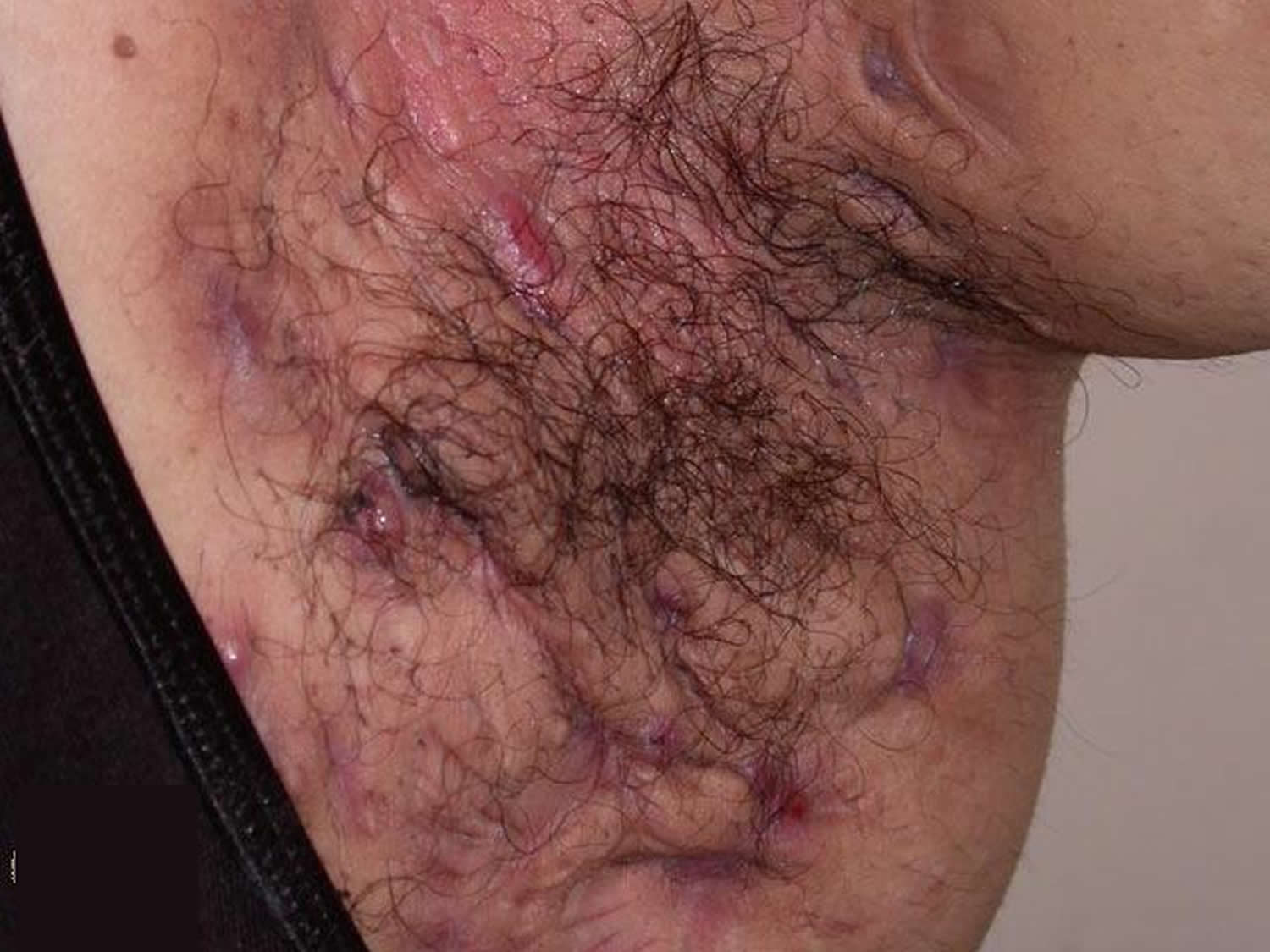
Hidradenitis suppurativa
Hidradenitis suppurativa is an inflammatory skin disease that affects apocrine gland-bearing skin in the armpits, in the groin, and under the breasts. It is characterised by recurrent boil-like nodules and abscesses that culminate in pus-like discharge, difficult-to-heal open wounds (sinuses) and scarring.
The term hidradenitis implies it starts as an inflammatory disorder of sweat glands, which is now known to be incorrect. Hidradenitis suppurativa is also known as acne inversa.
- Chronic disorder
- Boil-like follicular papules and nodules
- Discharging sinuses and scars
Figure 12. Armpit Hidradenitis suppurativa
Who gets hidradenitis suppurativa ?
Hidradenitis often starts at puberty and is most active between the ages of 20 and 40 years, and in women, can resolve at menopause. It is three times more common in females than in males. Risk factors include:
- Other family members with hidradenitis suppurativa
- Obesity and insulin resistance/metabolic syndrome
- Cigarette smoking
- Follicular occlusion disorders: acne conglobata, dissecting cellulitis, pilonidal sinus
- Inflammatory bowel disease (Crohn disease)
- Rare autoinflammatory syndromes associated with abnormalities of PSTPIP1 gene.*
* PAPA syndrome (pyogenic arthritis, pyoderma gangrenosum and acne), PASH syndrome (pyoderma gangrenosum, acne, suppurative hidradenitis) and PAPASH syndrome (pyogenic arthritis, pyoderma gangrenosum, acne, suppurative hidradenitis).
What causes hidradenitis suppurativa ?
Hidradenitis suppurativa is an autoinflammatory disorder. Although the exact cause is not yet understood, contributing factors include:
- Friction from clothing and body folds
- Aberrant immune response to commensal bacteria
- Abnormal cutaneous or follicular microbiome
- Follicular occlusion
- Release of pro-inflammatory cytokines
- Inflammation causing rupture of the follicular wall and destroying apocrine glands and ducts
- Secondary bacterial infection
- Certain drugs.
What are the clinical features of hidradenitis suppurativa ?
Hidradenitis can affect a single or multiple areas in the armpits, neck, submammary area, and inner thighs. Anogenital involvement most commonly affects the groin, mons pubis, vulva (in females), sides of the scrotum (in males), perineum, buttocks and perianal folds.
Signs include:
- Open and closed comedones
- Painful firm papules and larger nodules
- Pustules, fluctuant pseudocysts and abscesses
- Pyogenic granulomas
- Draining sinuses linking inflammatory lesions
- Hypertrophic and atrophic scars.
Many patients with hidradenitis suppurativa also suffer from other skin disorders, including acne, hirsutism and psoriasis.
The severity and extent of hidradenitis suppurativa is recorded at assessment and when determining the impact of a treatment. The Hurley system describes three distinct clinical stages:
- Solitary or multiple, isolated abscess formation without scarring or sinus tracts
- Recurrent abscesses, single or multiple widely separated lesions, with sinus tract formation
- Diffuse or broad involvement, with multiple interconnected sinus tracts and abscesses.
Severe hidradenitis (Hurley Stage 3) has been associated with:
- Male gender
- Armpit and perianal involvement
- Obesity
- Smoking
- Disease duration.
What is the treatment for hidradenitis suppurativa ?
General measures
- Weight loss; follow an anti-inflammatory, low-sugar, low-grain, low-dairy diet (mainly plants)
- Smoking cessation: this can lead to improvement within a few months
- Loose fitting clothing
- Daily unfragranced antiperspirants
- If prone to secondary infection, wash with antiseptics or take bleach baths
- Apply hydrogen peroxide solution or medical grade honey to reduce malodour
- Apply simple dressings to draining sinuses
- Analgesics, such as paracetamol (acetaminophen), for pain control.
Medical management of hidradenitis suppurativa
Medical management of hidradenitis suppurativa is difficult. Treatment is required long term. Effective options are listed below.
Antibiotics 2
- Topical clindamycin, with benzoyl peroxide to reduce bacterial resistance
- Short course of oral antibiotics for acute staphylococcal abscesses, eg flucloxacillin
- Prolonged courses (minimum 3 months) of tetracycline, metronidazole, cotrimoxazole, fluoroquinolones or dapsone for their anti-inflammatory action
- 6–12 week courses of the combination of clindamycin (or doxycycline) and rifampicin for severe disease.
Antiandrogens
- Long-term oral contraceptive pill; antiandrogenic progesterones drospirenone or cyproterone acetate may be more effective than standard combined pills.
- These are more suitable than progesterone-only pills or devices.
- Spironolactone and finasteride
- Response takes 6 months or longer.
Immunomodulatory treatments for severe disease
- Intralesional corticosteroids into nodules
- Systemic corticosteroids short-term for flares
- Methotrexate, ciclosporin, and azathioprine
- TNF-α inhibitors adalimumab and infliximab, used in higher dose than required for psoriasis, are the most successful treatments to date. Note that paradoxically, they may sometimes induce new-onset hidradenitis suppurativa
- Other biologics are under investigation, such as the IL-1β antagonist, canakinumab
Other medical treatments
- Metformin in patients with insulin resistance
- Acitretin (unsuitable for females of childbearing potential)
- Isotretinoin — effective for acne but appears unhelpful for most cases of hidradenitis
- Colchicine
Surgical management of hidradenitis suppurativa 3
- Incision and drainage of acute abscesses
- Curettage and deroofing of nodules, abscesses and sinuses
- Laser ablation of nodules, abscesses and sinuses
- Wide local excision of persistent nodules
- Radical excisional surgery of entire affected area
- Nd:YAG laser hair removal
Hailey-Hailey disease
Benign familial chronic pemphigus or Hailey–Hailey disease is a rare hereditary blistering skin disease first described by the Hailey brothers in 1939.
Benign familial pemphigus usually appears in the third or fourth decade, although it can occur at any age. It then tends to persist life-long. It can affect people of all races.
- Intermittent painful shallow blisters that quickly break down
- Rare inherited condition
- Often starts age 20–40 years
- Most troublesome during summer months
Figure 13. Armpit benign familial pemphigus (Hailey-Hailey disease)
What is the cause of benign familial pemphigus ?
Benign familial pemphigus is a inherited skin disorder, although occasionally sporadic cases arise without a family history. The defect responsible has now been identified on a gene called ATP2C1 found on chromosome 3q21-24. This gene codes for the protein SPCA1 (Secretory Pathway Calcium/manganese-ATPase), a calcium and manganese pump. The skin cells (keratinocytes) stick together via structures called desmosomes and it seems the desmosomes do not assemble properly if there is insufficient calcium.
The genetic defect in benign familial pemphigus causes the skin cells to become unstuck from one another. Normally the cells are packed together tightly in much the same way as bricks and mortar. Patients with Hailey-Hailey disease have defective ´mortar´ and the cells fall apart, like a dilapidated brick wall.
What are the complications of benign familial pemphigus ?
Secondary bacterial infection, which is not uncommon, can give rise to an unpleasant smell. Herpes simplex can infect blistered sites and may evolve to widespread viral infection (eczema herpeticum).
How is benign familial pemphigus diagnosed ?
Usually benign familial pemphigus is diagnosed by its appearance and the family history, but it is often is mistaken for other skin problems. Impetigo, thrush, tinea (jock itch) and other blistering conditions look similar.
Diagnosis may require a skin biopsy. The histology is characteristic, with layers of detached skin cells (‘acantholysis’) lining up like ‘a row of tombstones’. Unlike pemphigus vulgaris, the immunofluorescence test for antibodies is negative.
As yet there is no diagnostic test available to family members.
How is benign familial pemphigus treated ?
Unfortunately there is no cure for Hailey-Hailey disease. Treatment is aimed at reducing symptoms and preventing flares.
General advice
- Avoid trigger factors such as sunburn, sweating and friction where possible; eg when hot, stay indoors with fan or air conditioning, limit amount of exercise taken.
- Wash and dry skin folds carefully, once or twice daily using mild soap and water.
- Wear soft, loose clothing, with absorbant pads in underwear.
- If overweight, try to decrease body fat to minimise friction.
- Apply wet compresses, eg wtih 1:40 diluted aluminium acetate or vinegar, to dry up oozing patches.
- Take bleach baths twice weekly reduce superficial infections.
- Apply zinc paste to inflamed patches.
Topical prescriptions
- Corticosteroid (cortisone) creams used short-term (eg one to two weeks) are effective in treating inflamed lesions; they work best if started early.
- Topical antibiotics such as clindamycin or mupirocin are used short-term for localised infection but are best avoided long-term due to risk of inducing bacterial resistance, eg MRSA.
- Short-term use of combination corticosteroid/ antibiotic creams may also be helpful.
- Benzoyl peroxide is a useful antiseptic available as cream or wash.
- Ketoconazole cream can be used in case of fungal infection.
- Calcipotriol cream is useful in some patients.
- Fluorouracil cream has been reported effective in at least one patient.
- Topical calcineurin inhibitors such as pimecrolimus cream or tacrolimus ointment have been reported to reduce need for topical steroids.
Oral prescriptions
- Prolonged courses of oral antibiotics such as tetracycline may be useful.
- If herpes virus infection is a recurrent problem, oral antivirals such as aciclovir are prescribed.
- Anticholinergic medications such as glycopyrrolate may be prescribed to reduce hyperhidrosis (excessive sweating).
- A number of other oral medications (retinoids, ciclosporin, dapsone, and methotrexate) have been reported in single cases as partially effective, but large trials have not been performed.
Other treatments
- Corticosteroid injections into inflamed plaques
- Botulinum toxin to reduce sweating in axillae and groins, thus reducing colonisation by microorganisms and flare-ups
- Phototherapy (ultraviolet light) has also been used.
- Photodynamic therapy has had varying success.
- Lasers have been reported to be useful in one study, eg CO2 laser or Er:YAG laser vaporising the affected skin, or pulsed dye laser enhancing wound healing.
- In severe cases surgery can be performed to remove the affected skin. Skin grafts are usually necessary to repair the wounds.
- Dermabrasion has been reported to give excellent long-term results.
- Afamelanotide implants cleared Hailey-Hailey disease in 2 patients 4.
- Several patients have been reported that have had improvement in Hailey-Hailey disease when treated with low-dose naltrexone 5.
Are there any complications in benign familial pemphigus ?
For many patients benign familial pemphigus is a mild condition, but for others the pain and smell can be serious problems. If the lesions get infected with herpes virus a sudden severe flare can occur, which often needs prompt treatment (see above).
Will benign familial pemphigus improve in time ?
Many patients have long remissions and an improvement with age does occur.
Granular parakeratosis
Granular parakeratosis is an uncommon red and scaly skin condition that mainly affects body folds, most often the armpits. It has a characteristic appearance under the microscope.
It may be the same condition as recurrent flexural pellagroid dermatitis 6.
- Red brown scaly rash
- May be itchy
- Rare
- Biopsy essential for diagnosis.
What is the cause of granular parakeratosis ?
Granular parakeratosis is thought to be provoked by friction, occlusion and sweating.
A defect in filaggrin production may predispose to granular parakeratosis. This protein is normally present in the upper epidermis, but there are various genetic variants that lead to a weakness of the skin’s barrier function. These include a common cause of dry skin, ichthyosis vulgaris.
Cosmetic or laundry products containing the antiseptic, benzalkonium chloride, may be responsible for some cases. Acting as a surfactant, benzalkonium chloride may disrupt cellular lipid membranes and inactivate enzymes within the skin, inducing proliferation, abnormal development of the skin cells (keratinocytes), and irritant contact dermatitis. Recurrent flexural pellagroid dermatitis 6 was reported to be an irritant contact dermatitis to the anionic surfactant, sodium lauryl sulphate (SLS), in soapless cleansing bars.
This may induce proliferation and abnormal development of the skin cells (keratinocytes).
How is granular parakeratosis treated ?
Granular parakeratosis may resolve by itself. If due to a specific contact factor, it will settle down within a few weeks of recognising and avoiding it.
Treatments that have been reported to be useful include:
- Topical steroids
- Antiseptics
- Topical retinoids
- Keratolytic agents such as lactic acid
- Calcipotriol cream
- Cryotherapy
- Oral isotretinoin
- Botulinum toxin
Figure 14. Armpit Granular parakeratosis
Fox-Fordyce disease
Fox-Fordyce disease is a rare skin disorder that occurs mainly in women between the ages of 13 and 35 years. However, it sometimes affects males and children. The condition is also referred to as ‘apocrine duct occlusion’ and ‘sweat retention disease’.
The condition is characterised by the development of itchy bumps around the hair follicles of the underarm area, pubic region, and/or around the nipples. It results from inflammation of the aprocrine sweat glands, which are found only in these areas.
- Dome-shaped follicular papules in armpits
- Often persistent
- Asymptomatic or itchy
- Reduced sweating.
What causes Fox-Fordyce disease ?
The cause of Fox-Fordyce disease is unknown. For some reason apocrine sweat becomes trapped as a scaly plug forms in the hair follicle. The apocrine sweat ducts rupture, leak and become inflamed, resulting in intense itching.
Factors identified as playing a part in the development of the condition include:
- Emotional and/or hormonal influences
- Alterations in sweat components
How is the diagnosis made ?
Diagnosis is made on patient history and clinical appearance of the rash.
What is the treatment for Fox-Fordyce disease ?
There is no cure for Fox-Fordyce disease. Medical treatments that have been used with varying degrees of success include:
- Topical retinoids
- Topical steroids
- Oral antibiotics
- Clindamycin solution
- Antiandrogenic hormonal therapy
Other forms of treatment used are ultraviolet radiation (phototherapy), dermabrasion, liposuction and surgical excision.
Figure 15. Armpit Fox-Fordyce disease
- Erythrasma. Merck Manual. https://www.merckmanuals.com/professional/dermatologic-disorders/bacterial-skin-infections/erythrasma[↩]
- van der Zee HH, Boer J, Prens EP, Jemec GB. The effect of combined treatment with oral clindamycin and oral rifampicin in patients with hidradenitis suppurativa. Dermatology 2009; 219: 143–7. Epub 2009 Jul 8. https://www.ncbi.nlm.nih.gov/pubmed/19590174[↩]
- Slade DEM, Powell BW, Mortimer PS. Hidradenitis suppurativa: pathogenesis and management. The British Association of Plastic Surgeons 2003; 56: 451–61. https://www.ncbi.nlm.nih.gov/pubmed/12890458[↩]
- Biolcati G, Aurizi C, Barbieri L, Cialfi S, Screpanti I, Talora C. Efficacy of the melanocortin analogue Nle4-D-Phe7-α-melanocyte-stimulating hormone in the treatment of patients with Hailey–Hailey disease. Clinical and Experimental Dermatology. 2014;39(2):168-175. doi:10.1111/ced.12203. https://www.ncbi.nlm.nih.gov/pmc/articles/PMC4255790/[↩]
- http://www.omim.org/[↩]
- Segal R, Eskin-Schwartz M, Trattner A, Feinmesser M, Hodak E, Ingber A, David M. Recurrent flexural pellagroid dermatitis: an unusual variant of irritant contact dermatitis. Acta Derm Venereol. 2015 Jan;95(1):116-7. doi: 10.2340/00015555-1862.[↩][↩]

