What is maxilla
The maxillary bones, or maxillae are the largest facial bones and they form the upper jawbone and the central part of the facial skeleton (see Figure 1). The maxillae form the upper jawbone and meet each other at a median intermaxillary suture. They are considered the keystone bones of the face because they articulate with all other facial bones except the mandible (lower jawbone). The maxillae form part of the floors of the orbits, part of the lateral walls and floor of the nasal cavity, and most of the hard palate. The hard palate is the bony roof of the mouth, and is formed by the palatine processes of the maxillae and horizontal plates of the palatine bones. The hard palate separates the nasal cavity from the oral cavity. The maxilla supports the external nose structures, and morphological changes in the maxilla greatly affect the shape of the nose and face. Each maxilla contains a large maxillary sinus that empties into the nasal cavity (see Figure 6).
The alveolar process (alveol- = small cavity) of the maxilla is a ridgelike arch that contains the alveoli (sockets) for the maxillary (upper) teeth. The palatine process is a horizontal projection of the maxilla that forms the anterior three-quarters of the hard palate or bony roof of the mouth (Figure 4). The union and fusion of the maxillary bones normally is completed before birth. If this fusion fails, this condition is referred to as a cleft palate.
The frontal processes extend superiorly to reach the frontal bone, forming part of the lateral aspect of the bridge of the nose (Figure 1). The maxillae lie just lateral to the nasal cavity and contain the maxillary sinuses. These sinuses, the largest of the paranasal air sinuses, extend from the orbit down to the roots of the upper teeth. Laterally, the maxillae articulate with the zygomatic bones at the zygomatic processes.
The maxilla, along with several other bones, forms the borders of the inferior orbital fissure in the floor of the orbit (Figure 1). This fissure transmits several vessels and nerves, including the maxillary nerve (a branch of cranial nerve 5 or trigeminal nerve) or its continuation, the infraorbital nerve. The infraorbital nerve proceeds anteriorly to enter the face through the infraorbital foramen (Figure 1). The infraorbital foramen (infra- = below; -orbital = orbit), an opening in the maxilla inferior to the orbit, allows passage of the infraorbital blood vessels and nerve, a branch of the maxillary division of the trigeminal (V) nerve. Another prominent foramen in the maxilla is the incisive foramen (= incisor teeth) just posterior to the incisor teeth (see Figure 4). It transmits branches of the greater palatine blood vessels and nasopalatine nerve. A final structure associated with the maxilla and sphenoid bone is the inferior orbital fissure, located between the greater wing of the sphenoid and the maxilla (see Figure 1).
The maxillary bones important structures:
- Alveolar process: sockets for teeth
- Zygomatic process: helps form the zygomatic arches
- Palatine process: forms the anterior part of the hard palate; the two processes meet medially in the intermaxillary suture
- Frontal process: forms part of lateral aspect of bridge of nose
- Incisive fossa: passageway for blood vessels and nerves through hard palate (fused palatine processes)
- Inferior orbital fissure: passageway for maxillary branch of cranial nerve V, the zygomatic nerve, and blood vessels
- Infraorbital foramen: passageway for infraorbital nerve to skin of face
The bones surrounding the nasal cavity—the frontal, ethmoid, sphenoid, and both maxillary bones—contain hollow spaces internally. These air-filled sinuses are called paranasal sinuses (para = near) because they cluster around the nasal cavity (Figure 6). In fact, the paranasal sinuses are extensions of the nasal cavity, lined by the same mucous membrane, and help warm, moisten, and filter inhaled air. The paranasal sinuses also lighten the skull, giving the bones they occupy a moth-eaten appearance in an X-ray image. These sinuses connect to the nasal cavity through small openings, most of which occur at the meatuses inferior to the conchae.
The maxillary sinus is the largest of the four bilateral air‐filled cavities in the skull. The maxillary sinus is located in the body of the maxilla and is a pyramidal‐shaped structure having as its base the medial wall (Figure 6). The pyramid has three main processes or projections: (1) the alveolar process inferiorly (bounded by the alveolar ridge), (2) the zygomatic recess (bounded by the zygomatic bone), and (3) the infraorbital process pointing superiorly. The microscopic anatomy of the sinuses reveals four basic cell types: namely, pseudostratified ciliated columnar epithelium, nonciliated columnar cells, goblet cells, and basal cells.
Figure 1. Maxilla and the skull
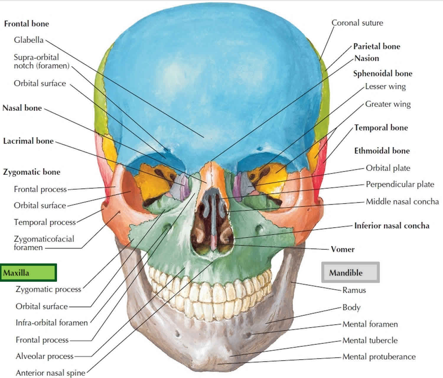
Figure 2. Maxilla and skull lateral view
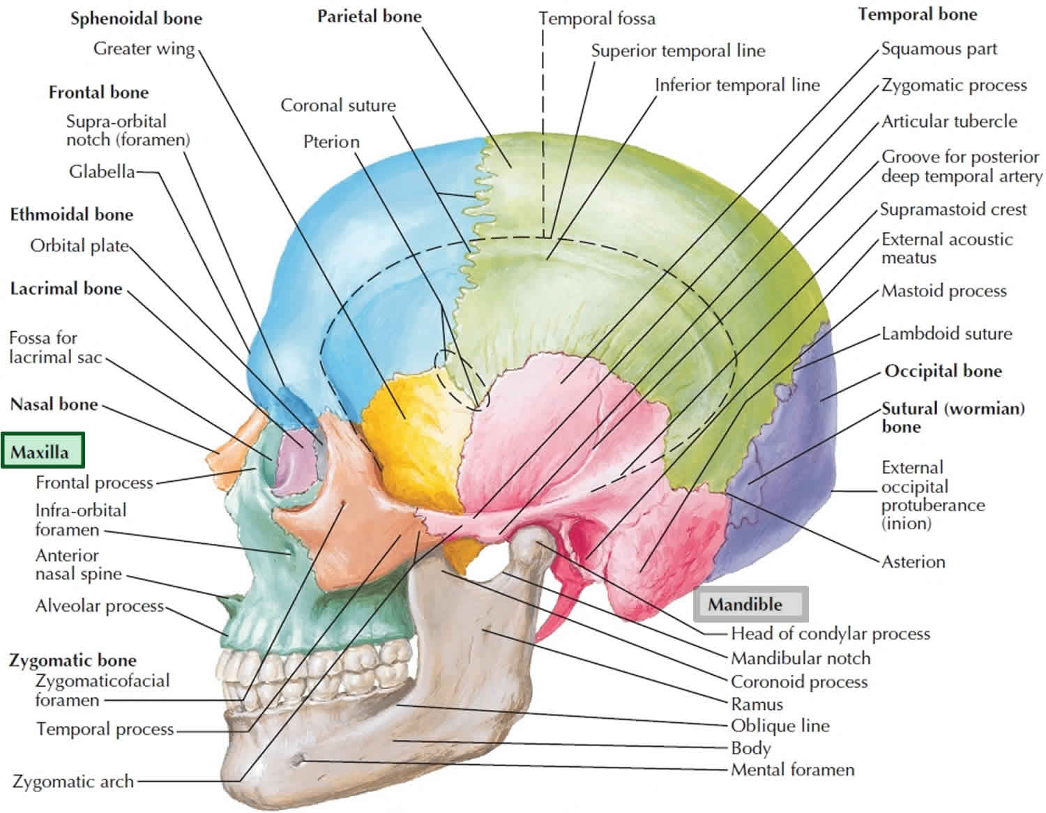
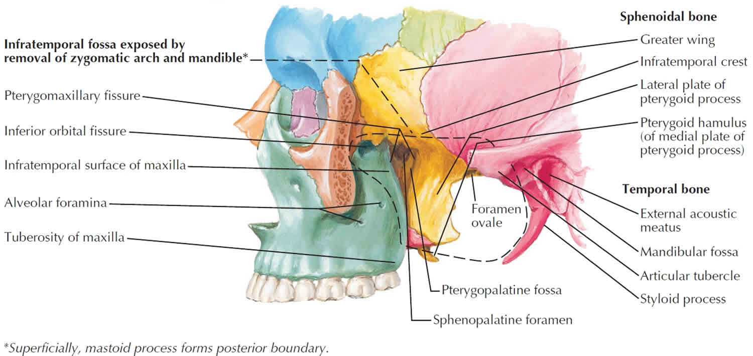
Figure 3. Maxilla median section
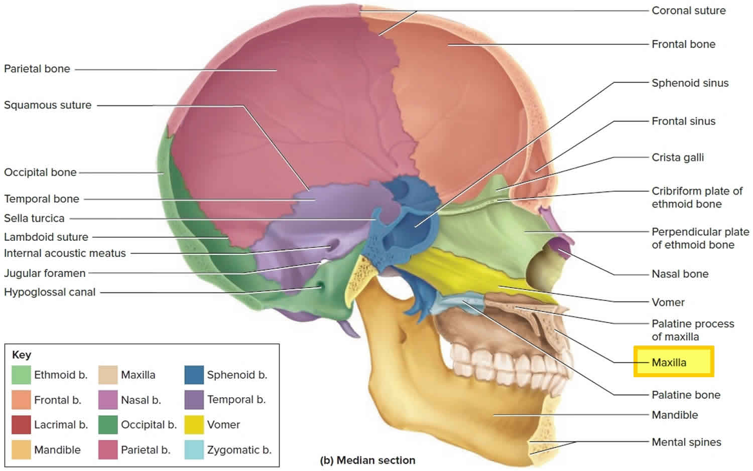
Figure 4. Maxilla anatomy
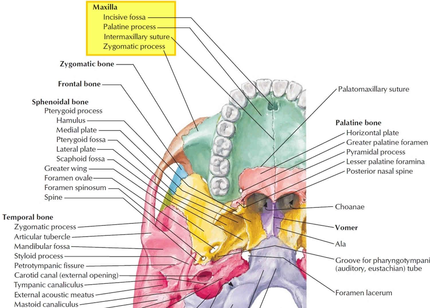
Figure 5. Maxilla and lateral wall of nasal cavity
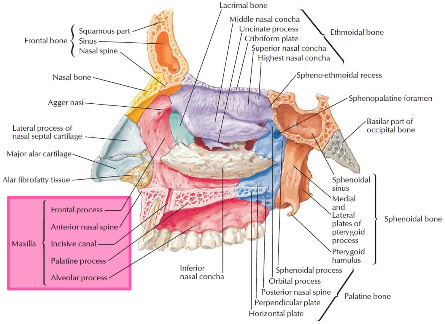 Figure 6. Maxillary sinuses
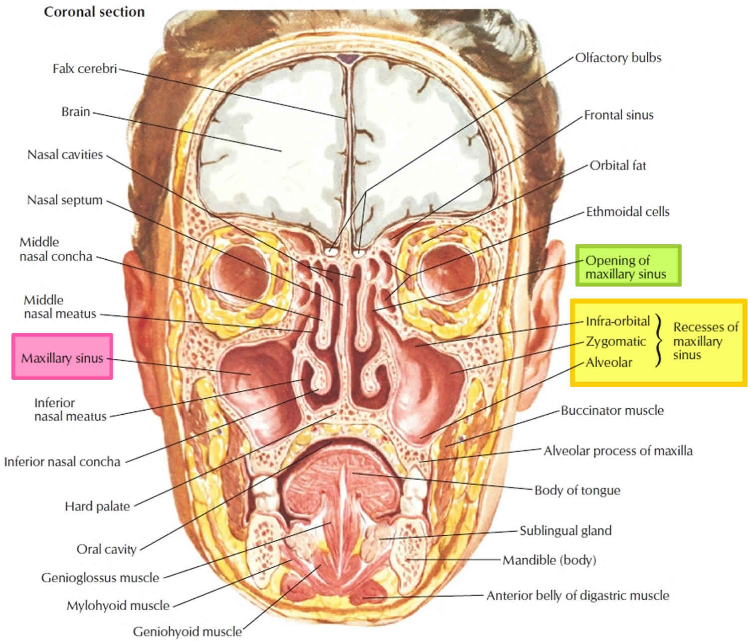
Maxilla function
The function of the maxilla is to provide protection of the face, support of the orbits, hold the top half of the teeth in place, and form the floor of the nose.
The functions of the maxillary sinuses:
- Imparts resonance to the voice
- Increases the surface area and lightens the skull
- Moistens and warms inspired air
- Filters the debris from the inspired air
- Mucus production and storage
- Limit extent of facial injury from trauma
- Provides thermal insulation to important tissues
- Serves as accessory olfactory organs.


