
Contents
What is mitral valve prolapse
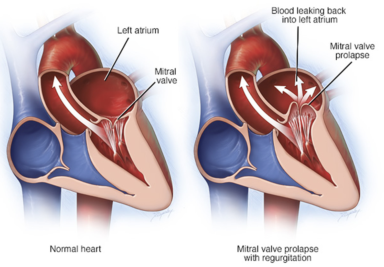
Mitral valve prolapse is a condition where the two valve flaps of the mitral valve do not close smoothly or evenly, but instead bulge (prolapse) upward into the left atrium (see Figure 5 below). Mitral valve prolapse is also known as click-murmur syndrome, Barlow’s syndrome or floppy valve syndrome. In the United States, the leading cause of mitral regurgitation is mitral valve prolapse. Mitral valve prolapse is caused by extra tissue in the valve that keeps it from closing tightly. As much as four to five percent of the population has extra mitral valve tissue, and the majority of those people never require surgery. However, it should be tracked, and if leaks become severe, they can nearly always be corrected with surgery.
In mitral valve prolapse when your heart pumps (contracts) part of one or both mitral valve flaps collapse backward into the left atrium. In some cases, the prolapsed mitral valve lets a small amount of blood leak backward through the valve, which may cause a heart murmur.
The most common cause of mitral valve prolapse is abnormally stretchy valve leaflets (called myxomatous valve disease). Mitral valve prolapse occurs in around 2.4 percent of the population 1. There is no gender differences and an equal distribution from 30 to 80 years of age 1. A person can be born with the genetic risk of developing mitral valve prolapse or it can be caused by other health problems, such as some connective tissue diseases.
Many people with a mitral valve prolapse don’t have symptoms and it may only be spotted during a heart scan (echocardiogram) carried out for another reason.
Mitral valve prolapse can sometimes cause:
- dizziness
- breathlessness
- tiredness and fatigue
- an irregular heartbeat (arrhythmia) or bursts of rapid heartbeat (palpitations)
- mitral regurgitation
- chest discomfort or chest pain that’s not caused by a heart attack or coronary artery disease
Because most patients with mitral valve prolapse do not experience symptoms, a murmur may be detected during a routine physical examination when a healthcare provider uses a stethoscope to listen to the heart.
Even for those who are not experiencing symptoms, if a murmur is detected suggesting mitral valve prolapse, an echocardiogram, or echo, is usually recommended. The echo uses ultrasound to evaluate the characteristics of the valve cusps and how much blood may be leaking (regurgitation) from the valve when the heart contracts. Other tests may include magnetic resonance imaging (MRI), or angiogram. The test results and extent of your symptoms will guide your healthcare provider in determining if further testing is indicated.
In many instances, having mitral valve prolapse will not impact your health and requires no treatment. Talk with your healthcare provider or cardiologist about how best to prevent complications from mitral valve prolapse based on your level of risk. If you are prescribed medication, take it as directed.
Mitral valve prolapse very rarely becomes a highly serious condition. However, in the most serious cases, mitral valve prolapse can cause abnormal heartbeats (arrhythmias) that may eventually become life-threatening.
When mitral valve prolapse is severe enough to cause significant mitral valve leakage, it can lead to serious complications like stroke. This happens because a mitral valve leaking (regurgitating) a significant amount of blood can cause blood clots to form. When clots travel from the heart to the arteries or the brain, it can lead to a stroke or heart attack.
The routine use of antibiotics before having a dental procedure is no longer recommended by the American Heart Association for patients with mitral valve prolapse regardless of whether or not they have any associated symptoms 2. Still, if you’ve been told to take antibiotics before any procedures in the past, check with your doctor whether that’s still necessary.
Mitral valve prolapse and exercise
Most people with mitral valve prolapse lead normal, productive and symptom-free lives.
Doctors generally won’t recommend restrictions on your lifestyle or any limitations on your personal exercise or dietary program. However, ask your doctor if he or she recommends any changes to your lifestyle. If you have severe mitral valve regurgitation, your doctor may recommend certain exercise limitations.
Your doctor may recommend regular follow-up visits to evaluate your condition.
Mitral valve prolapse complications
Although most people with mitral valve prolapse never have problems, complications can occur. They may include:
- Mitral valve regurgitation. The most common complication is a condition in which the valve leaks blood back into the left atrium (mitral valve regurgitation). Being male or having high blood pressure increases your risk of mitral valve regurgitation. If mitral valve regurgitation is severe, you may need surgery to repair or replace the valve in order to prevent heart failure.
- Heart rhythm problems (arrhythmias). Irregular heart rhythms most commonly occur in the upper chambers of the heart. They may be bothersome, but aren’t usually life-threatening. People with severe mitral valve regurgitation or severe deformity of their mitral valve are most at risk of having rhythm problems, which can affect blood flow through the heart.
- Heart valve infection (endocarditis). The inside of your heart is lined by a thin membrane called the endocardium. Endocarditis is an infection of this inner lining. An abnormal mitral valve increases your chance of getting endocarditis from bacteria, which can further damage the mitral valve. People at high risk of endocarditis may be prescribed antibiotics before certain dental and medical procedures, to reduce the risk of infection. However the routine use of antibiotics before having a dental procedure is no longer recommended by the American Heart Association for patients with mitral valve prolapse regardless of whether or not they have any associated symptoms 2. Still, if you’ve been told to take antibiotics before any procedures in the past, check with your doctor whether that’s still necessary.
What are heart valves ?
Your heart is a strong muscle about the size of the palm of your hand. Your body depends on the heart’s pumping action to deliver oxygen- and nutrient-rich blood to the body’s cells. When the cells are nourished properly, the body can function normally. Just like an engine makes a car go, the heart keeps your body running. The heart has two pumps separated by an inner wall called the septum. The right side of the heart pumps blood to the lungs to pick up oxygen. The left side of the heart receives the oxygen-rich blood from the lungs and pumps it to the body.
The heart has four chambers 3, two on the right and two on the left:
- Two upper chambers are called atrium (two is called an atria). The atria collect blood as it flows into the heart.
- Two lower chambers are called ventricles. The ventricles pump blood out of the heart to the lungs or other parts of the body.
The heart also has four valves that open and close to let blood flow from the atria to the ventricles and from the ventricles into the two large arteries connected to the heart in only one direction when the heart contracts (beats). The four heart valves are:
- Tricuspid valve, located between the right atrium and right ventricle
- Pulmonary or pulmonic valve, between the right ventricle and the pulmonary artery. This artery carries blood from the heart to the lungs.
- Mitral valve, between the left atrium and left ventricle
- Aortic valve, between the left ventricle and the aorta. This aorta carries blood from the heart to the body.
Each valve has a set of flaps (also called leaflets or cusps). The mitral valve has two flaps; the others have three. Valves are like doors that open and close. They open to allow blood to flow through to the next chamber or to one of the arteries. Then they shut to keep blood from flowing backward. Blood flow occurs only when there’s a difference in pressure across the valves, which causes them to open. Under normal conditions, the valves permit blood to flow in only one direction.
The heart four chambers and four valves and is connected to various blood vessels. Veins are blood vessels that carry blood from the body to the heart. Arteries are blood vessels that carry blood away from the heart to the body.
The heart pumps blood to the lungs and to all the body’s tissues by a sequence of highly organized contractions of the four chambers. For the heart to function properly, the four chambers must beat in an organized way.
When the heart’s valves open and close, they make a “lub-DUB” sound that a doctor can hear using a stethoscope 4.
- The first sound—the “lub”—is made by the mitral and tricuspid valves closing at the beginning of systole. Systole is when the ventricles contract, or squeeze, and pump blood out of the heart.
- The second sound—the “DUB”—is made by the aortic and pulmonary valves closing at the beginning of diastole. Diastole is when the ventricles relax and fill with blood pumped into them by the atria.
Figure 1. The anatomy of the heart valves
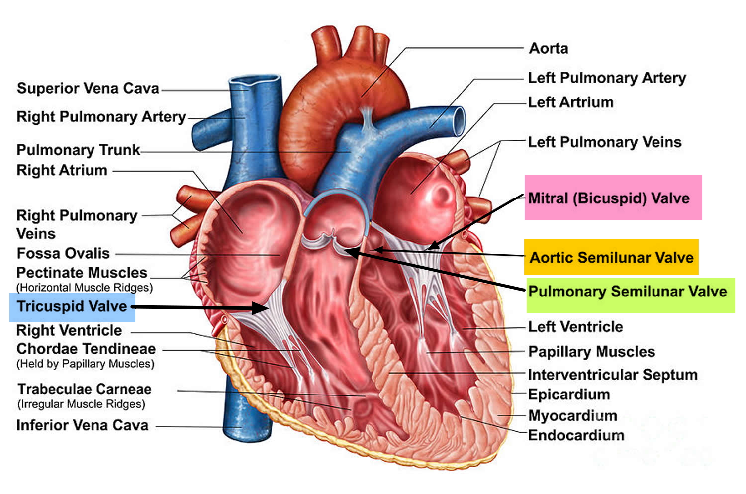
Figure 2. Top view of the 4 heart valves
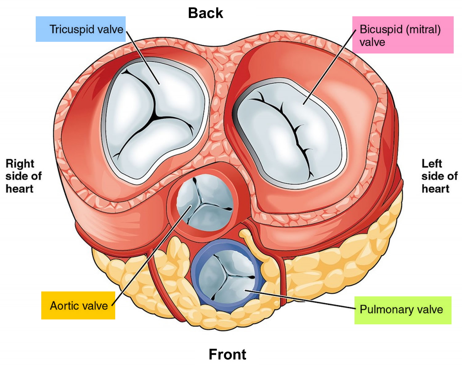
Figure 3. Normal heart blood flow

Figure 4. Heart valves function
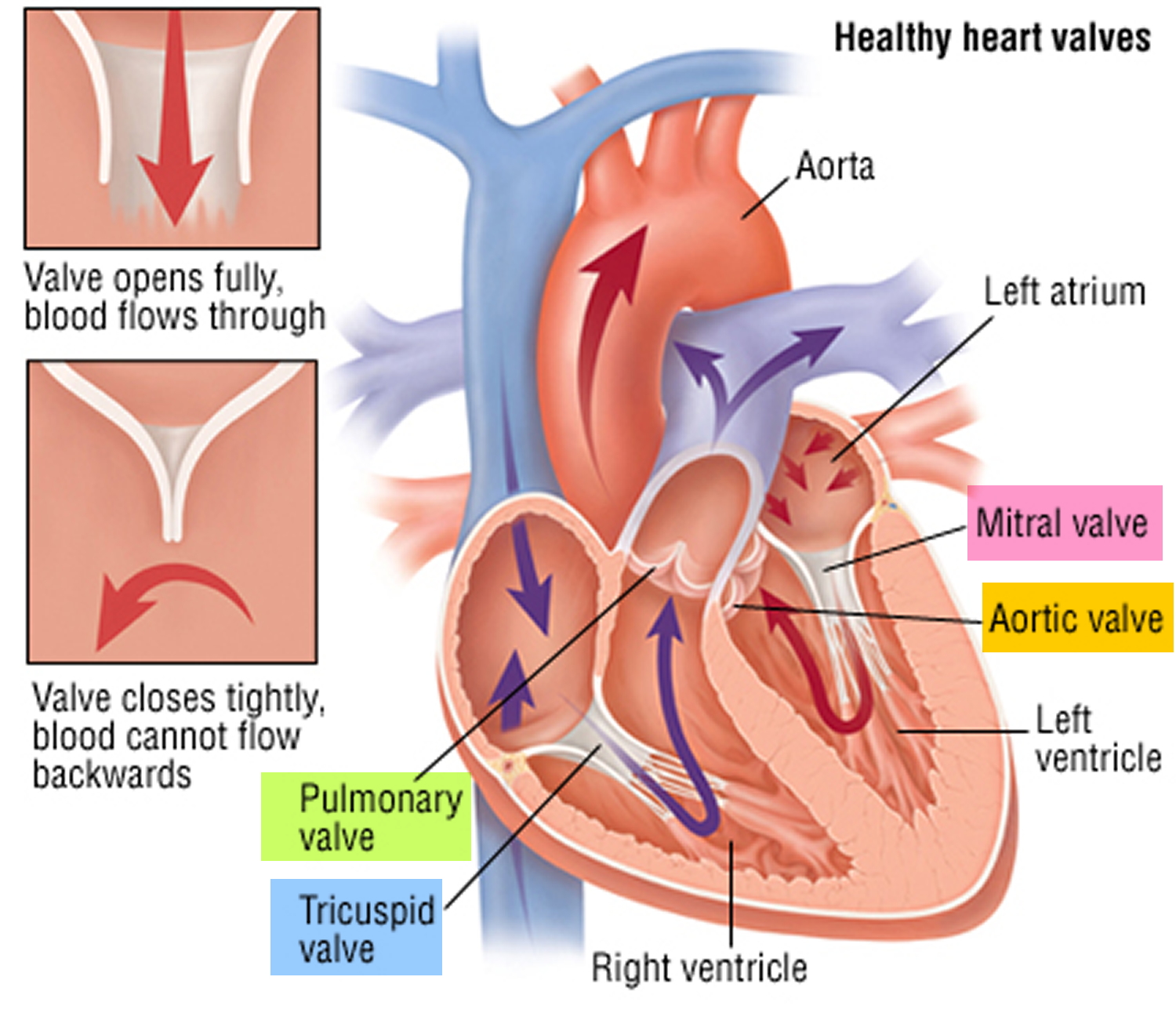
Figure 5. Mitral valve prolapse
Mitral valve prolapse and anxiety
Mitral valve prolapse is also seen in concordance with mental disorders including depression (23.1%), anxiety (26.7–35.9%) and panic disorder (0–50%) but no causal link has been identified for these coincidences 5. Some studies reported higher prevalence of symptoms of anxiety disorder in patents with mitral valve prolapse compared to healthy individuals and that these symptoms were reduced after psychotherapy or medication 6. Contrary to these studies, recent studies have shown no difference in presentation on anxiety disorder symptoms in patients with mitral valve prolapse 5. Although there is doubt about the relationship between psychological disorders and mitral valve prolapse, it was previously shown that patients with mitral valve prolapse and anxiety comorbidity were found to have more intense emotional experiences compared with patients with mitral valve prolapse who do not express anxiety symptoms, and therefore might benefit from medical treatment for their condition 6. On the other hand, induced psychological stress was found to result in changes in rhythm (based on electrocardiogram) and amplitude of click in phonocardiogram 7. Regarding the fact that a number of symptoms of anxiety, including increased tachycardia, chest pain and blood pressure lability might aggravate mitral valve prolapse symptoms 8, it is hypothesized that treatment of the underlying or concomitant psychological disease might have a palliative effect on mitral valve prolapse signs and symptoms 9. Previous studies have shown improvements in mitral valve prolapse symptoms and depth of the prolapse by psychotherapy or medical treatment of anxiety (benzodiazepines, β-blockers) 10. Selective serotonin reuptake inhibitors (SSRIs) are considered as a treatment option for depression, anxiety and stress. However, the findings of this study 11 suggest the use of fluoxetine with propranolol may aggravate the effects of propranolol in mitral valve prolapse patients. More research is required to identify the mechanism of the observed effect of fluoxetine and also on the effects of other SSRI drugs on the echocardiographic and symptomatic characteristics of mitral valve prolapse patients with anxiety.
Mitral valve prolapse causes
Mitral valve prolapse is usually caused by problems with the tissues that join the mitral valve to the heart muscles. Some people with the condition are born with it, and it’s more common in people with connective tissue disorders, such as Marfan syndrome. Rarely, it can be caused by damage to the heart muscles themselves – for example, as the result of a heart attack.
When your heart is working properly, the mitral valve closes completely during contraction of the left ventricle and prevents blood from flowing back into your heart’s upper left chamber (left atrium).
But in some people with mitral valve prolapse, one or both of the mitral valve’s flaps (leaflets) have extra tissue bulging (prolapsing) like a parachute into the left atrium each time the heart contracts.
The bulging may keep the valve from closing tightly. When blood leaks backward through the valve, it’s called mitral valve regurgitation.
This may not cause problems if only a small amount of blood leaks back into the atrium. More-severe mitral valve regurgitation can cause symptoms such as shortness of breath, fatigue or lightheadedness.
Another name for mitral valve prolapse is click-murmur syndrome. When a doctor listens to your heart using a stethoscope, he or she may hear a clicking sound as the valve’s leaflets billow out, followed by a murmur resulting from blood flowing back into the atrium. Other names to describe mitral valve prolapse include:
- Barlow’s syndrome
- Floppy valve syndrome
- Billowing mitral valve syndrome
- Myxomatous mitral valve disease
Risk factors for mitral valve prolapse
Mitral valve prolapse can develop in any person at any age.
Serious symptoms of mitral valve prolapse tend to occur most often in men older than 50.
Mitral valve prolapse can run in families and may be linked to several other conditions, such as:
- Marfan syndrome
- Ehlers-Danlos syndrome
- Ebstein’s anomaly
- Muscular dystrophy
- Graves’ disease
- Scoliosis
Mitral valve prolapse prevention
You can’t prevent mitral valve prolapse. However, you can lower your chances of developing the complications associated with it by making sure you take your medications, if any, as directed.
Mitral valve prolapse symptoms
Although mitral valve prolapse is usually a lifelong disorder, many people with this condition never have symptoms. When diagnosed, people may be surprised to learn that they have a heart condition.
When signs and symptoms do occur, it may be because blood is leaking backward through the mitral valve (mitral regurgitation). Mitral valve prolapse symptoms can vary widely from one person to another. They tend to be mild and develop gradually.
Symptoms of mitral valve prolapse may include:
- A racing or irregular heartbeat (arrhythmia)
- Dizziness or lightheadedness
- Difficulty breathing or shortness of breath, often when lying flat or during physical activity
- Fatigue
- Chest pain that’s not caused by a heart attack or coronary artery disease
Mitral valve prolapse diagnosis
Doctors may diagnose mitral valve prolapse at any age. Your doctor is most likely to diagnose mitral valve prolapse while listening to your heart with a stethoscope during a physical examination.
If you have mitral valve prolapse, your doctor may hear a clicking sound, which is common with this condition. Your doctor may detect a heart murmur, which would be due to mitral regurgitation if it is present.
Other tests that may be used to evaluate your heart may include:
- Echocardiogram. An echocardiogram is usually done to confirm the diagnosis and determine the severity of your condition. An echocardiogram is a noninvasive ultrasound evaluation of your heart. This test uses high-frequency sound waves to create images of your heart. It helps doctors see the flow of blood through your mitral valve and measure the amount of blood leakage (regurgitation). You may have a transesophageal echocardiogram (TEE). In this test, your doctor inserts a flexible tube with a small device (transducer) attached into your throat and down into your esophagus — the tube that connects the back of your mouth to your stomach. From there, the transducer can be positioned to obtain more-detailed images of your heart.
- Chest X-ray. A chest X-ray shows a picture of your heart, lungs and blood vessels and can help your doctor make a diagnosis. It can help show if your heart is enlarged.
- Electrocardiogram (ECG). In this noninvasive test, a technician will place probes on your chest that record the electrical impulses that make your heart beat. An ECG records these electrical signals and can help your doctor detect irregularities in your heart’s rhythm and structure, including mitral valve prolapse.
- Stress test. Your doctor may order a stress test to see if mitral valve regurgitation limits your ability to exercise. In a stress test, you exercise or take certain medications to increase your heart rate and make your heart work harder. You may also have a stress test if your doctor is trying to determine if you have another condition such as coronary artery disease.
- Coronary angiogram. This type of cardiac catheterization uses X-ray imaging to see your heart’s blood vessels. It isn’t generally used to diagnose mitral valve prolapse, but may reveal the condition when you’re being testing for another suspected diagnosis. In some cases, your doctor may recommend a coronary angiogram and cardiac catheterization to gather more information about the severity of your condition. In a cardiac catheterization procedure, doctors insert a catheter in an artery in your groin (femoral artery) or in your wrist (radial artery). The catheter is then threaded through your blood vessels to your heart.
Mitral valve prolapse treatment
You probably won’t need treatment if you don’t have any symptoms. Your doctor may suggest:
- making lifestyle changes, such as giving up cigarettes, caffeine and alcohol – these things can cause your heart to become overworked
- having regular check-ups to monitor your condition
If you have symptoms or your mitral valve is very floppy, your doctor may recommend:
- medication to relieve your symptoms, such as beta-blockers for an irregular heartbeat
- mitral valve surgery to repair or replace the mitral valve
Medications
If you develop symptoms, your doctor might prescribe certain medications to treat mitral valve prolapse-related chest pain, heart rhythm abnormalities or other complications. Some medications you might be prescribed include:
- Beta blockers. These drugs help prevent irregular heartbeats by making your heart beat more slowly and with less force, which reduces your blood pressure. Beta blockers also help blood vessels relax and open up to improve blood flow.
- Diuretics. Your doctor may prescribe water pills (diuretics) to drain fluid from your lungs.
- Heart rhythm medications. Your doctor may prescribe an antiarrhythmic medication, such as flecainide (Tambocor), amiodarone (Cordarone, Pacerone), and propafenone (Rythmol, Rythmol SR). Antiarrhythmics help control your heart rhythm by normalizing electrical signals in heart tissue.
- Aspirin. If you have mitral valve prolapse and a history of strokes, your doctor might prescribe aspirin to reduce the risk of blood clots.
- Prescription anticoagulants (blood thinners). These medications — such as warfarin (Coumadin), heparin, dabigatran (Pradaxa), rivaroxaban (Xarelto), apixaban (Eliquis) and edoxaban (Savaysa) — prevent your blood from clotting if you have had irregular heart rhythms, such as atrial fibrillation. If you have atrial fibrillation, a history of heart failure or a history of strokes, your doctor may suggest these drugs. They can have dangerous side effects, however, and must be taken exactly as prescribed.
Mitral valve prolapse surgery
Though most people with mitral valve prolapse don’t need surgery, your doctor may suggest surgical treatment if you have severe mitral valve regurgitation with or without symptoms.
Severe mitral valve regurgitation can eventually cause heart failure, preventing your heart from effectively pumping blood. If regurgitation goes on too long, your heart may be too weak for surgery.
If your doctor suggests surgery, he or she may suggest repairing or replacing the mitral valve. Valve repair and replacement may be performed using open-heart surgery or minimally invasive surgery. Minimally invasive surgery involves smaller incisions and may have less blood loss and a quicker recovery time than open surgery.
- Valve repair. Mitral valve repair is a surgery that preserves your own valve. For most people with mitral valve prolapse, this is the preferred surgical treatment to correct the condition. Your mitral valve consists of two triangular-shaped flaps of tissue called leaflets. The leaflets of the mitral valve connect to the heart muscle through a ring called the annulus. The surgeon can modify the original valve (valvuloplasty) to eliminate backward blood flow. Surgeons can also repair the valve by reconnecting valve leaflets or by removing excess valve tissue so that the leaflets can close tightly. Sometimes repairing the valve includes tightening or replacing the annulus. This is called an annuloplasty. It is important to ensure that your surgeon is experienced in performing mitral valve repair.
- Valve replacement. Your surgeon may perform a valve replacement if valve repair isn’t possible. In valve replacement surgery, the damaged mitral valve is replaced by an artificial (prosthetic) valve. Artificial valves are mechanical or tissue valves. Mechanical valves may last a long time. However, if you have a mechanical valve, you must use an anticoagulant medication, such as warfarin (Coumadin), for the rest of your life to prevent blood clots from forming on the valve. If a blood clot forms on the valve and breaks free, it could travel to your brain and cause a stroke. Tissue valves are made from animal tissue such as a pig or cow valve. These kinds of valves are called bioprostheses. They may wear out over time and need replacement. However, an advantage of the tissue valve is that you don’t have to use long-term anticoagulant medication.
- Prevalence and clinical outcome of mitral-valve prolapse. Freed LA, Levy D, Levine RA, Larson MG, Evans JC, Fuller DL, Lehman B, Benjamin EJ. N Engl J Med. 1999 Jul 1; 341(1):1-7. http://www.nejm.org/doi/full/10.1056/NEJM199907013410101[↩][↩]
- http://www.heart.org/HEARTORG/Conditions/More/HeartValveProblemsandDisease/Problem-Mitral-Valve-Prolapse_UCM_450441_Article.jsp[↩][↩]
- American Heart Association. About Arrhythmia. http://www.heart.org/HEARTORG/Conditions/Arrhythmia/AboutArrhythmia/About-Arrhythmia_UCM_002010_Article.jsp[↩]
- Centers for Disease Control and Prevention. Division of Birth Defects and Developmental Disabilities. Congenital Heart Defects (CHDs). https://www.cdc.gov/ncbddd/heartdefects/index.html[↩]
- Filho AS, Maciel BC, Romano MM, Lascala TF, Trzesniak C, Freitas-Ferrari MC, et al. Mitral valve prolapse and anxiety disorders. Br J Psychiatry. 2011;199(3):247–8. doi: 10.1192/bjp.bp.111.091934. https://www.ncbi.nlm.nih.gov/pubmed/21881100[↩][↩]
- Zinchenko YP, Pervichko EI, Martynov AI. Psychological underpinning of personalized approaches in modern medicine: syndrome analysis of mitral valve prolapsed patients. Psychol Russ State Art. 2013;6(2):89–102. doi: 10.11621/pir.2013.0208.[↩][↩]
- Combs RL, Shah PM, Klorman RS, Klorman R. Effects of induced psychological stress on click and rhythm in mitral valve prolapse. Am Heart J. 1980;99(6):714–21. doi: 10.1016/0002-8703(80)90620-1. https://www.ncbi.nlm.nih.gov/pubmed/7377092[↩]
- Hu X, Zhao Q. Autonomic dysregulation as a novel underlying cause of mitral valve prolapse: A hypothesis. Med Sci Monit. 2011;17(9):27–31. doi: 10.12659/msm.881918. https://www.ncbi.nlm.nih.gov/pmc/articles/PMC3560509/[↩]
- Lung FW, Cheng CT, Chang WT, Shu BC. Anxiety and mood disorder in young males with mitral valve prolapse. J Multidiscip Healthc. 2008;1:89–92. https://www.ncbi.nlm.nih.gov/pmc/articles/PMC3004540/[↩]
- Pervichko E, Zinchenko Y, Martynov A. EPA-0581 – Long-term integrative psychotherapy of anxiety disorders in mitral valve prolapse patients: the factors of mental health improvement. Eur Psychiatry. 2014;29(1):1. doi: 10.1016/S0924-9338(14)77970-2.[↩]
- Esfehani RJ, Kamranian H, Jalalyazdi M. Effect of Fluoxetine Administration on Clinical and Echocardiographic Findings in Patients with Mitral Valve Prolapse and Generalized Anxiety Disorder: Randomized Clinical Trial. Electronic Physician. 2017;9(1):3483-3491. doi:10.19082/3483. https://www.ncbi.nlm.nih.gov/pmc/articles/PMC5308485/[↩]


