Contents
What is pyoderma gangrenosum
Pyoderma gangrenosum is a rare, destructive inflammatory skin disease of which a painful nodule or pustule breaks down to form a progressively enlarging very painful ulcer. Pyoderma gangrenosum is not an infection (pyoderma), nor does it cause gangrene 1. Lesions may occur either in the absence of any apparent underlying disorder or in association with other diseases, such as ulcerative colitis, Crohn’s disease, polyarthritis (an inflammation of several joints together), gammopathy, vasculitis, leukemia, and other conditions 2. Pyoderma gangrenosum usually occurs on the legs, although it can affect any area of skin. Pyoderma gangrenosum sometimes develops around an injury or surgical wound, for example, it may develop around a wound, a needle prick, a biopsy or an insect bite.
Pyoderma gangrenosum skin reaction isn’t passed down to children from their parents through genes. Pyoderma gangrenosum is also not contagious, so can’t be transferred from or to another person.
Each year in the United States, pyoderma gangrenosum occurs in about 1 person per 100,000 people 2.
Pyoderma gangrenosum is an uncommon disease that affects males and females of any age, but is more common in females aged between 20 and 50 years. Pyoderma gangrenosum rarely affects infants and children. A review of 13 children with pyoderma gangrenosum found that the skin lesions have a more varied anatomic distribution (i.e., legs, trunk, arms, head and neck), a greater predominance of pustular lesions and a strong association with inflammatory bowel disease–Crohn’s disease being the most common 3.
Pyoderma gangrenosum is thought to be a reaction to an internal disease or condition. Known associations include:
- Inflammatory bowel disease (ulcerative colitis and Crohn disease)
- Rheumatoid arthritis
- Myeloid blood dyscrasias including leukaemia
- Monoclonal gammopathy (usually IgA)
- Chronic active hepatitis
- Granulomatosis with polyangiitis
- PAPA syndrome
- Use of levamisole-adulterated cocaine
- Miscellaneous less common associations.
About 50% of those affected by pyoderma gangrenosum have none of the associated risk factors. An ulcer or ulcers about an ostomy site may represent pyoderma gangrenosum (peristomal pyoderma gangrenosum). If Crohn’s disease is the reason for the ostomy, involvement of the gastrointestinal tract adjacent to the ostomy site should be considered. Often, there are multiple holes and fistulous tracts that characterize pyoderma gangrenosum as opposed to a periostomal ulcer caused by a poorly fitting ostomy apparatus.
Pyoderma gangrenosum is a serious condition that can progress quickly, so it’s important to get it diagnosed and treated as soon as possible.
Pyoderma gangrenosum belongs to a group of autoinflammatory skin diseases called neutrophilic dermatoses 1. Neutrophils are a type of white blood cell or leukocyte which form an early line of defense against bacterial infections. Ulcerations associated with pyoderma gangrenosum may occur after trauma or injury to the skin, a process called pathergy. Treatment involves wound care and the use of anti-inflammatory agents, including antibiotics, corticosteroids, immunosuppressants, and biologics.
What does pyoderma gangrenosum look like?
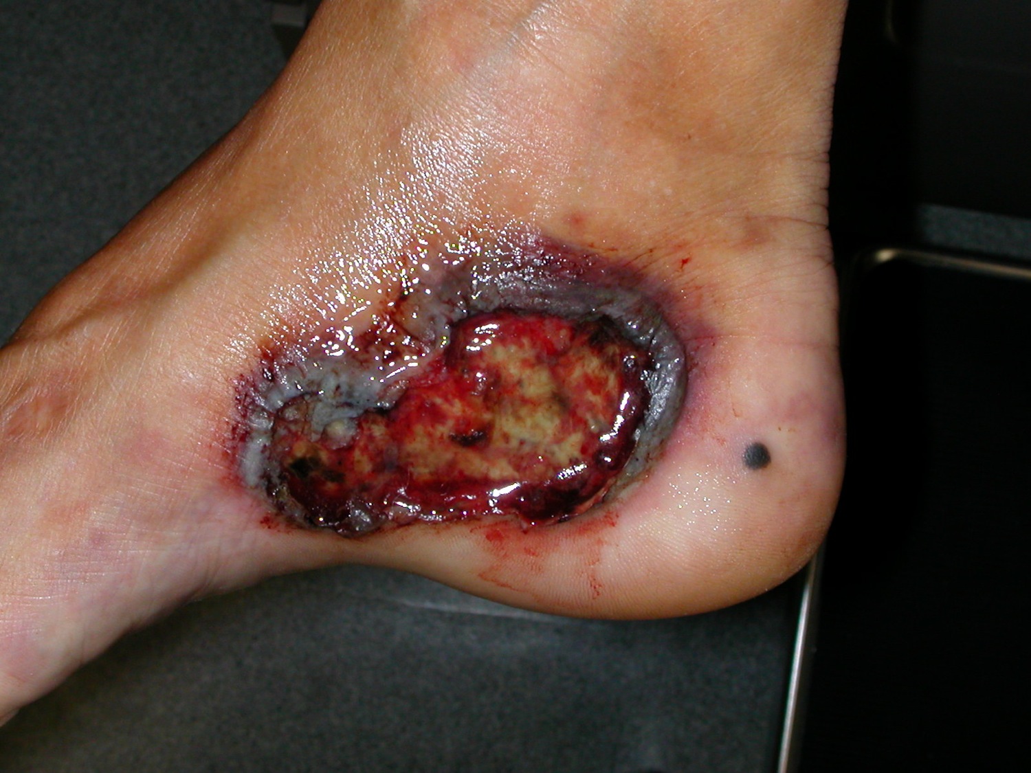
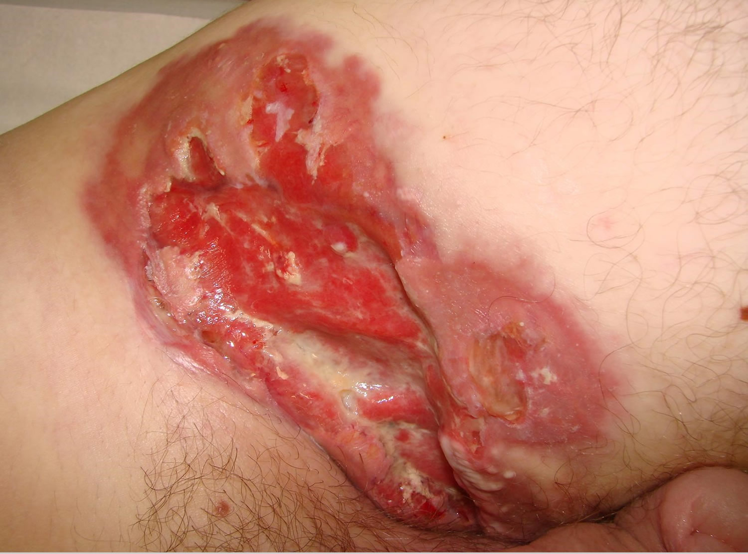
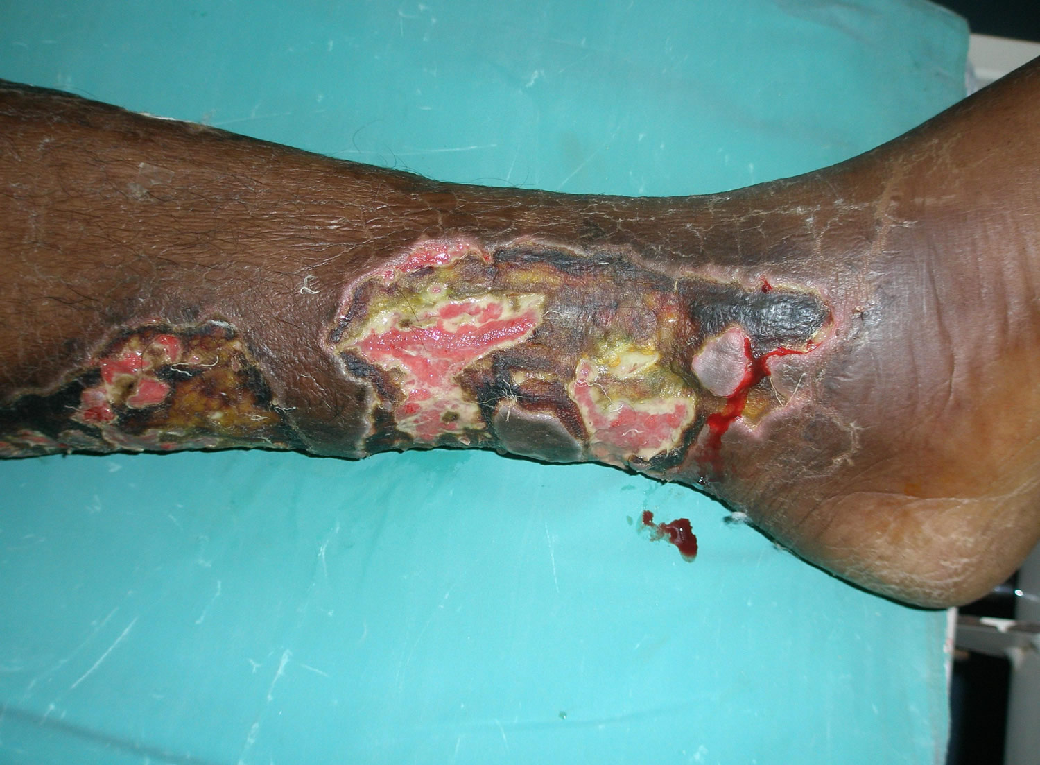
The appearance of pyoderma gangrenosum varies from person to person. Lesions appear as small pus-filled pimples, red bumps or blood-filled blisters. These lesions turn into painful ulcers with pus and grow in size very quickly. There is usually a single large lesion. However, multiple lesions can also appear simultaneously. They can appear with other symptoms such as fever, muscle aches and joint pains. Although lesions can appear on any part of the skin, the lower legs and hands are most commonly involved.
Figure 1. Pyoderma gangrenosum
Pyoderma gangrenosum prognosis
Pyoderma gangrenosum is a treatable condition but it may take months to heal completely and it requires expert wound care. When the underlying disease is treated, the lesions tend to improve. As affected individuals have different medical illnesses and lesions differ in their severity, the outcome of pyoderma gangrenosum differs from person to person.
Regular follow-up with the treating doctor is crucial for close monitoring of the condition. The adverse effects of oral therapy should be discussed thoroughly with a doctor prior to and during treatment.
Pyoderma gangrenosum complications
Possible complications of pyoderma gangrenosum include:
- Infection
- Scarring
- Uncontrolled pain
- Depression
- Loss of mobility
Pyoderma gangrenosum causes
Pyoderma gangrenosum is an autoinflammatory disease (excessive response to an internal antigen) due to some form of neutrophil dysfunction. T lymphocytes and cytokines are involved. There may be a genetic predisoposition.
When the affected skin tissue is tested, it usually has a high concentration of neutrophils (white blood cells involved in inflammation). This means pyoderma gangrenosum may be related to overactivity of the immune system.
Drugs are occasionally implicated as triggers of pyoderma gangrenosum, especially cocaine, isotretinoin, propylthiouracil and sunitinib.
Risk factor for pyoderma gangrenosum
People with the following health conditions are most likely to develop pyoderma gangrenosum, even if the condition is mild or well controlled:
- Inflammatory bowel disease – including ulcerative colitis and Crohn’s disease
- Rheumatoid arthritis – a long-term condition causing pain, swelling and stiffness in the joints
- Blood dyscrasia – a blood disorder
- Hepatitis – inflammation of the liver
- Granulomatosis with polyangiitis – a rare condition in which the walls of blood vessels become inflamed
- Cancer affecting the blood cells – including acute myeloid leukemia
- PAPA syndrome – a rare genetic disorder
- Surgical procedures. In one study of 601 surgical procedures in patients with a known history of pyoderma gangrenosum, 5.5% led to recurrence of pyoderma gangrenosum 4. More invasive procedures and chronically present pyoderma gangrenosum was associated with higher risk. In a different study of 18 patients, the average time to symptoms was 11 days 5.
- Medications. Drug-induced pyoderma gangrenosum is rare but does occur. Most are colony-stimulatings factor (e.g., granulocyte colony-stimulating factor) or small-molecule tyrosine kinase inhibitors (e.g., gefitinib, imatinib, simitinib and pazopanib) 6.
Pyoderma gangrenosum prevention
You can’t totally prevent pyoderma gangrenosum. If you have the condition, try to avoid injuring your skin. Injury or trauma to your skin, including from surgery, can provoke new ulcers to form. It may also help to control any underlying condition that may be causing the ulcers.
Pyoderma gangrenosum symptoms
Pyoderma gangrenosum usually starts quite suddenly, often at the site of a minor injury. It may start as a small pustule, red bump or blood-blister. The skin then breaks down resulting in an ulcer. The ulcer can deepen and widen rapidly. Characteristically, the edge of the ulcer is purple and undermined. Pyoderma gangrenosum is usually very painful. Several ulcers may develop at the same time or over a period of months to years. If the ulcer gets infected, you may also feel unwell and develop a high temperature (fever).
Untreated, the ulcers may continue to enlarge, persist unchanged or may slowly heal. Treatment is usually successful in arresting the process, but complete healing may take months. This is particularly true if there is underlying venous disease, another reason for leg ulcers.
Deep ulcers heal with scarring and this is sometimes with a characteristic cribriform or criss-cross pattern. A rare superficial bullous variant of pyoderma gangrenosum may heal without leaving a scar. This may be similar to or confused with acute febrile neutrophilic dermatosis (Sweet disease).
Extra-cutaneous pyoderma gangrenosum (lung, intestine, cornea, spleen, etc.) involvement is rare. Pulmonary involvement is the most common, although there are only few reported cases 7.
Pyoderma gangrenosum diagnosis
Pyoderma gangrenosum is diagnosed by its characteristic appearance. There is no specific test. The wound should be swabbed and cultured for micro-organisms, but these are not the cause of pyoderma gangrenosum. Biopsy may be necessary to rule out other causes of ulceration. Pyoderma gangrenosum characteristically results in a neutrophilic inflammatory infiltrate but this is not always present.
Mostly, blood tests are not particularly helpful. Some patients may have a positive ANCA (antineutrophil cytoplasmic antibody).
The pathergy test is usually positive (a skin prick test causing a papule, pustule or ulcer).
Sue et al 8 proposed that pyoderma gangrenosum can be diagnosed if there are 2 major crtieria and 2 minor criteria as follows, but this is unconfirmed.
Major criteria
- Rapid progression of painful, necrolytic, cutaneous ulcer with an irregular, violaceous and undermined border
- Other causes of cutaneous ulceration have been excluded
Minor criteria
- History suggestive of pathergy or clinical finding of cribriform scarring
- Systemic diseases associated with pyoderma gangrenosum
- Histopathological findings (sterile dermal neutrophilia +/- mixed inflammation +/- lymphocytic vasculitis
- Treatment response (rapid response to systemic steroid treatment)
Pyoderma gangrenosum treatment
Pyoderma gangrenosum often heals with relatively simple treatments. However, it may take some time and often leaves some scarring in the affected area. Treatment is non-surgical. The necrotic tissue should be gently removed. Wide surgical debridement should be avoided during the active stage of pyoderma gangrenosum because it may result in enlargement of the ulcer. Skin grafting and other surgical procedures may be performed when the active disease phase has settled, with care to minimize trauma.
Some people heal very slowly, over months or years. Others may find the condition clears up within a few weeks. In some cases, it returns after treatment.
There are a number of treatment options, but there’s no clear evidence to suggest which the best one is.
Often conventional antibiotics such as flucloxacillin are prescribed prior to making the correct diagnosis. These may be continued if bacteria are cultured in the wound (secondary wound infection) or there is surrounding cellulitis (red hot painful skin), but they are not helpful for uncomplicated pyoderma gangrenosum.
Anemia is very common in patients with pyoderma gangrenosum 9. A 30-year-old man with Crohn’s disease, anemia, and recalcitrant pyoderma gangrenosum cleared completely after iron sucrose infusion 10.
Small ulcers are often treated with:
- Potent topical steroid ointment
- Tacrolimus ointment
- Intralesional steroid injections
- Ciclosporin solution
- Special dressings
- Oral anti-inflammatory antibiotics such as doxycycline or minocycline
- If tolerated, careful compression bandaging to reduce swelling.
Strong steroid creams or ointments are applied on and around ulcers either daily or every other day. They can help the ulcers heal quickly, particularly if the ulcers are small and diagnosed early. Patients with solitary pyoderma gangrenosum or smaller lesions may be candidates for potent topical steroid therapy alone. In one study of such patients, 44% healed within 6 months 11. Patients may also keep the topical steroid around to use as early as possible if their lesions recur.
Alternatively, a steroid called triamcinolone may be injected into the edge of the ulcer. In more severe cases, steroids may be injected into a vein (intravenously).
Tacrolimus ointment has also proved useful in treating pyoderma gangrenosum ulcers.
Systemic treatment for larger ulcers due to pyoderma gangrenosum may include:
- Oral prednisone for several weeks or longer, or intermittent intravenous methyl prednisolone for 3–5 days
- Ciclosporin. Cyclosporin (orally e.g., 5 mg/kg/day) is dramatically effective and many consider it the drug of choice. It is an excellent alternative to prednisone. Cyclosporin at lower doses (e.g., 3 mg/kg/d) has also been used 12 but required 3-6 months for healing. Cyclosporin can be very effective alone, but if the patient is already on prednisone, the cyclosporin may be added without stopping the steroid.
- Biologic agents: success with infliximab, adalimumab, etanercept and ustekinumab is reported in a small number of cases (used off-label).
Infliximab (Remicade) has been very helpful in treating pyoderma gangrenosum including in children as young as 17 months of age 13. It may be used in combination with topical tacrolimus and/or prednisone. One protocol suggested is infusions of infliximab at zero, two, and eight weeks using a dose of 4-5 mg/kg. Often the prednisone may be tapered once the Remicade is started and then maintenance of infliximab may be given. In some cases, topical tacrolimus is sufficient to maintain control.
Canakinumab is a human monoclonal antibody that selectively blocks IL-1β. It has been studied in 5 patients with pyoderma gangrenosum 14. Canakinumab 150 mg was administered subcutaneously to all patients at week 0 with more doses administered depending upon clinical response. Four of five patients showed a decrease in target-lesion size, and three of five achieved complete remission.
Most people with pyoderma gangrenosum need to take steroid tablets, either on their own or with antibiotics. These reduce inflammation and help the ulcers to heal. However, the long-term use of steroids is associated with serious side effects such as bone thinning (osteoporosis), so they need to be used with caution.
Either systemic steroids or cyclosporin are usually prescribed initially. Both of these may be combined with a potent topical steroid daily (e.g., clobetasol). Alternatively, tacrolimus ointment may be employed. Prednisone 1 mg/kg or higher (e.g., 40 to 120 mg/day) is common but pulse methylprednisolone has been used. This high-dose therapy should be continued until the ulcers are healed. Tapering too quickly can cause a flare. Low-dose therapy may be needed to prevent recurrence.
In severe, recalcitrant cases, combination therapy may be needed. For example, a 13-year-old girl was treated with prednisone and cyclosporine but remission was not achieved until the addition of adalimumab 15.
Other therapies may include:
- Mycophenolate mofetil. Mycophenolate mofetil appears quite effective and is often given with prednisone 16.
- Dapsone. Dapsone 100-200 mg/day can be of considerable benefit.
- Potassium iodide solution
- Methotrexate
- Cyclophosphamide
- Intravenous immunoglobulins and plasmapheresis.
Pyoderma gangrenosum is thought to be caused by an overactive immune system. Immunosuppressants are able to reduce pain and help the ulcers to heal.
However, immunosuppressants can have unpleasant side effects, and need to be given and monitored by a specialist.
Only take immunosuppressants if they’re prescribed to you by a doctor.
Once a medical regimen has controlled the pain and stopped progression, gentle wound debridement may be helpful along with attention to dressings and controlling any wound infection. Without control of the pyoderma gangrenosum, pathergy may occur.
Topical cromolyn sodium (e.g., 4% stock solution sprayed or poured and occluded with a hydrocolloid dressing changed every 2-3 days) has been reported effective within 2 weeks. Local wound care and oral antibiotics should be given if bacterial superinfection occurs. Other drugs that have been used include supersaturated potassium iodide, minocycline, clofazimine, azathioprine, cyclophosphamide, IV Cytoxan, and pulse steroids with chlorambucil. Healing may leave a cribriform scar.
Coping and support
With treatment you’re likely to recover from pyoderma gangrenosum. You may feel depressed if the process takes a long time and is painful. Or you may feel stressed about the possibility of recurrence or about how your skin looks. You may find it helpful to talk with a counselor, medical social worker or other people who have or had pyoderma gangrenosum.
If you want counseling or support, ask your doctor for a referral to a mental health professional or contact information for a support group in your area.
- Amanda Oakley. Pyoderma gangrenosum. DermNet NZ. https://www.dermnetnz.org/topics/pyoderma-gangrenosum[↩][↩]
- http://emedicine.medscape.com/article/1123821-overview[↩][↩]
- PD 2017;34;39[↩]
- JAAD 2018;78;310[↩]
- JAAD 2015;73;615[↩]
- Br J Dermatol 2017;177;72[↩]
- JEADV 2017;31;e214[↩]
- Su WP, Davis MD, Weenig RH, Powell FC, Perry HO. Pyoderma gangrenosum: clinicopathologic correlation and proposed diagnostic criteria. Int J Dermatol. 2004 Nov;43(11):790-800. Review. https://www.ncbi.nlm.nih.gov/pubmed/15533059[↩]
- Orphanet J Rare Dis. 2013;8:136[↩]
- JAAD Case Reports March 2015 Volume 1, Issue 2, Pages 54–55[↩]
- JAAD 2016;75;940[↩]
- Ht 1995;46;697[↩]
- Treatment of refractory pyoderma gangrenosum with infliximab in a 17-month-old boy. https://escholarship.org/uc/item/9cj6f62c[↩]
- BJD 2015 173;1216–1223[↩]
- Multimodal therapy of idiopathic pyoderma gangrenosum. Dermatol Online J. 2014 Jun 15;20(6). pii: 13030/qt5s5397rd. https://escholarship.org/uc/item/5s5397rd[↩]
- JAAD 2013;69:565-9[↩]


