Contents
- What is Stevens Johnson syndrome
- Who gets Stevens-Johnson syndrome/toxic epidermal necrolysis?
- How does Stevens Johnson Syndrome start
- Stevens–Johnson syndrome / toxic epidermal necrolysis prognosis
- Complications of Stevens–Johnson syndrome / toxic epidermal necrolysis
- Stevens Johnson syndrome causes
- How can Stevens–Johnson syndrome / toxic epidermal necrolysis be prevented?
- Stevens Johnson syndrome symptoms
- Stevens Johnson syndrome SJS diagnosis
- Stevens Johnson syndrome treatment
What is Stevens Johnson syndrome
Stevens-Johnson syndrome/toxic epidermal necrolysis (SJS/TEN) is a rare, acute, serious, and potentially fatal skin reaction in which there is sheet-like skin and mucosal loss most often triggered by particular medications. Stevens-Johnson syndrome is a medical emergency that usually requires hospitalization.
Although Stevens-Johnson syndrome (SJS) and toxic epidermal necrolysis were once thought to be separate conditions, they are now considered part of a continuum. Stevens-Johnson syndrome represents the less severe end of the disease spectrum and toxic epidermal necrolysis represents the more severe end.
Stevens-Johnson syndrome/toxic epidermal necrolysis often begins with a fever and flu-like symptoms. Within a few days, the skin begins to blister and peel, forming very painful raw areas called erosions that resemble a severe hot-water burn. The skin erosions usually start on the face and chest before spreading to other parts of the body. In most affected individuals, the condition also damages the mucous membranes, including the lining of the mouth and the airways, which can cause trouble with swallowing and breathing. The painful blistering can also affect the urinary tract and genitals. Stevens-Johnson syndrome/toxic epidermal necrolysis often affects the eyes as well, causing irritation and redness of the conjunctiva, which are the mucous membranes that protect the white part of the eye and line the eyelids, and damage to the clear front covering of the eye (the cornea).
Severe damage to the skin and mucous membranes makes Stevens-Johnson syndrome/toxic epidermal necrolysis a life-threatening disease. Because the skin normally acts as a protective barrier, extensive skin damage can lead to a dangerous loss of fluids and allow infections to develop. Serious complications can include pneumonia, overwhelming bacterial infections (sepsis), shock, multiple organ failure, and death. About 10 percent of people with Stevens-Johnson syndrome die from the disease, while the condition is fatal in up to 50 percent of those with toxic epidermal necrolysis.
Among people who survive, long-term effects of Stevens-Johnson syndrome/toxic epidermal necrolysis can include changes in skin coloring (pigmentation), dryness of the skin and mucous membranes (xerosis), excess sweating (hyperhidrosis), hair loss (alopecia), and abnormal growth or loss of the fingernails and toenails. Other long-term problems can include impaired taste, difficulty urinating, and genital abnormalities. A small percentage of affected individuals develop chronic dryness or inflammation of the eyes, which can lead to increased sensitivity to light (photophobia) and vision impairment.
Treatment focuses on eliminating the underlying cause, controlling symptoms and minimizing complications as your skin regrows.
Recovery after Stevens-Johnson syndrome can take weeks to months, depending on the severity of your condition. If it was caused by a medication, you’ll need to permanently avoid that drug and others closely related to it.
Figure 1. Stevens Johnson syndrome on face
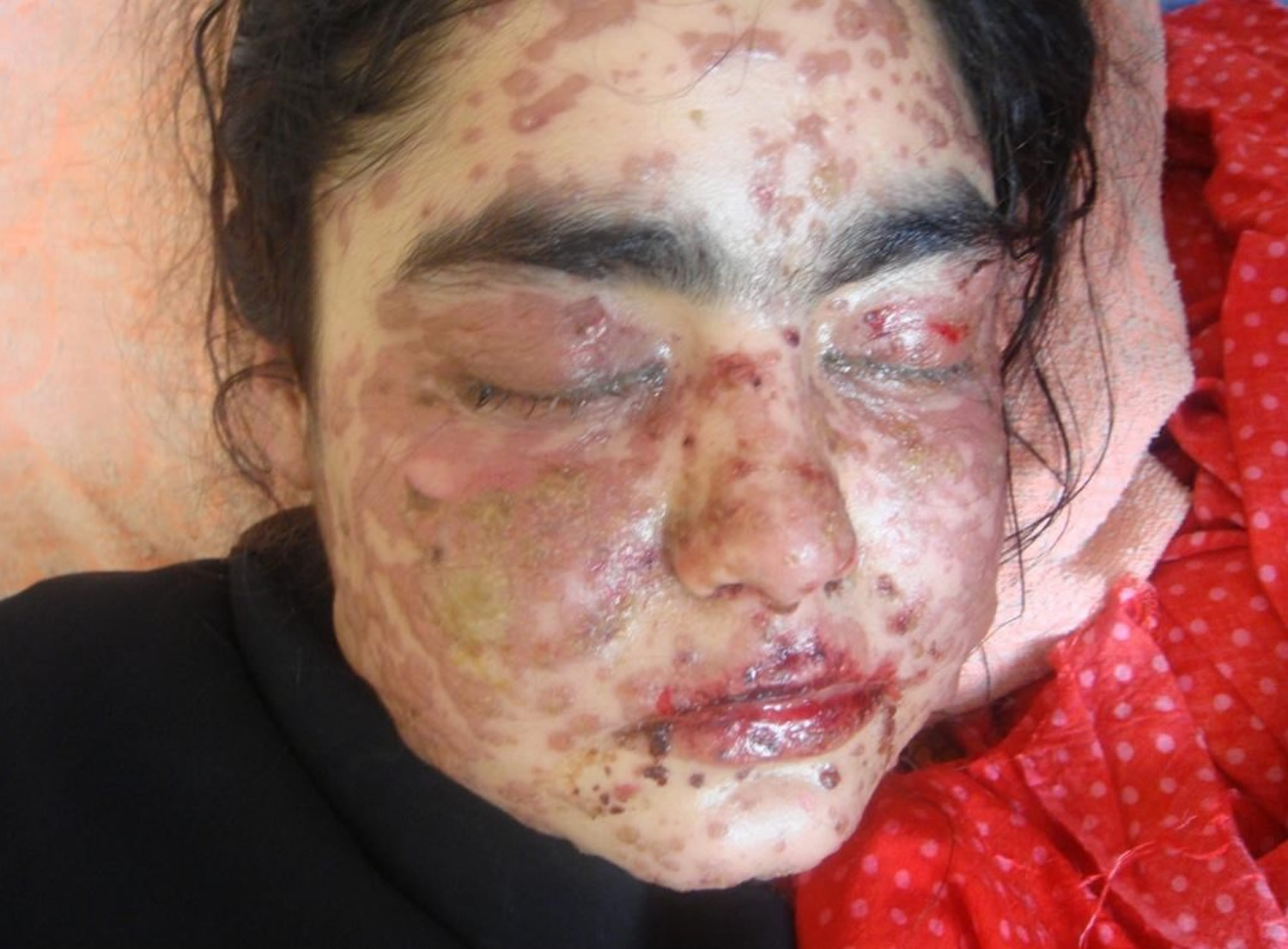
Figure 2. Stevens Johnson syndrome affecting the eye
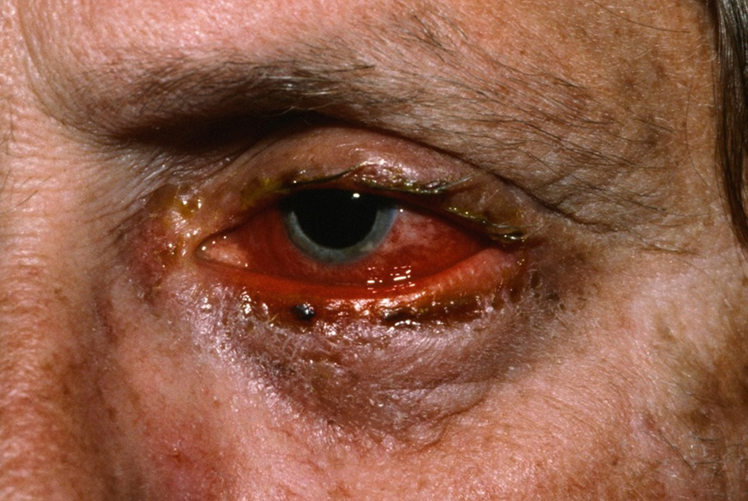
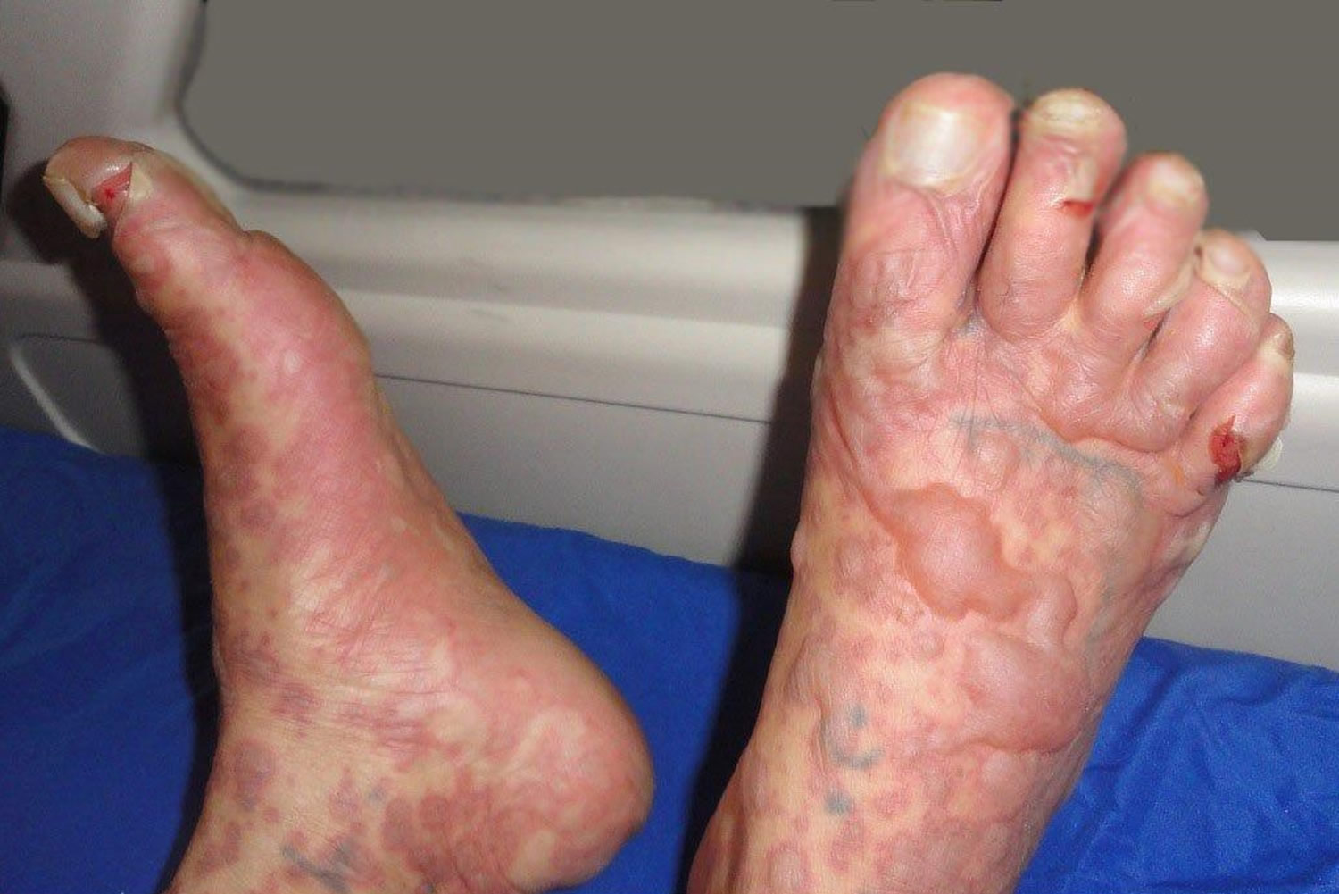
Figure 3. Stevens Johnson syndrome on feet
Who gets Stevens-Johnson syndrome/toxic epidermal necrolysis?
Stevens-Johnson syndrome/toxic epidermal necrolysis is a very rare complication of medication use (estimated at 1–2/million each year for SJS, and 0.4–1.2/million each year for toxic epidermal necrolysis).
- Anyone on medication can develop Stevens-Johnson syndrome/toxic epidermal necrolysis unpredictably.
- It can affect all age groups and all races.
- It is slightly more common in females than in males.
- It is 100 times more common in association with human immunodeficiency virus infection (HIV).
Genetic factors are important
- There are HLA associations in some races to anticonvulsants and allopurinol
- Polymorphisms to specific genes have been detected (eg, CYP2C coding for cytochrome P450 in patients reacting to anticonvulsants).
More than 200 medications have been reported in association with Stevens-Johnson syndrome/toxic epidermal necrolysis.
- It is more often seen with drugs with long half-lives compared to even a chemically similar related drug with a short half-life. A half-life of a medication is the time that half of the delivered dose remains circulating in the body.
- The medications are usually systemic (taken by mouth or injection) but TEN has been reported after topical use.
- No drug is implicated in about 20% of cases
- Stevens-Johnson syndrome/toxic epidermal necrolysis has rarely been associated with vaccination and infections such as mycoplasma and cytomegalovirus. Infections are generally associated mucosal involvement and less severe cutaneous disease than when drugs are the cause.
The drugs that most commonly cause Stevens-Johnson syndrome/toxic epidermal necrolysis are:
- Sulfonamides: cotrimoxizole;
- Beta-lactam: penicillins, cephalosporins
- Anti-convulsants: lamotrigine, carbamazepine, phenytoin, phenobarbitone
- Allopurinol
- Paracetamol/acetominophen
- Nevirapine (non-nucleoside reverse-transcriptase inhibitor)
- Nonsteroidal anti-inflammatory drugs (NSAIDs) (oxicam type mainly)
How does Stevens Johnson Syndrome start
Stevens–Johnson syndrome / toxic epidermal necrolysis usually develops within the first week of antibiotic therapy but up to 2 months after starting an anticonvulsant. For most drugs the onset is within a few days up to 1 month.
Before the rash appears, there is usually a prodromal illness of several days duration resembling an upper respiratory tract infection or ‘flu-like illness. Symptoms may include:
- Fever > 39 °C (102.2 °F)
- Sore throat, difficulty swallowing
- Runny nose and cough
- Sore red eyes, conjunctivitis
- General aches and pains.
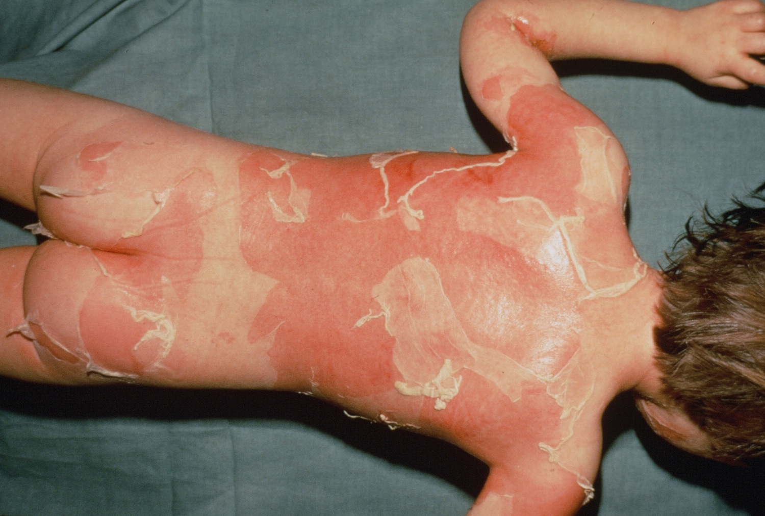
There is then an abrupt onset of a tender/painful red skin rash starting on the trunk and extending rapidly over hours to days onto the face and limbs (but rarely affecting scalp, palms or soles). The maximum extent is usually reached by 4 days.
The skin lesions may be:
- Macules — flat, red and diffuse (measles-like spots) or purple (purpuric) spots
- Diffuse erythema (redness)
- Targetoid — as in erythema multiforme (target like skin lesions)
- Blisters — flaccid (i.e not tense).
The blisters then merge to form sheets of skin detachment, exposing red, oozing dermis. The Nikolsky sign is positive in areas of skin redness. This means that blisters and erosions appear when the skin is rubbed gently.
Nikolsky sign
Your health care provider may use a pencil eraser to test for Nikolsky sign. The eraser is placed on your skin and gently twirled back and forth.
If the test result is positive, a blister will form in the area, usually within minutes.
A positive result is usually a sign of a blistering skin condition. People with a positive sign have loose skin that slips free from the underlying layers when rubbed. The area beneath is pink and moist, and usually very tender.
Figure 4. Stevens–Johnson syndrome – Bullous erythema multiforme
Figure 5. Toxic epidermal necrolysis – severe form of Stevens Johnson Syndrome (SJS)
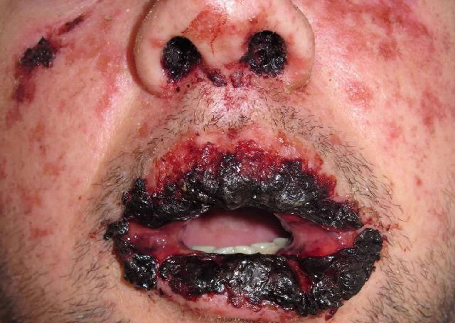
Mucosal involvement is prominent and severe, although not forming actual blisters. At least 2 mucosal surfaces are affected including:
- Eyes (conjunctivitis, less often corneal ulceration, anterior uveitis, panophthalmitis) — red, sore, sticky, photosensitive eyes
- Lips/mouth (cheilitis, stomatitis) — red crusted lips, painful mouth ulcers
- Pharynx, oesophagus — causing difficulty eating
- Genital area and urinary tract — erosions, ulcers, urinary retention
- Upper respiratory tract (trachea and bronchi) — cough and respiratory distress
- Gastrointestinal tract — diarrhea.
The patient is very ill, extremely anxious and in considerable pain. In addition to skin/mucosal involvement, other organs may be affected including liver, kidneys, lungs, bone marrow and joints.
Figure 6. Toxic epidermal necrolysis – lips and mouth
Stevens–Johnson syndrome / toxic epidermal necrolysis prognosis
The acute phase of Stevens–Johnson syndrome / toxic epidermal necrolysis lasts 8–12 days.
Repithelialisation of denuded areas takes several weeks, and is accompanied by peeling of the less severely affected skin. Survivors of the acute phase have increased on-going mortality especially if aged or sick.
Long-term sequelae include:
- Pigment change — patchwork of increased and decreased pigmentation
- Skin scarring, especially at sites of pressure or infection
- Loss of nails with permanent scarring (pterygium) and failure to regrow
- Scarred genitalia — phimosis (constricted foreskin which cannot retract) and vaginal adhesions (occluded vagina)
- Joint contractures
- Lung disease — bronchiolitis, bronchiectasis, obstructive disorders.
Eye problems can lead to blindness:
- Dry and/or watery eyes, which may burn and sting when exposed to light
- Conjunctivitis: red, crusted, or ulcerated conjunctiva
- Corneal ulcers, opacities and scarring
- Symblepharon: adhesion of conjunctiva of eyelid to eyeball
- Ectropion or entropion: turned-out or turned-in eyelid
- Trichiasis: inverted eyelashes
- Synechiae: iris sticks to cornea.
It may take weeks to months for symptoms and signs to settle.
Complications of Stevens–Johnson syndrome / toxic epidermal necrolysis
Stevens–Johnson syndrome / toxic epidermal necrolysis can be fatal due to complications in the acute phase. The mortality rate is up to 10% for Stevens Johnson syndrome SJS and at least 30% for toxic epidermal necrolysis.
During the acute phase, potentially fatal complications include:
- Dehydration and acute malnutrition
- Infection of skin (cellulitis), mucous membranes, lungs (pneumonia), septicemia (blood poisoning)
- Acute respiratory distress syndrome
- Gastrointestinal ulceration, perforation and intussusception
- Shock and multiple organ failure including kidney failure
- Thromboembolism and disseminated intravascular coagulopathy
- Eye problems. The rash caused by Stevens-Johnson syndrome can lead to inflammation in your eyes. In mild cases, this may cause irritation and dry eyes. In severe cases, it can lead to extensive tissue damage and scarring that results in visual impairment and, rarely, blindness.
- Lung involvement. The condition may lead to acute respiratory failure.
- Permanent skin damage. When your skin grows back following Stevens-Johnson syndrome, it may have abnormal bumps and coloring. And you may have scars. Lasting skin problems may cause your hair to fall out, and your fingernails and toenails may not grow normally.
- Problems with internal organs – organs can become inflamed – for example, the lungs (pneumonia), heart (myocarditis), kidneys (nephritis) or liver (hepatitis); the oesophagus may also become narrowed and scarred (oesophageal stricture)
- Problems with the sexual organs, such as vaginal stenosis (narrowing of the vagina caused by a build-up of scar tissue), and scarring of the penis, is also a possible complication of Stevens-Johnson syndrome.
Stevens Johnson syndrome causes
Stevens–Johnson syndrome / toxic epidermal necrolysis is a rare and unpredictable reaction to medication. The mechanism has still not been understood and is complex.
Drug specific CD8+ cytotoxic lymphocytes can be detected in the early blister fluid. They have some natural killer cell activity and can probably kill keratinocytes by direct contact. Cytokines implicated include perforin/granzyme, Fas-L and tumour necrosis factor alpha (TNFα).
There are probably two major pathways involved:
- Fas-Fas ligand pathway of apoptosis has been considered a pivotal step in the pathogenesis of TEN. The Fas ligand (FasL), a form of tumour necrosis factor, is secreted by blood lymphocytes and can bind to the Fas ‘death’ receptor expressed by keratinocytes.
- Granule-mediated exocytosis via perforin and granzyme B resulting in cytotoxicity (cell death). Perforin and granzyme B can be detected in early blister fluid and it has been suggested that levels may be associated with disease severity.
Triggers for Stevens–Johnson syndrome / toxic epidermal necrolysis
In children, Stevens-Johnson syndrome is usually triggered by a viral infection, such as:
- Mumps
- Flu or influenza
- Herpes-simplex virus, which causes cold sores
- Coxsackie virus, which causes Bornholm disease
- Epstein-Barr virus, which causes glandular fever
Less commonly, bacterial infections can also trigger the syndrome.
In adults, Stevens-Johnson syndrome is often caused by an adverse reaction to medication. The medications associated with a high risk of Stevens-Johnson syndrome are:
Medications (listed alphabetically)
This list of drugs known to cause Stevens–Johnson syndrome / toxic epidermal necrolysis is not exclusive.
- Abacavir
- Acetominophen
- Allopurinol
- Antibiotics
- Anticonvulsants
- Antifungals
- Antivirals
- Atenolol
- Azathioprine
- Beta-lactam antibiotics
- Captopril
- Carbamazepine
- Cephalosporins
- Clomipramine
- Cotrimoxazole
- Dapsone
- Diltiazem
- Gold salts
- Ibuprofen
- Imidazole antifungals, eg ketoconazole, itraconazole, fluconazole
- Isoniazid
- Lamotrigine
- Mexiletine
- Minocycline
- Naproxen
- Nevirapine (non-nucleoside reverse-transcriptase inhibitor)
- Nonsteroidal anti-inflammatory drugs (NSAIDs) (oxicam type mainly)
- Paracetamol
- Penicillins
- Phenobarbitone
- Phenytoin
- Sulfasalazine
- Sulfonamides
- Trimethoprim
- Valproic acid
- Vemurafenib
As a result of the associated risk, a thorough evaluation of the expected benefits of treatment is required when prescribing the above medications. Doctors are advised to carefully consider using these medications as first-line treatments, particularly if there are safer alternatives.
It’s important to emphasise that Stevens-Johnson syndrome is rare, and the overall risk of getting the syndrome is low, even for people using “high risk” medications (one in 1,000 to one in 100,000).
Other causes of Stevens–Johnson syndrome / toxic epidermal necrolysis
- Mycoplasma pneumoniae and cytomegalovirus infections
- Vaccinations
- Cancer, especially haematological cancers
- Systemic diseases
- Contrast medium
- External chemical exposure
- Herbal medicines
- Foods
- Bone marrow transplantation
- Radiotherapy
Risk factors for Stevens Johnson syndrome
Factors that increase your risk of developing Stevens-Johnson syndrome include:
- An HIV infection. Among people with HIV, the incidence of Stevens-Johnson syndrome is about 100 times greater than among the general population.
- A weakened immune system. If you have a weakened immune system, you may have an increased risk of Stevens-Johnson syndrome. Your immune system can be affected by an organ transplant, HIV/AIDS and autoimmune diseases.
- A history of Stevens-Johnson syndrome. If you’ve had a medication-related form of this condition, you are at risk of a recurrence if you use that drug again.
- A family history of Stevens-Johnson syndrome. If an immediate family member has had Stevens-Johnson syndrome or a related condition called toxic epidermal necrolysis, you may be more susceptible to developing Stevens-Johnson syndrome too.
- The HLA-B 1502 and HLA B 1508 gene. If you have a gene called HLA-B 1502 and HLA B1508, you have an increased risk of Stevens-Johnson syndrome, particularly if you take certain drugs for seizures, gout or mental illness. Families of Chinese, Southeast Asian or Indian descent are more likely to carry this gene. For example, Chinese people with the HLA B1502 gene have experienced Stevens-Johnson syndrome after taking carbamazepine, and allopurinol has also triggered the syndrome in Chinese people with the HLA B1508 gene.
How can Stevens–Johnson syndrome / toxic epidermal necrolysis be prevented?
People who have survived Stevens–Johnson syndrome / toxic epidermal necrolysis must be educated to avoid taking the causative drug or structurally related medicines as Stevens–Johnson syndrome / toxic epidermal necrolysis may recur. Cross-reactions can occur between:
- The anticonvulsants carbamazepine, phenytoin, lamotrigine and phenobarbital
- Beta-lactam antibiotics penicillin, cephalosporin and carbapenem
- Nonsteroidal anti-inflammatory drugs
- Sulfonamides: sulfamethoxazole, sulfadiazine, sulfapyridine.
If you’ve had this condition, avoid the medication that triggered it. If you’ve had Stevens-Johnson syndrome and your doctor told you it was caused by a medication, avoid that drug and others like it. This is key to preventing a recurrence, which is usually more severe than the first episode and can be fatal.
Your family members also might want to avoid this drug because some forms of this condition have a genetic risk factor.
If you have had Stevens-Johnson syndrome, be sure to:
- Know what caused your reaction. If your condition was caused by a medication, learn its name and that of closely related medications.
- Inform your health care providers. Tell all your health care providers that you have a history of Stevens-Johnson syndrome. If the reaction was caused by a medication, tell them which one.
- Wear a medical information bracelet or necklace. Have information about your condition and what caused it inscribed on a medical information bracelet or necklace. Always wear it.
In the future, doctors may be able to predict who is at risk of Stevens–Johnson syndrome / toxic epidermal necrolysis using genetic screening.
- Consider genetic testing before taking certain drugs. If you are of Chinese, Southeast Asian or Indian descent, talk with your doctor before taking carbamazepine (Carbatrol, Tegretol). This drug is useful to treat epilepsy, bipolar disorder and other conditions. But people with a gene called HLA-B1502 and HLA B1508 have an increased risk of Stevens-Johnson syndrome if they take this drug.
Allopurinol should be prescribed for good indications (e.g, gout with hyperuricaemia) and commenced at low dose (100 mg/day), as Stevens–Johnson syndrome / toxic epidermal necrolysis is more likely at doses > 200 mg/day.
Stevens Johnson syndrome symptoms
Stevens-Johnson syndrome signs and symptoms include:
- Fever
- Unexplained widespread skin pain
- A red or purple skin rash that spreads
- Blisters on your skin and the mucous membranes of your mouth, nose, eyes and genitals
- Shedding of your skin within days after blisters form
If you have Stevens-Johnson syndrome, several days before the rash develops you may experience:
- Fever
- Sore mouth and throat
- Fatigue
- Cough
- Burning eyes.
Stevens Johnson syndrome SJS diagnosis
Stevens–Johnson syndrome / toxic epidermal necrolysis is suspected clinically and classified based on the skin surface area detached at maximum extent.
Stevens–Johnson syndrome SJS
- Skin detachment < 10% of body surface area (BSA)
- Widespread erythematous or purpuric macules or flat atypical targets
Overlap Stevens–Johnson syndrome / toxic epidermal necrolysis
- Detachment between 10% and 30% of BSA
- Widespread purpuric macules or flat atypical targets
Toxic epidermal necrolysis with spots
- Detachment > 30% of BSA
- Widespread purpuric macules or flat atypical targets
Toxic epidermal necrolysis without spots
- Detachment of > 10% of BSA
- Large epidermal sheets and no purpuric macules
The category cannot always be defined with certainty on initial presentation. The diagnosis may therefore change during the first few days in hospital.
Investigations in Stevens–Johnson syndrome / toxic epidermal necrolysis
If the test is available, elevated levels of serum granulysin taken in the first few days of a drug eruption may be predictive of Stevens–Johnson syndrome / toxic epidermal necrolysis.
Skin biopsy is usually required to confirm the clinical diagnosis and to exclude Staphylococcal scalded skin syndrome and other generalized rashes with blisters.
The histopathology shows keratinocyte necrosis (death of individual skin cells), full thickness epidermal/epithelial necrosis (death of an entire layer of skin), minimal inflammation (very mild lymphocytic infiltrate of the superficial dermis). The direct immunofluoresence test on the skin biopsy is negative, indicating the disease is not due to deposition of antibodies in the skin.
Blood tests do not help to make the diagnosis but are essential to make sure fluid and vital nutrients have been replaced, to identify complications and to assess prognostic factors.
Abnormalities may include:
- Anemia occurs in virtually all cases (reduced hemoglobin).
- Leucopenia (reduced white cells), especially lymphopenia (reduced lymphocytes) is very common (90%).
- Neutropenia (reduced neutrophils), if present, is a bad prognostic sign.
- Eosinophilia (raised eosinophil count) and atypical lymphocytosis (odd-looking lymphocytes) do not occur.
- Mildly raised liver enzymes are common (30%) and approximately 10% develop overt hepatitis.
- Mild proteinuria (protein leaking into urine) occurs in about 50%. Some changes in kidney function occur in the majority.
Patch testing rarely identifies the culprit in Stevens–Johnson syndrome / toxic epidermal necrolysis following recovery, and is not recommended.
SCORTEN
SCORTEN is an illness severity score that has been developed to predict mortality in SJS and toxic epidermal necrolysis cases. One point is scored for each of seven criteria present at the time of admission. The SCORTEN criteria are:
- Age > 40 years
- Presence of a malignancy (cancer)
- Heart rate > 120
- Initial percentage of epidermal detachment > 10%
- Serum urea level > 10 mmol/L
- Serum glucose level > 14 mmol/L
- Serum bicarbonate level < 20 mmol/L.
The risk of dying from Stevens–Johnson syndrome / toxic epidermal necrolysis depends on the score.
- SCORTEN 0-1 > 3.2%
- SCORTEN 2 > 12.1%
- SCORTEN 3 > 35.3%
- SCORTEN 4 > 58.3%
- SCORTEN 5 or more > 90%
Differential diagnosis of Stevens–Johnson syndrome / toxic epidermal necrolysis
In the differential diagnosis of Stevens–Johnson syndrome / toxic epidermal necrolysis consider:
- Other severe cutaneous adverse reactions to drugs (e.g, drug hypersensitivity syndrome)
- Staphylococcal scalded skin syndrome and toxic shock syndrome
- Erythema multiforme, particularly erythema multiforme major (with mucosal involvement)
- Mycoplasma infections
- Bullous systemic lupus erythematosus
- Paraneoplastic pemphigus.
Stevens Johnson syndrome treatment
Stevens-Johnson syndrome requires hospitalization, often in an intensive care unit or a burn unit.
Stopping nonessential medications
The first and most important step in treating Stevens-Johnson syndrome is to discontinue any medications that may be causing it. Because it’s difficult to determine exactly which drug may be causing the problem, your doctor may recommend that you stop taking all nonessential medications.
The prognosis of Stevens–Johnson syndrome / toxic epidermal necrolysis should be determined during the first 24 hours. SCORTEN is an illness severity score that has been developed to predict mortality in SJS/TEN. One point is scored for each of seven criteria present at the time of admission. The SCORTEN criteria are:
- Age > 40 years
- Presence of a malignancy
- Heart rate > 120
- Initial percentage of epidermal detachment > 10%
- Serum urea level > 10 mmol/L
- Serum glucose level > 14 mmol/L
- Serum bicarbonate level < 20 mmol/L.
The risk of dying from Stevens–Johnson syndrome / toxic epidermal necrolysis depends on the score. Depending on local protocols, if SCORTEN is more than 1, the patient is managed in intensive care, a burns unit or a specialist dermatology unit of a regional hospital.
Estimate total body surface with epidermal detachment
Use the Wallace rule of 9 to estimate the affected body surface area.
Percentages of the total body surface area for an adult or child over 10 years
- Head and neck (front and back): 9%
- Each upper limb (front and back): 9%
- Chest and front of abdomen: 18%
- Back: 18%
- Perineum: 1%
- Each lower limb (front and back): 18%
Percentages of the total body surface area for a child under the age of 1
- Head and neck (front and back): 18%
- Each upper limb (front and back): 9%
- Chest and front of abdomen: 18%
- Back: 18%
- Perineum: 1%
- Each lower limb (front and back): 14%
Over 1 year and below 10 years, the percentage of body surface area changes
- Head decreases by 1% per year
- Lower limbs each increase by 0.5% per year
Care of a patient with Stevens–Johnson syndrome / toxic epidermal necrolysis
- Cessation of suspected causative drug(s) — the patient is less likely to die and complications are less if the culprit drug is on or before the day that blisters/erosions appear
- Hospital admission — preferably immediately to an intensive care and/or burns unit with specialist nursing care, as this improves survival, reduces infection and shortens hospital stay
- Consider fluidized air bed
- Nutritional and fluid replacement (crystalloid) by intravenous and nasogastric routes — reviewed and adjusted daily
- Temperature maintenance — as body temperature regulation is impaired, patient should be in a warm room (30–32C)
- Pain relief — as pain can be extreme
- Sterile handling and reverse isolation procedures.
- Wound care.
- Eye care.
Skin care
- Examine daily for extent of detachment and for infection (take swabs for bacterial culture)
- Topical antiseptics (eg, silver nitrate, chlorhexidine [but not silver sulfadiazine as it is a sulfa drug])
- Dressings such as gauze with petrolatum, non-adherent nanocrystalline-containing silver gauze or biosynthetic skin substitutes can reduce pain
- Avoid using adhesive tapes and unnecessary removal of dead skin; leave the blister roof as a ‘biological dressing’
Bathing
- Daily bathing should not exceed 15 minutes. It may contain either;
- Antiseptic solution, e.g, chlorhexidine 4% for 1.5L of water, if suspicious of infection; or;
- Oatmeal : 3 packets for one bath, if there are dry lesions or crusts.
- Check the water temperature of bath and hand shower.
- Motivate the patient to move by himself to avoid injury during carrying.
- Put the patient under a nursing board.
- Carefully immerse the patient in the bathwater.
- Gently remove dressings, crust, and exudate; avoid scrubbing.
- To clean the skin, tap with a wash cloth rather than rub.
- Rinse with the hand shower, lifting the nursing board above the bath.
- Gently tap to dry using dry wash cloths.
If a bath is contraindicated or unavailable, perform a gentle bed-bath using aqueous cream, warm water and a soft cloth.
Eye care
- Daily assessment by ophthalmologist
- Frequent eye drops/ointments (antiseptics, antibiotic, corticosteroid)
- Eye care is undertaken 3–6 times each day depending on severity of eye involvement.
- Apply vitamin A or other sterile ocular lubricant ointment generously under the upper and the lower eyelid using one quarter of tube for one eye on each occasion.
- Synechiae can be released by applying the eye ointment.
- Ask the patient to open his/her eyes frequently to avoid synechia formation.
- Do not use saline eye drops or sticks.
- Only use other eye preparations that have been prescribed by an ophthalmologist (including prior prescriptions).
- Put some petroleum jelly on the eyelids if there is crust or erosions
Mouth care
- Mouthwashes
- Topical oral anesthetic
- Mouth care is undertaken 6 times a day.
- Put the mouthwash solution in glass or syringe.
- Ask the patient to gargle with the mouthwash.
- Repeat the gargle three times.
- Spit the solution into the bean-shaped bowl.
- Presoak the stick with the mouthwash solution.
- Use the stick to delicately clean the mucosa of cheek, gum, tongue; change sticks frequently and avoid injury to mucosal lesions.
- Make sure the patient doesn’t swallow the solution.
- Patients can do this care themselves.
Genital care
If ulcerated, prevent vaginal adhesions using intravaginal steroid ointment, soft vaginal dilators.
Genital care in females
- During the bath
- Clean the genitals delicately with a compress to remove exudate and necrotic mucosa.
- Avoiding injuring the mucosa.
- Sever any synechia between labia minora and labia majora.
- After the bath
- Put a sterile compress into the finger of a sterile glove. Apply petroleum jelly to the outside of the glove.
- The patient or the health provider must put the petroleum-jelly-wrapped compress/glove into the vagina and gently remove it so that the jelly lubricates the lining of the vagina.
Genital care in males
- During the bath
- Pull back the foreskin.
- Clean the genitals delicately with a compress to remove exudate and necrotic mucosa.
- Sever any synechia.
- Avoid injuring the mucosa.
- Replace the foreskin.
- After the bath and 4–6 times a day
- Tap gently with soft wash cloth to dry; do not rub.
- Pull back the foreskin to apply petroleum jelly.
- Replace foreskin.
Lung care
- Consider aerosols, bronchial aspiration, physiotherapy
- May require intubation and mechanical ventilation if trachea and bronchi are involved
Urinary care
- Catheter because of genital involvement and immobility
- Culture urine for bacterial infection
General
- Psychiatric support for extreme anxiety and emotional lability
- Physiotherapy to maintain joint movement and reduce risk of pneumonia
- Regular assessment for staphylococcal or gram negative infection
- Appropriate antibiotic should be given if infection develops; prophylactic antibiotics are not recommended and may even increase the risk of sepsis
- Consider heparin to prevent thromboembolism (blood clots).
The role of systemic corticosteroids (cortisone) remains controversial. Some clinicians prescribe high doses of corticosteroids for a short time at the start of the reaction, e.g prednisone 1–2 mg/kg/day for 3–5 days. However concerns have been raised that they may increase the risk of infection, impair wound healing and other complications, and they have not been proven to have any benefit. They are not effective later in the course of the illness.
Case reports and small patient series have reported benefit from active adjuvant treatments delivered during the first 24–48 hours of illness. As Stevens–Johnson syndrome / toxic epidermal necrolysis is fortunately a rare condition, controlled trials of therapies in large numbers of patients are difficult.
Ciclosporin 3–5 mg/kg/day is reported to reduce mortality by 60% compared to patients with similar SCORTEN score on admission that were not treated with ciclosporin.
Other options include:
- Anti-TNFα monoclonal antibodies (eg, infliximab, etanercept)
- Cyclophosphamide
- Intravenous immunoglobulin (IVIG) 2–3 g/kg given over 2–3 days
- Plasmapheresis.
Thalidomide, trialled because of its anti-TNFα effect, increased mortality, and should not be used.


