Contents
What is cerebrospinal fluid
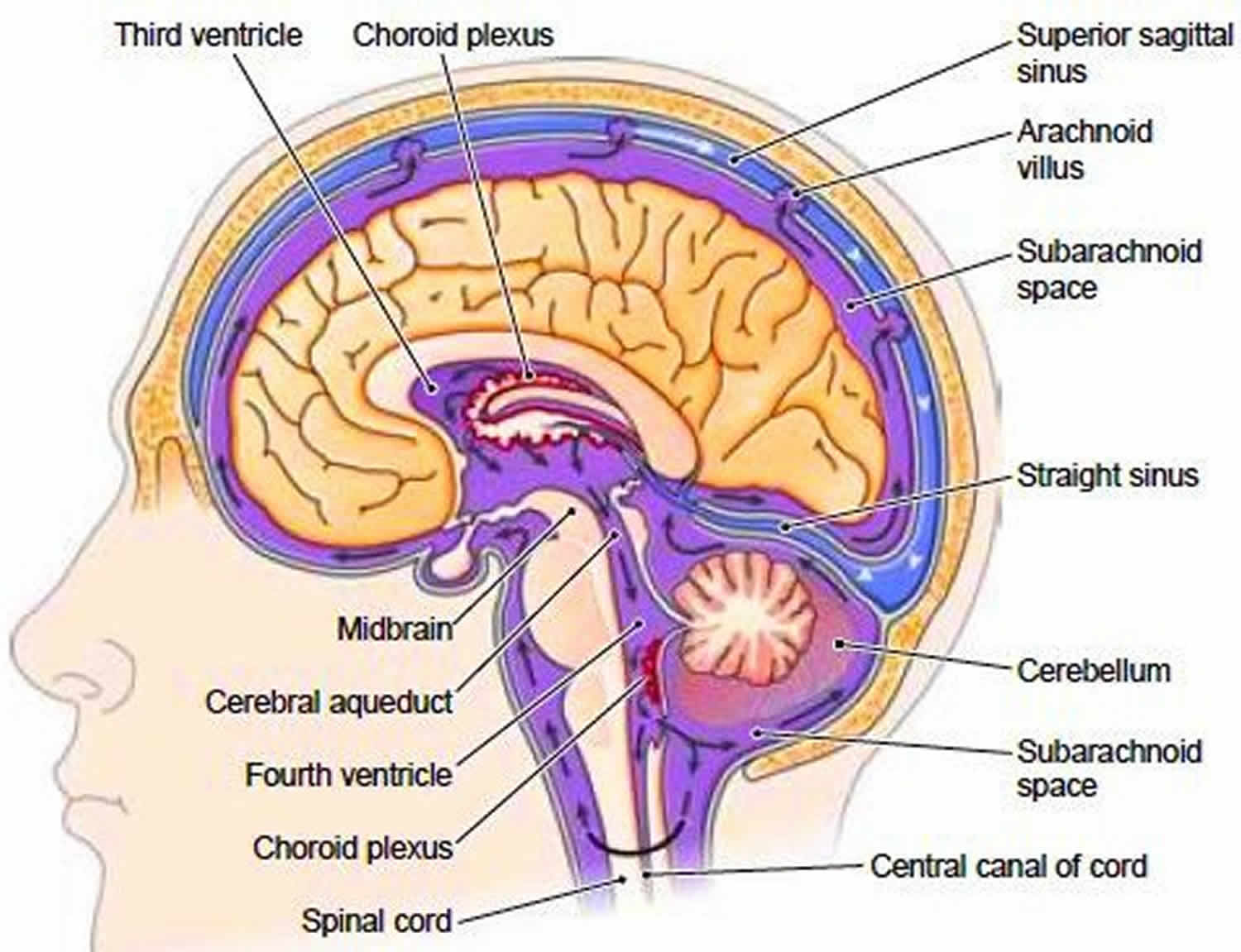
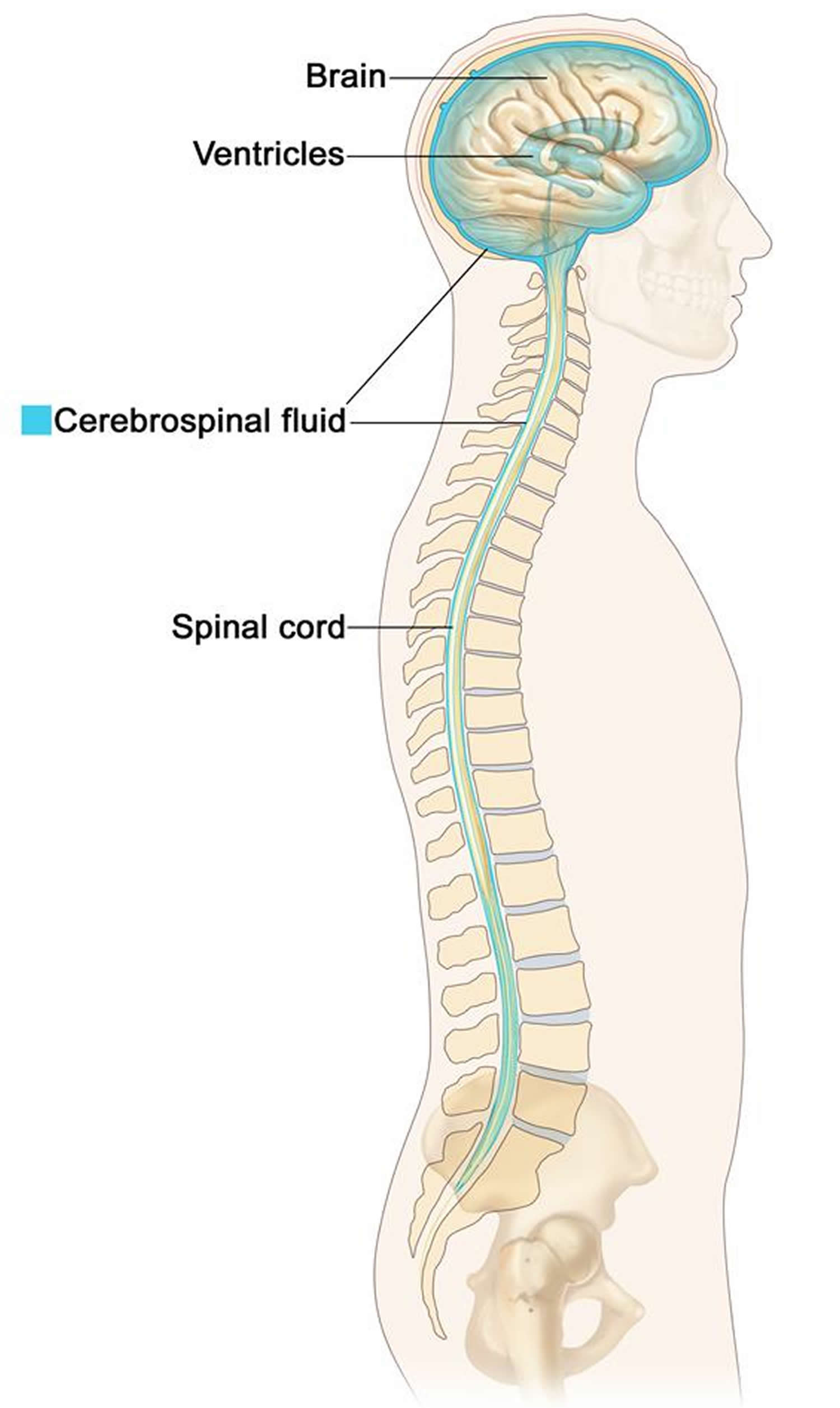
Cerebrospinal fluid (CSF) is a clear, colorless liquid that can be found around and within the ventricles and canals of the central nervous system (brain and spinal cord) and bathes its external surface. The brain produces about 500 mL of cerebrospinal fluid per day, but the cerebrospinal fluid is constantly reabsorbed at the same rate and only 100 to 160 mL is normally present at one time 1. It should be noted that cerebrospinal fluid is continuously generated and reabsorbed. Depending on the rate of production and absorption (which varies person to person), the supply of cerebrospinal fluid can be replaced about every 7.5 hours. Most this cerebrospinal fluid is produced in the ventricles of the brain by the choroid plexus. However, a smaller portion is produced by the ependymal cells which line the ventricles. After production, the cerebrospinal fluid travels through the ventricles and then around the brain and spinal cord. Cerebrospinal fluid (CSF) is then reabsorbed directly into the blood through structures in the arachnoid mater called arachnoid villi (arachnoid granulations). Importantly, cerebrospinal fluid can be examined clinically through a lumbar puncture. With a lumbar puncture, physicians can look for abnormalities in the cerebrospinal fluid, which can be helpful when creating a differential diagnosis 2.
Figure 1. Cerebrospinal fluid
Where is cerebrospinal fluid produced?
Cerebrospinal fluid is made by tissue called the choroid plexus that lines the ventricles (hollow spaces) in the brain. On the floor or wall of each ventricle is a spongy mass of blood capillaries called a choroid plexus, named for its histological resemblance to a fetal membrane called the chorion. Ependyma, a type of neuroglia that resembles a cuboidal epithelium, lines the ventricles and canals and covers the choroid plexuses. It produces cerebrospinal fluid (CSF).
About 40% of it is formed in the subarachnoid space external to the brain, 30% by the general ependymal lining of the brain ventricles, and 30% by the choroid plexuses. Cerebrospinal fluid production begins with the filtration of blood plasma through the capillaries of the brain. Ependymal cells modify the filtrate as it passes through them, so the cerebrospinal fluid has more sodium chloride than blood plasma, but less potassium, calcium, and glucose and very little protein.
It is not currently known when the production of cerebrospinal fluid (CSF) first begins. However, the choroid plexus can be seen developing at around 32 days of development. Also, the arachnoid villi are beginning to form around the 35 weeks with continued development during the first 18 months of life.
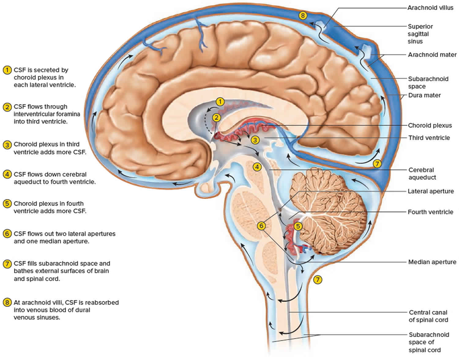
Figure 2. Cerebrospinal fluid formation, absorption and circulation around and within the brain
Cerebrospinal fluid circulation
The cerebrospinal fluid continually flows in and around the hollow spaces of the brain and spinal cord, and between two of the meninges (the thin layers of tissue that cover and protect the brain and spinal cord), driven partly by its own pressure, partly by the beating of ependymal cilia, and partly by rhythmic pulsations of the brain produced by each heartbeat. The cerebrospinal fluid of the lateral ventricles flows through the interventricular foramina into the third ventricle, then down the cerebral aqueduct to the fourth ventricle. The third and fourth ventricles and their choroid plexuses add more cerebrospinal fluid along the way. A small amount of cerebrospinal fluid fills the central canal of the spinal cord, but ultimately, all of it escapes through three pores in the fourth ventricle—a median aperture and two lateral apertures. These lead into the subarachnoid space on the brain and spinal cord surface. From here, cerebrospinal fluid is reabsorbed by arachnoid granulations, extensions of the arachnoid meninx shaped like little sprigs of cauliflower, protruding through the dura mater into the superior sagittal sinus. Cerebrospinal fluid penetrates the walls of the granulations and mixes with blood in the sinus.
Cerebrospinal fluid function
Cerebrospinal fluid is thought to have several important functions. One of these is functions is to provide a buoyant force to support the brain. The brain has a significant amount of mass (approximately 1400 gm) while at the same time being relatively malleable. However, since the cerebrospinal fluid surrounds the brain, it dissipates a lot of the downward force that would normally act on the organ. This reduces the stress on the brain and allows for it to maintain its shape. Another function of cerebrospinal fluid that comes from its physical properties as fluid is to protect the brain from the damage that would result from a sudden movement of the skull. Any rapid acceleration/deceleration of the head has the potential to injure the delicate contents that are contained within. The cerebrospinal fluid helps reduce the potential damage in such an event by acting as a cushion and a shock absorber. Since cerebrospinal fluid is being produced and absorbed continuously, it is also thought to help clear waste products from around the brain and regulate intracranial pressures. This dynamic process can be controlled by either increasing or reducing production to match the needs of the central nervous system at any given time. A similar regulatory mechanism can also reduce ischemia in the central nervous system (CNS) tissues. In times of ischemia, it is possible for the volume of cerebrospinal fluid to decrease, and therefore, decrease intracranial pressures. This decrease in intracranial pressure reduces the mechanical stress on blood vessels and may reduce ischemia 3.
Cerebrospinal fluid serves three functions for the brain:
- Buoyancy. Because the brain and cerebrospinal fluid are similar in density, the brain neither sinks nor floats in the cerebrospinal fluid. It hangs from delicate specialized fibroblasts of the arachnoid meninx. A human brain removed from the body weighs about 1.5 kg, but when suspended in cerebrospinal fluid its effective weight is only about 50 g. This buoyancy allows the brain to attain considerable size without being impaired by its own weight. If the brain rested heavily on the floor of the cranium, the pressure would kill the nervous tissue.
- Protection. Cerebrospinal fluid also protects the brain from striking the cranium when the head is jolted. If the jolt is severe, however, the brain still may strike the inside of the cranium or suffer shearing injury from contact with the angular surfaces of the cranial floor. This is one of the common findings in head injuries (concussions) from contact sports like NFL, rugby and boxing.
- Chemical stability. Cerebrospinal fluid rinses metabolic wastes from the nervous tissue and regulates its chemical environment. Slight changes in cerebrospinal fluid composition can cause malfunctions of the nervous system. For example, a high glycine concentration disrupts the control of body temperature and blood pressure, and a high pH causes dizziness and fainting.
Cerebrospinal fluid clinical significance
Lumbar Puncture
An important clinical test that clinicians use to monitor the cerebrospinal fluid is the lumbar puncture (spinal tap). In this procedure, a long, hollow needle is typically inserted into the subarachnoid space of the lumbar spine just below where the spinal cord ends (usually below L2). The needle can then be uncapped, and samples of cerebrospinal fluid can be obtained. The first thing that is noted when doing a lumbar puncture is the opening cerebrospinal fluid pressure. A high opening pressure can suggest various conditions including hydrocephalus, intracranial mass, and idiopathic intracranial hypertension. When there is a high cerebrospinal fluid pressure, there is a risk of causing damage to the brain by reducing the pressure too quickly. For this reason, care is often taken to make sure that there are no masses pressing on the brain before performing this procedure. A low cerebrospinal fluid pressure can also be clinically significant. This can be seen in conditions that allow for cerebrospinal fluid to leak into surrounding tissues. cerebrospinal fluid can leak due to physical trauma, previous lumbar punctures, or without an obvious physical cause called spontaneous cerebrospinal fluid leak.
Several analyses can be done on the contents of cerebrospinal fluid obtained from a lumbar puncture. Since cerebrospinal fluid should be transparent, the color is often noted. A cloudy appearance can suggest an infectious cause, and red color can suggest the presence of blood. Cultures can be taken from this fluid to look for different infectious agents that can cause meningitis. Additionally, molecular analyses can be done to look for protein abnormalities. These tests can be used to diagnose a wide variety of diseases affecting the central nervous system 4.
Hydrocephalus
Hydrocephalus is one important clinical disorder of cerebrospinal fluid. In this disorder, there is increased intracranial pressure due to an overabundance of cerebrospinal fluid. This abundance is usually caused by an obstruction in the flow of cerebrospinal fluid through the brain. In an adult, this can be caused by several different things. A few examples of possible culprits include neoplastic growths, infections (meningitis), traumatic brain injury, and hemorrhage. Hydrocephalus can also occur at birth causing an acute enlargement of the child’s head, or it can be acquired later in life. Increased intracranial pressure is not as dangerous in an infant since the cranial bones fuse later in life. For this reason, an infant with hydrocephalus can present with an enlarged head. When a child is born with this condition, it is usually due to a defect in the formation of the neural tube or stenosis of the cerebral aqueduct.
The treatment of hydrocephalus involves removing whatever is causing the obstruction of cerebrospinal fluid flow. In cases where there is recurrence or difficulty in ensuring a non-obstructed flow of cerebrospinal fluid, a shunt can be placed to help drain excess fluid from the intracranial cavity. These shunts most often drain the cerebrospinal fluid into the peritoneal cavity, but they can also be placed in other locations such as the pleural cavity, gallbladder, and the right atrium of the heart 5.
Idiopathic Intracranial Hypertension
Idiopathic intracranial hypertension is characterized by an increased intracranial pressure (usually detected through lumbar puncture) that has no discernable cause. In other words, it is unclear what is causing the increased cerebrospinal fluid pressures. Patients often experience symptoms such as a headache, vision changes, pulsatile tinnitus, weakness, nausea, vomiting, etc. The diagnosis is generally made based on the history and physical exam. However, neuroimaging investigations and lumbar puncture are often done to rule out other potential causes.
Chiari Malformations
Arnold-Chiari, also known as Chiari malformation, is the name given to a group of deformities of the hindbrain (cerebellum, pons and medulla oblongata) 6. There are two types recognized:
Chiari Type I Malformation
- Occurs in 1 in 1,000 births; more common than type II malformations
- Genetic syndromes related or more commonly idiopathic reduced volume of the posterior fossa leads to a displacement of the cerebellar tonsils into the spinal canal.
Chiari Type II Malformation
- Invariably results in the setting of myelomeningocele. Leakage of cerebrospinal fluid (cerebrospinal fluid) to the myelomeningocele yields herniation of the hindbrain into the spinal canal which disturbs the cerebrospinal fluid normal flow and subarachnoid adhesions.
Cerebrospinal fluid leak
A cerebrospinal fluid leak is an escape of the fluid that surrounds the brain and spinal cord.
Cerebrospinal fluid leak causes
Any tear or hole in the membrane that surrounds the brain and spinal cord (dura) can allow the cerebrospinal fluid that surrounds those organs to leak. When cerebrospinal fluid leaks out, the pressure around the brain and spinal cord drops.
Causes of cerebrospinal fluid leakage through the dura include:
- Certain head, brain, or spinal surgeries
- Head injury
- Placement of tubes for epidural anesthesia or pain medicines
- Spinal tap (lumbar puncture)
Sometimes, no cause can be found. This is called a spontaneous cerebrospinal fluid leak.
Cerebrospinal fluid leak symptoms
Cerebrospinal fluid leak symptoms may include:
- A headache that is worse when you sit up and improves when you lie down. It may be associated with light sensitivity, nausea, and neck stiffness.
- Drainage of cerebrospinal fluid from the ear (cerebrospinal fluid otorrhea) (rarely).
- Drainage of cerebrospinal fluid from the nose cerebrospinal fluid rhinorrhea (rarely).
Cerebrospinal fluid leak diagnosis
Your health care provider will perform a physical exam and ask about your symptoms. Tests may include:
- CT scan of the head with contrast dye
- CT myelogram of the spine
- MRI of the head or spine
- Radioisotope test of the CSF to track the leakage
Cerebrospinal fluid leak treatment
Depending on the cause of the leak, many symptoms improve on their own after a few days. Complete bed rest for several days is usually recommended. Drinking more fluids, especially drinks with caffeine, can help slow or stop the leak and may help with headache pain.
Headache may be treated with pain relievers and fluids. If the headache lasts longer than a week after a lumbar puncture, a procedure may be done to block the hole that may be leaking fluid. This is called a blood patch, because a blood clot can be used to seal the leak. In most cases, this makes symptoms go away. In rare cases, surgery is needed to repair the tear in the dura and stop the headache.
If symptoms of infection (fever, chills, change in mental status) are present, they need to be treated with antibiotics.
Cerebrospinal fluid leak prognosis
Outlook is usually good depending on the cause. Most cases heal by themselves with no lasting symptoms.
If the cerebrospinal fluid leak keeps coming back, high pressure of the cerebrospinal fluid (hydrocephalus) might be the cause and should be treated.
Cerebrospinal fluid leak possible complications
Complications may occur if the cause is surgery or trauma. Infections after surgery or trauma can lead to meningitis and serious complications, such as swelling of the brain, and need to be treated right away.
- Huff T, Tadi P, Varacallo M. Neuroanatomy, Cerebrospinal Fluid. [Updated 2019 May 1]. In: StatPearls [Internet]. Treasure Island (FL): StatPearls Publishing; 2019 Jan-. Available from: https://www.ncbi.nlm.nih.gov/books/NBK470578[↩]
- Javed K, Lui F. StatPearls [Internet]. StatPearls Publishing; Treasure Island (FL): Feb 18, 2019. Neuroanatomy, Choroid Plexus.[↩]
- Pormohammad A, Nasiri MJ, McHugh TD, Riahi SM, Bahr NC. A systematic review and meta-analysis of the diagnostic accuracy of nucleic acid amplification tests in cerebrospinal fluid for tuberculous meningitis. J. Clin. Microbiol. 2019 Apr 03[↩]
- Özütemiz C, Rykken JB. Lumbar puncture under fluoroscopy guidance: a technical review for radiologists. Diagn Interv Radiol. 2019 Mar;25(2):144-156[↩]
- Graff-Radford NR, Jones DT. Normal Pressure Hydrocephalus. Continuum (Minneap Minn). 2019 Feb;25(1):165-186[↩]
- Mohammad SA, Osman NM, Ahmed KA. The value of CSF flow studies in the management of CSF disorders in children: a pictorial review. Insights Imaging. 2019 Jan 28;10(1):3[↩]