Contents
What is upper motor neuron
Upper motor neurons originate in the gray matter of the brain and send long axons which carry the impulses for movement down a descending tract of the spinal cord. These axons synapse with lower motor neurons in the ventral horn of the spinal cord. The axons of the lower motor neurons exit the ventral root of the spinal cord to innervate the muscles or glands of the body. An upper motor neuron is actually an interneuron and not a true motor neuron: it is so named because the cell originates in the upper part of the central nervous system (brain) and regulates the activity of lower motor neurons. Only a lower motor neuron is a true motor neuron because it conveys action potentials from the central nervous system (CNS) to skeletal muscles in the periphery.
Upper motor neurons from the cerebral cortex are essential for the planning and execution of voluntary movements of the body. Other upper motor neurons originate in motor centers of the brainstem: the vestibular nuclei, reticular formation, superior colliculus, and red nucleus. Upper motor neurons from the brainstem help regulate posture, balance, muscle tone, and reflexive movements of the head and trunk.
The primary tract which carries signals for voluntary movement is known as the pyramidal tract. The pyramidal tract divides further into the corticospinal tract and the corticobulbar tract. Injury or lesions to upper motor neuron’s are common because of the vast areas covered by the motor neuron pathways. upper motor neuron lesions are designated as any damage to the motor neurons that reside above nuclei of cranial nerves or the anterior horn cells of the spinal cord. Damage to upper motor neuron’s lead to a characteristic set of clinical symptoms known as the upper motor neuron syndrome. These symptoms can include weakness, spasticity, clonus, and hyperreflexia. upper motor neuron’s lesions have a wide differential diagnosis which ranges from cerebrovascular accidents, traumatic brain injury, malignancy, infections, inflammatory disorders, neurodegenerative disorders, and metabolic disorders.
Figure 1. Upper motor neuron
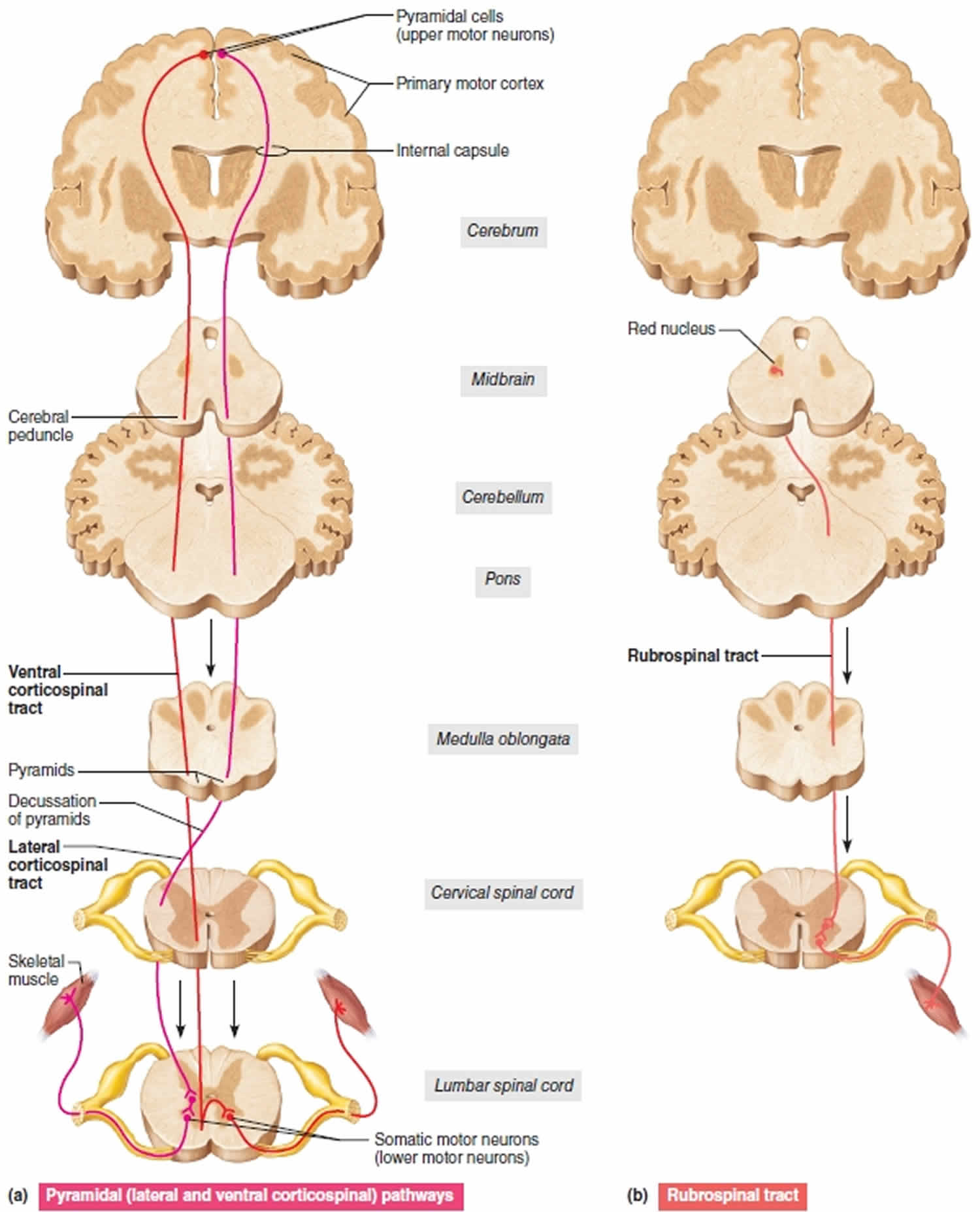
Footnote: Three upper motor neuron descending pathways by which the brain influences movement. Cross sections up to the cerebrum, which is shown in frontal section. (a) Pyramidal (lateral and ventral corticospinal pathways) originate from the cerebral cortex and control skilled, voluntary movements. (b) The rubrospinal tract, one of the indirect pathways, helps regulate muscle tone.
Figure 2. Upper and lower motor neuron
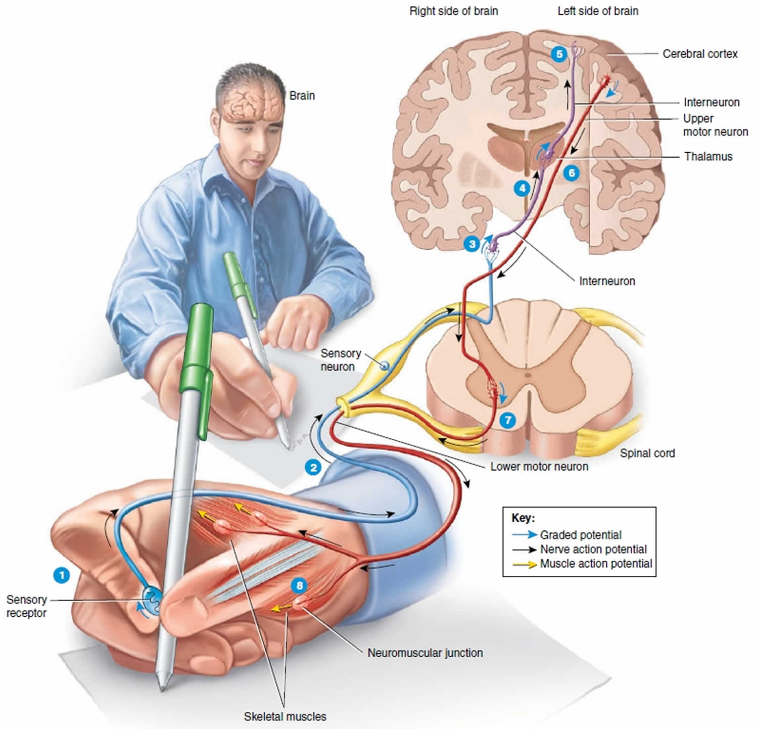
Footnotes: 1) As you touch the pen, a sensory receptor in the skin of the fingers is stimulated. 2) The sensory receptor in the skin of the fingers triggers the axon of the sensory neuron to send electrical signal which travels along the axon into the spinal cord and the brain and ultimately causes the release of neurotransmitter at a synapse with an interneuron. 3) The neurotransmitter stimulates the interneuron to form a graded potential in its dendrites and cell body. 4) In response to the graded potential, the axon of the interneuron forms a nerve action potential. The nerve action potential travels along the axon, which results in neurotransmitter release at the next synapse with another interneuron. 5) This process of neurotransmitter release at a synapse followed by the formation of a graded potential and then a nerve action potential occurs over and over as interneurons in higher parts of the brain (such as the thalamus and cerebral cortex) are activated. Once interneurons in the cerebral cortex, the outer part of the brain, are activated, perception occurs and you are able to feel the smooth surface of the pen touch your fingers. The conscious awareness of a sensation, is primarily a function of the cerebral cortex.
Suppose that you want to use the pen to write a letter.
The nervous system would respond in the following way:
6) A stimulus in the brain causes a graded potential to form in the dendrites and cell body of an upper motor neuron, a type of motor neuron that synapses with a lower motor neuron farther down in the CNS (central nervous system) in order to contract a skeletal muscle. The graded potential subsequently causes a nerve action potential to occur in the axon of the upper motor neuron, followed by neurotransmitter release. 7) The neurotransmitter generates a graded potential in a lower motor neuron, a type of motor neuron that directly supplies skeletal muscle fibers. The graded potential triggers the formation of a nerve action potential and then release of the neurotransmitter at neuromuscular junctions formed with skeletal muscle fibers that control movements of the fingers. 8) The neurotransmitter stimulates the muscle fibers that control finger movements to form muscle action potentials. The muscle action potentials cause these muscle fibers to contract, which allows you to write with the pen.
The axons of upper motor neurons extend from the brain to lower motor neurons via two types of pathways—direct and indirect pathways. Direct motor pathways (pyramidal tracts) provide input to lower motor neurons via axons that extend directly from the cerebral cortex. Indirect motor pathways (extrapyramidal tracts) provide input to lower motor neurons from motor centers in the brainstem. Direct and indirect pathways both govern generation of action potentials in the lower motor neurons, the neurons that stimulate contraction of skeletal muscles.
Pyramidal Tracts (Direct pathways)
The direct pathways extend without synapsing from the pyramidal cells in the cerebral cortex to the spinal cord. The pyramidal cells are the large neurons found in the primary motor cortex of the brain. The direct motor pathways consist of corticospinal pathways and the corticobulbar pathway. The long axons of pyramidal cells form the pyramidal tracts (Figure 1a), also called corticospinal tracts, which control precise and skilled voluntary movements.
In the pyramidal tracts:
- Pyramidal cells are upper motor neurons that have pyramid-shaped cell bodies. They are the main output cells of the cerebral cortex. The pyramidal pathways (the upper motor neurons), the direct motor pathways consist of axons that descend from pyramidal cells of the primary motor area and premotor area in the cerebral cortex descend through the brain stem to the spinal gray matter—mostly to the ventral horns.
- In the ventral horn, the axons either synapse with short interneurons that activate somatic motor neurons or synapse directly on somatic motor neurons, the lower motor neurons.
In this way, signals that travel along the pyramidal pathways exert influence over the limb muscles, especially muscles that move the hand and fingers. The axons of the pyramidal tracts decussate along their course: In the lateral corticospinal tract, this occurs in the medulla within the decussation of the pyramids; in the ventral corticospinal tract, the axons decussate in the spinal cord.
Corticospinal pathways
The corticospinal pathways conduct impulses for the control of muscles of the limbs and trunk. Axons of upper motor neurons in the cerebral cortex form the corticospinal tracts, which descend through the internal capsule of the cerebrum and the cerebral peduncle of the midbrain. In the medulla oblongata, the axon bundles of the corticospinal tracts form the ventral bulges known as the pyramids. About 90% of the corticospinal axons decussate (cross over) to the contralateral (opposite) side in the medulla oblongata and then descend into the spinal cord where they synapse with a local circuit neuron or a lower motor neuron. The 10% that remain on the ipsilateral (same) side eventually decussate at the spinal cord levels where they synapse with a local circuit neuron or lower motor neuron. Thus, the right cerebral cortex controls most of the muscles on the left side of the body, and the left cerebral cortex controls most of the muscles on the right side of the body. There are two types of corticospinal tracts: the lateral corticospinal tract and the anterior corticospinal tract.
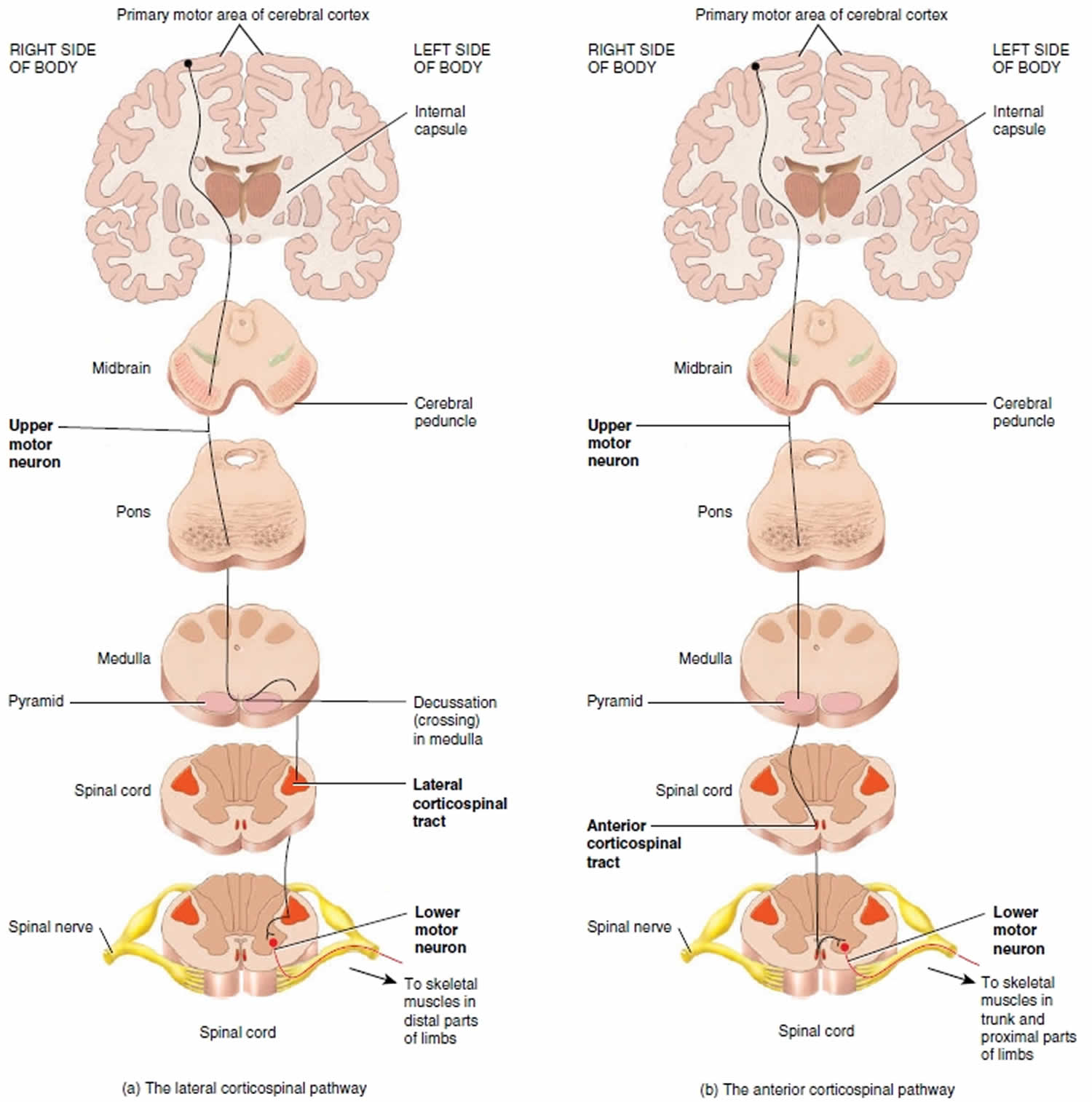
- Lateral corticospinal tract. Corticospinal axons that decussate in the medulla form the lateral corticospinal tract in the lateral white column of the spinal cord (Figure 3a). These axons synapse with local circuit neurons or lower motor neurons in the anterior gray horn of the spinal cord. Axons of these lower motor neurons exit the cord in the anterior roots of spinal nerves and terminate in skeletal muscles that control movements of the distal parts of the
limbs. The distal muscles are responsible for precise, agile, and highly skilled movements of the hands and feet. Examples include the movements needed to button a shirt or play the piano. - Anterior corticospinal tract. Corticospinal axons that do not decussate in the medulla form the anterior corticospinal tract in the anterior white column of the spinal cord (Figure 3b). At each spinal cord level, some of these axons decussate via the anterior white commissure. Then, they synapse with local circuit neurons or lower motor neurons in the anterior gray horn. Axons of these lower motor neurons exit the cord in the anterior roots of spinal nerves. They terminate in skeletal muscles that control movements of the trunk and proximal parts of the limbs.
Figure 3. Corticospinal pathways
Corticobulbar pathway
The corticobulbar pathway conducts impulses for the control of skeletal muscles in the head. Axons of upper motor neurons from the cerebral cortex form the corticobulbar tract, which descends along with the corticospinal tracts through the internal capsule of the cerebrum and cerebral peduncle of the midbrain. Some of the axons of the corticobulbar tract decussate; others do not. The axons terminate in the motor nuclei of nine pairs of cranial nerves in the brain-stem: the oculomotor (III), trochlear (IV), trigeminal (V), abducens (VI), facial (VII), glossopharyngeal (IX), vagus (X), accessory (XI), and hypoglossal (XII). The lower motor neurons of the cranial nerves convey impulses that control precise, voluntary movements of the eyes, tongue, and neck, plus chewing, facial expression, speech, and swallowing.
Extrapyramidal Tracts (Indirect pathways)
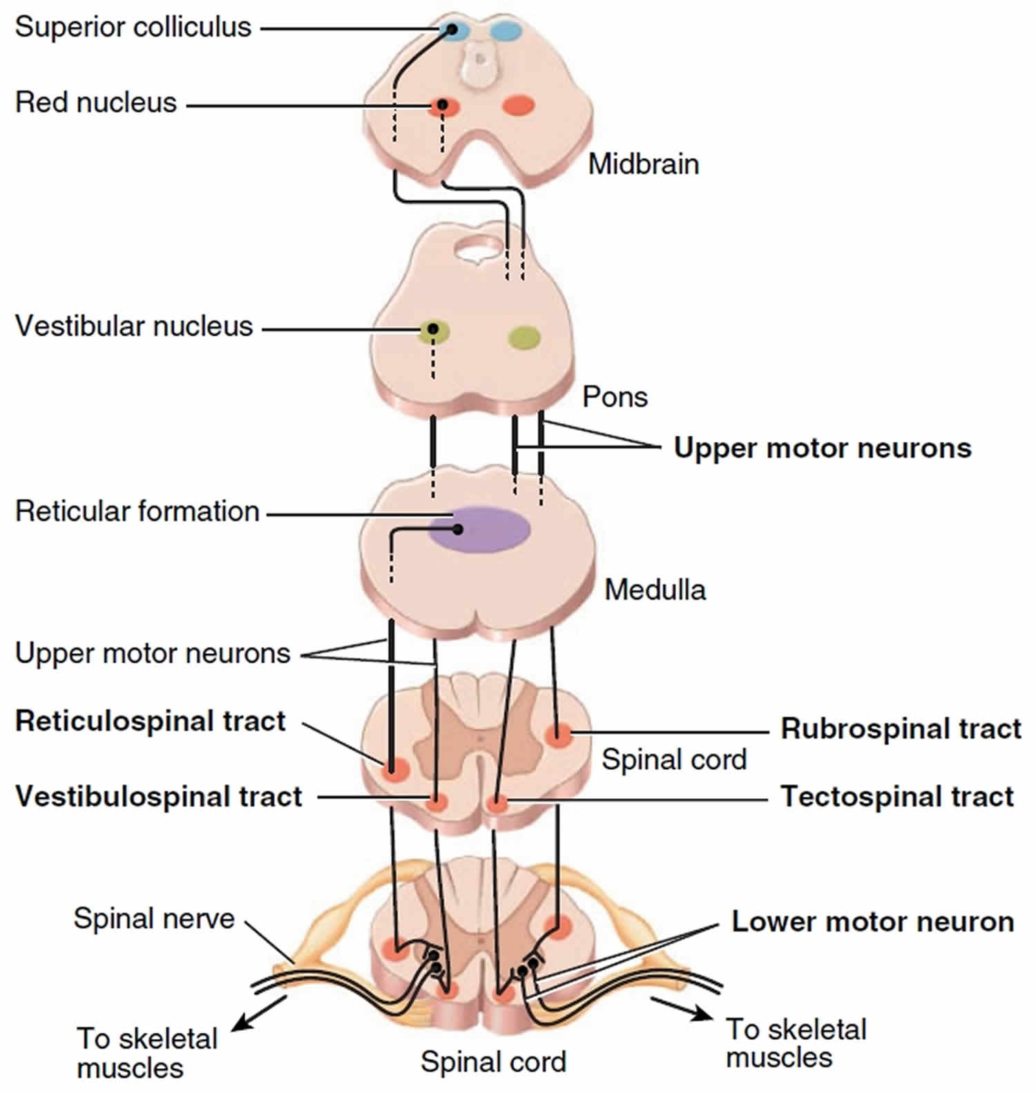
The brainstem motor centers give rise to the indirect motor pathways, also known as extrapyramidal pathways, which include all somatic motor tracts other than the corticospinal and corticobulbar tracts. Axons of upper motor neurons descend from the brainstem motor centers into five major tracts of the spinal cord and terminate on local circuit neurons or lower motor neurons. These tracts are the rubrospinal, tectospinal, vestibulospinal, lateral reticulospinal and medial reticulospinal tracts (Figure 4). In general, the indirect motor pathways convey action potentials from the brainstem to cause involuntary movements that regulate posture, balance, muscle tone, and reflexive movements of the head and trunk. An exception is the rubrospinal tract, which plays an ancillary role to the lateral corticospinal tract in the regulation of voluntary movements of the upper limbs.
Historically, these pathways were thought to be independent of the pyramidal cells and were called the extrapyramidal tracts, a term still used clinically. It is now known, however, that the pyramidal cells do project to and influence these pathways; thus, these pathways are referred to as indirect or multineuronal pathways. The indirect motor pathways include the tectospinal tract from the superior colliculus (the tectum of the midbrain), the vestibulospinal tract from the vestibular nuclei, the rubrospinal tract from the red nucleus (rubro = red) (Figures 1b and 4), and the reticulospinal tract from the reticular formation.
- Rubrospinal pathway: Conveys nerve impulses from red nucleus (which receives input from cerebral cortex and cerebellum) to contralateral skeletal muscles that govern precise, voluntary movements of distal parts of upper limbs.
- Tectospinal pathway: Conveys nerve impulses from superior colliculus to contralateral skeletal muscles that reflexively move head, eyes, and trunk in response to visual or auditory stimuli.
- Vestibulospinal pathway: Conveys nerve impulses from vestibular nucleus (which receives input about head movements from inner ear) to ipsilateral skeletal muscles of trunk and proximal parts of limbs for maintaining posture and balance in response to head movements. Lateral and medial reticulospinal pathways: Conveys nerve impulses from reticular formation to ipsilateral skeletal muscles of trunk and proximal parts of limbs for maintaining posture and regulating muscle tone in response to ongoing body movements.
Figure 4. Extrapyramidal tracts
Upper motor neuron vs Lower motor neuron
Lower motor neurons have their cell bodies in the lower parts of the central nervous system (brainstem and spinal cord). From the brainstem, axons of lower motor neurons extend through cranial nerves to innervate skeletal muscles of the face and head. From the spinal cord, axons of lower motor neurons extend through spinal nerves to innervate skeletal muscles of the limbs and trunk. Only lower motor neurons provide output from the central nervous system to skeletal muscle fibers. For this reason, they are also called the final common pathway.
Input arrives at lower motor neurons from upper motor neurons and nearby interneurons called local circuit neurons. Most upper motor neurons synapse with local circuit neurons, which in turn synapse with lower motor neurons. The local circuit neurons are located close to the lower motor neuron cell bodies in the brainstem and spinal cord. Local circuit neurons receive input from somatic sensory receptors, such as nociceptors and muscle spindles, as well as from higher centers in the brain. They help coordinate rhythmic activity in specific muscle groups, such as alternating flexion and extension of the lower limbs during walking. A few upper motor neurons synapse directly with lower motor neurons.
Lower motor neurons damage or disease produces flaccid paralysis of muscles on the same side of the body. There is neither voluntary nor reflex action of the innervated muscle fibers, muscle tone is decreased or lost, and the muscle remains limp or flaccid. Injury or disease of upper motor neurons in the cerebral cortex removes inhibitory influences that some of these neurons have on lower motor neurons, which causes spastic paralysis of muscles on the opposite side of the body. In this condition muscle tone is increased, reflexes are exaggerated, and pathological reflexes such as the Babinski sign appear.
Upper motor neuron function
Upper motor neurons are first-order neurons which are responsible for carrying the electrical impulses that initiate and modulate movement. Various descending upper motor neuron tracts are responsible for the coordination of movement. The major upper motor neuron tract that initiates voluntary movement is the pyramidal tract. The pyramidal tract provides a direct pathway between the cerebral cortex and the spinal cord, in contrast with extra-pyramidal tracts which provide indirect pathways for the coordination of movement. The pyramidal tract divides into the corticospinal tract and the corticobulbar tract. Corticospinal tract fibers synapse with spinal nerves while corticobulbar fibers synapse with cranial nerves. The cell bodies of the pyramidal tract concentrate around the motor area of the cerebral cortex. In general, the motor areas of the left and right hemispheres will innervate the musculature on the contralateral side of the body. The motor areas are somatotopically organized. This means that control of facial musculature is situated on the most lateral area of the cerebral hemisphere while control of the legs takes a more medial position. The mapping of different parts of the motor area for specific body parts is called the cortical homunculus 1.
Upper motor neurons of the pyramidal tract have the majority of their cell bodies located in the precentral motor cortex (Brodmann area 4) and the premotor area (Brodmann area 6). Cell bodies are also present in the supplementary motor area, primary somatosensory cortex, and the superior parietal lobe. upper motor neuron axons radiate out into the corona radiata and converge at the posterior limb of the internal capsule. The pathway of the corticospinal tract descends through cerebral peduncle in the midbrain, ventral pons, and the pyramids of the medulla. At the inferior aspect of the medulla, the majority of corticospinal tract axons decussate at the pyramidal decussation. The axons continue their descent contralateral from their cell bodies of origin and enter the spinal cord at the lateral funiculus. The tract is now the lateral corticospinal tract. The axons terminate throughout the spinal cord in the ventral gray column and the base of the dorsal column. The lateral corticospinal tract axons that control distal extremities synapse directly on lower motor neurons. These direct connections are presumed to be necessary for the fine control of the fingers and hands. The rest of the lateral corticospinal tract axons will synapse on premotor interneurons. At the pyramidal decussation, approximately 10% of the corticospinal tract axons do not decussate and continue their descent down the brain ipsilateral to their cell bodies of origin. These fibers enter the ventral aspect of the spinal cord and are known as the anterior corticospinal tract. As the fibers descend the spinal cord, most of them will decussate through the anterior white commissure before synapsing with interneurons. A small percentage of corticospinal axons will not decussate anywhere along their descent the brainstem or the spinal cord. These axons provide the impulses which control axial musculature necessary to maintain body posture 2.
The corticobulbar tract fibers originate from the parts of the motor cortex that represent the face. The axons share a similar trajectory to the corticospinal tract descending through the corona radiata and the internal capsule. At the level of the brainstem, the axons will synapse with each cranial nerve nuclei at their respective levels. The upper motor neuron innervation of most cranial nerves is bilateral which means that each cranial nerve receives impulses from the left and right hemisphere. This bilateral innervation pertains to the muscles of the eyes, jaw, pharynx, upper face, larynx, and neck. Two exceptions to this rule are CN VII and XII. The control of tongue protrusion by CN XII and the control of the muscles of the lower face by CN VII only receive contralateral innervation from the pyramidal tract 3.
Knowledge about the pathways of the pyramidal tracts is paramount to understanding the clinical presentation of upper motor neuron lesions. Lesions above or below the pyramidal decussation will have symptoms on different parts of the body. upper motor neuron lesions rostral to the pyramidal decussation will result in symptoms contralateral to the site of the lesion. For example, a unilateral lesion on the right corticospinal tract before the pyramidal decussation would cause weakness and spasticity of musculature on the left side of the body. upper motor neuron lesions caudal to the decussation will cause symptoms ipsilateral to the site of the lesion. This presentation would generally be with lesions to the spinal cord. For example, left-sided lesions of the corticospinal tract in the spinal cord will cause left-sided weakness and spasticity. Unilateral upper motor neuron lesions innervating cranial nerves do not manifest with clinically significant symptoms due to their bilateral innervation from the left and right motor areas. Hence, only bilateral lesions to the upper motor neuron of cranial nerves would create deficits. Lesions of upper motor neuron’s to CN VII and XII are the exceptions because of their unilateral innervation from contralateral motor areas. For example, a right-sided upper motor neuron lesion to the motor area that controls CN VII would manifest as a weakness on the left lower face 4.
Upper motor neuron disease
Upper motor neuron lesions can arise from a variety of injuries to the brain or spinal cord. Some diseases that can damage upper motor neurons include cerebrovascular accidents (strokes), traumatic brain injury, amyotrophic lateral sclerosis, primary lateral sclerosis, multiple sclerosis, Brown-Sequard Syndrome, vitamin B12 deficiency, anoxic brain injury, malignancy, infections, inflammatory disorders, neurodegenerative disorders, and metabolic disorders.
Cerebrovascular accidents
Cerebrovascular accidents or strokes are the sudden cessations of blood flow to areas of the brain leading to cell death. Strokes fall into two etiologic categories, being either ischemic or hemorrhagic. Ischemic strokes are the sudden interruption of blood flow to the brain which can be due to thrombi, emboli, or compression. Hemorrhagic strokes are characterized by bleeding into the brain due to rupture of a blood vessel. The cerebral blood supply has many branches which supply different areas of the brain. Occlusion of the middle cerebral artery or anterior cerebral artery can damage the motor areas of the cerebral cortex. Given the extensive areas of the brain supplied by the middle cerebral and anterior cerebral artery, strokes in those regions are likely to present with sensory, language, perceptual, and visual deficits in addition to upper motor neuron signs. Occlusion of the lenticulostriate arteries can damage the internal capsule. A stroke that targets the posterior limb of the internal capsule presents with pure motor deficits of the contralateral face, arm, and leg. Occlusion of the various branches of the vertebral artery or basilar artery can lead to strokes in different areas of the brainstem. Notable brainstem strokes that damage the corticospinal tract are medial medullary syndrome, medial pontine syndrome, and Weber Syndrome 5.
Amyotrophic Lateral Sclerosis
Amyotrophic lateral sclerosis (ALS) is the most prevalent neurodegenerative disease that is characterized by its involvement of both upper and lower motor neurons. Hence, the clinical presentation is a combination of upper motor signs and lower motor neuron signs. Nerve conduction studies and electromyography are utilized to confirm the diagnosis. Labs are generally used to rule out other disease processes that can manifest with weakness in patients. ALS is currently incurable however various treatments have been developed to extend life in patients. Riluzole is a glutamate pathway antagonist that is the only current drug shown to extend life in patients with ALS 6.
Primary Lateral Sclerosis
Primary lateral sclerosis is a neurodegenerative disorder that targets upper motor neurons. Primary lateral sclerosis is generally seen in adults and is sporadic in nature, though hereditary variants have been observed. Compared to amyotrophic lateral sclerosis (ALS), primary lateral sclerosis has a slower progression and lacks lower motor neuron signs. However, some individuals with primary lateral sclerosis do develop lower motor neuron signs as their disease progresses. The condition would then be considered upper motor neuron onset ALS. There are no cures for primary lateral sclerosis and treatment is aimed at alleviating symptoms of spasticity and weakness through medications and physical therapy 7.
Brown-Sequard syndrome
Brown-Sequard Syndrome is a spinal cord lesion caused by a hemisection injury to the spinal cord. The most common etiology is from penetrating trauma to the spine. However, other etiologies include blunt trauma, hematoma, tumors, or disc herniation. As a result of the hemisection of the spinal cord, the symptoms are manifestations of damage to the lateral corticospinal tract, dorsal column, and the lateral spinothalamic tract. Patients present with upper motor neuron signs ipsilateral and below the level of the lesion. Patients will also present with ipsilateral loss of fine touch, vibration, and proprioception in addition to the contralateral loss of pain and temperature sensation 8.
Multiple Sclerosis
Multiple Sclerosis (MS) is an immune-mediated, inflammatory demyelinating disease. The symptomatology of MS is characterized by episodes that occur in different anatomic locations in the central nervous system and occur months or years apart. The presenting symptoms of patients are highly variable. Symptoms can include cognitive disturbance, visual changes, hemiparesis, ataxia, and sensory deficits. The upper motor neuron signs of MS are due to the demyelination of upper motor neurons. MRI is the imaging test of choice used to diagnose MS. CSF studies may also be used to aid in diagnosis. Oligoclonal bands and intrathecal immunoglobulin G are classically seen in the CSF of MS patients 9.
Vitamin B12 Deficiency
The most prevalent etiologies of vitamin B12 deficiency are pernicious anemia, bariatric surgery, small intestine surgery, and gastritis. Other etiologies include pancreatic insufficiency, inadequate dietary intake, and drug side effects. Vitamin B12 deficiency causes degeneration of the dorsal column and lateral white matter of the spinal cord. This can lead to degeneration of the lateral corticospinal tract with subsequent upper motor neuron signs. Degeneration of the dorsal column manifests as sensory ataxia. Deficiency also leads to macrocytic anemia. Supplementation with vitamin B12 generally corrects the anemia and stops the progression of degeneration of the spinal cord 10.
Upper motor neuron symptoms
The clinical manifestation of a upper motor neuron lesion is known as upper motor neuron syndrome. The symptoms of upper motor neuron damage require differentiation from damage to lower motor neurons which would manifest with weakness, muscle atrophy, hypotonia, hyporeflexia, fasciculations, and fibrillation 11.
Upper Motor Neuron Syndrome
The upper motor neuron syndrome is a collective term that refers to different types of motor behaviors produced by patients who have lesions of the descending corticospinal system 11. Lesions involving the upper motor neuron, its pathways, and its connections can occur at the level of the cerebral cortex, internal capsule, brain stem, or spinal cord. The clinical features of upper motor neuron syndrome have traditionally been classified as “positive” symptoms, referring to overt behaviors generated by various forms of muscle overactivity, and “negative” symptoms, referring to loss of overt behaviors secondary to impaired muscle activation, impaired control of motor behavior, weakness, decreased motor control, easy fatigability and impaired motor performance. The clinical impact of upper motor neuron syndrome on patients is broad and tends to limit functional capacity. The negative and positive signs lead to reduced mobility and limb usage. A unique characteristic of upper motor neuron syndrome is its tendency to affect specific muscle groups. The weakness caused by upper motor neuron syndrome will predominantly affect the extensors of the arm and flexors of the leg. Positive symptoms are those involving increased muscle activity.
The symptoms of upper motor neuron syndrome can separate into negative and positive symptoms. Negative symptoms include weakness, decreased motor control, and easy fatigability.
Upper motor neuron symptoms can include:
Spasticity
The definition of spasticity is a velocity-dependent increase in a muscle’s resistance to a passive stretch. Slow passive movements of the arms or legs will not elicit the increased resistance. Brisk stretches of muscles will cause an abrupt increase in tone followed by a decrease in muscular resistance with continued stretch. This phenomenon is called clasp-knife rigidity. The antigravity muscles of the arms and legs are most affected. These include the flexors of the arms and the extensors of the leg. Because of the decreased modulation of spinal reflexes in upper motor neuron syndrome, patients will often exhibit flexor and extensor spasms.
Clonus
Clonus is a sequence of rhythmic, involuntary muscle contractions. These contractions occur at a frequency of 5 to 7 Hz and are a response to abruptly applied stretch stimuli. Clonus is most easily elicited at the ankle with brisk dorsiflexion and plantar-flexion movements. Clonus can also be observed during voluntary movement or through cutaneous stimulation.
Hyperreflexia of deep tendon reflexes
Patients can be seen to have abnormally brisk reflexes which are due to decreased modulation by descending inhibitory pathways. Radiation of reflexes is a regular observation with the hyperreflexia of upper motor neuron lesions. For example, tapping of the supra-patellar tendon would elicit a knee-jerk reflex.
Hyporeflexia of superficial reflexes
The superficial abdominal reflex and the cremasteric reflex are seen to be decreased or abolished following upper motor neuron lesions. The superficial abdominal reflex is the tensing of abdominal by stroking the overlying skin while the cremasteric reflex is the elevation of the scrotum in response to stroking the medial thigh.
Synkinesias
Synkinesias are involuntary movements in a limb that have associations with the voluntary movements in other limbs. For example, flexion of the arm may result in flexion of the leg. These involuntary movements can also occur with yawning or sneezing. Volitional movements of one arm or leg may also result in mirror movements of the opposite limb.
Co-contraction
Co-contraction is defined as the simultaneous contraction of agonist and antagonist muscles around a joint. This increases the stiffness and stabilization around the joint which prepares it for activity in healthy individuals. The pathological co-contraction in upper motor neuron lesions causes a decreased rate of rapid alternating movements and creates greater fatigability for voluntary movements in weakened muscles.
Babinski sign and other reflexes
The Babinski sign can be elicited by stroking the sole of the foot with a firm stimulus. The normal adult response is plantar-flexion. The sign is positive when the application of the stimulus elicits extension of the large toe and fanning of the other toes. The Babinski sign is known to be a normal response in infants before full maturation of the corticospinal tract. However, in adults, a positive sign is indicative of underlying upper motor neuron damage. Other reflexes exist which represent lesions of the corticospinal tract. The Brissaud reflex is linked with the extensor response of the Babinski sign and is positive when stroking the sole of the foot elicits contraction of the tensor fascia latae of the ipsilateral leg. The Hoffman sign is an analog of the Babinski reflex for the upper limbs. The test is performed by loosely holding the patient’s middle finger and quickly flicking the fingernail downward. A positive sign is the flexion and adduction of the thumb 12.
Pseudo-Bulbar Palsy
As previously stated, most cranial nerves have bilateral innervation from the brain with the exception of CN VII and CN XII. The muscles of cranial nerves with bilateral innervation include the eyes, jaw, pharynx, upper face, larynx, and neck. These muscles would only show deficits with bilateral upper motor neuron lesions. Bilateral damage of upper motor neuron’s to cranial nerves is known as a pseudobulbar palsy. Slurred speech is often the first presenting symptom. Other characteristic deficits include dysphagia, dysarthria, brisk jaw jerk, spastic tongue, and pseudobulbar affect 13.
CN VII and CN XII upper motor neuron lesions
CN VII and CN XII innervate muscles of the lower face and the tongue, respectively. These cranial nerves receive unilateral innervation from the pyramidal tract. Unilateral lesions of upper motor neuron’s to CN VII or CN XII would manifest as a lower facial droop or tongue deviation away from the side of the lesion, respectively.
Spinal Shock
Spinal shock refers to the period of acute flaccid paralysis following spinal cord injury. Hypotonia and hyporeflexia are the most characteristic symptoms. The paralysis is most evident in the arms and legs with preservation of truncal musculature. The duration can range from a few days to weeks after which spasticity and hyperreflexia replace the prior symptoms. The symptoms of spinal shock are most pronounced with lesions of the spinal cord versus cerebral lesions 14.
- Hooks BM, Papale AE, Paletzki RF, Feroze MW, Eastwood BS, Couey JJ, Winnubst J, Chandrashekar J, Gerfen CR. Topographic precision in sensory and motor corticostriatal projections varies across cell type and cortical area. Nat Commun. 2018 Sep 03;9(1):3549.[↩]
- Dall’Orso S, Steinweg J, Allievi AG, Edwards AD, Burdet E, Arichi T. Somatotopic Mapping of the Developing Sensorimotor Cortex in the Preterm Human Brain. Cereb. Cortex. 2018 Jul 01;28(7):2507-2515[↩]
- Canedo A. [Functional heterogeneity of the piramidal system: corticobulbar and corticospinal tracts]. Rev Neurol. 2003 Mar 1-15;36(5):438-52[↩]
- Armand J. [The pyramidal tract. Recent anatomic and physiologic findings]. Rev. Neurol. (Paris). 1984;140(5):309-29[↩]
- Lee SJ, Lee DG, Moon HJ, Lee TK. Lesion Pattern, Mechanisms, and Long-Term Prognosis in Patients with Monoparetic Stroke: A Comparison with Nonmonoparetic Stroke. Biomed Res Int. 2017;2017:9373817[↩]
- de Carvalho M, Poliakov A, Tavares C, Swash M. Interplay of upper and lower motor neuron degeneration in amyotrophic lateral sclerosis. Clin Neurophysiol. 2017 Nov;128(11):2200-2204[↩]
- Le Forestier N, Meininger V. [Primary lateral sclerosis: the era of international diagnosis criteria]. Rev. Neurol. (Paris). 2009 May;165(5):415-29[↩]
- Zeng Y, Ren H, Wan J, Lu J, Zhong F, Deng S. Cervical disc herniation causing Brown-Sequard syndrome: Case report and review of literature (CARE-compliant). Medicine (Baltimore). 2018 Sep;97(37):e12377[↩]
- Kamińska J, Koper OM, Piechal K, Kemona H. Multiple sclerosis – etiology and diagnostic potential. Postepy Hig Med Dosw (Online). 2017 Jun 30;71(0):551-563.[↩]
- Langan RC, Goodbred AJ. Vitamin B12 Deficiency: Recognition and Management. Am Fam Physician. 2017 Sep 15;96(6):384-389[↩]
- Mayer NH, Esquenazi A. Muscle overactivity and movement dysfunction in the upper motoneuron syndrome. Phys Med Rehabil Clin N Am. 2003 Nov;14(4):855-83, vii-viii[↩][↩]
- Damodaran O, Rizk E, Rodriguez J, Lee G. Cranial nerve assessment: a concise guide to clinical examination. Clin Anat. 2014 Jan;27(1):25-30[↩]
- Chu J, Liu X, Chen F, Hong F, Bao Y. [Effects of GAO’s neck acupuncture on swallowing function and quality of life in patients with post-stroke pseudobulbar palsy:a randomized controlled trial]. Zhongguo Zhen Jiu. 2017 Jul 12;37(7):691-695.[↩]
- Hiersemenzel LP, Curt A, Dietz V. From spinal shock to spasticity: neuronal adaptations to a spinal cord injury. Neurology. 2000 Apr 25;54(8):1574-82[↩]