Contents
What is urticarial vasculitis
Urticarial vasculitis is a condition characterized by inflammation of the small blood vessels (small vessel vasculitis) in the skin. Urticarial vasculitis is characterised by inflamed and reddened patches, hives or weals on the skin that appear to resemble urticaria, due to swelling of the small blood vessels, but when the skin is examined closely under a microscope, a vasculitis is found (inflamed blood vessels). Furthermore, in contrast to classic urticaria, urticarial vasculitis lesions persist at the same location for more than 24 hours.
Urticarial vasculitis is not a specific disease but instead a clinical finding. A search for a cause of the vasculitis should be undertaken. The most common associated condition is systemic lupus erythematosus. Other possibilities include Sjögren’s syndrome, serum sickness, viral infections (e.g. hepatitis C, infectious mononucleosis), mixed cryoglobulins, a monoclonal IgM gammopathy (e.g. Schnitzler syndrome), IgA multiple myeloma, fluoxetine administration, and rarely neoplasm. Hypocomplementemia is common and when it does occur, the association with systemic lupus erythematosus (SLE) is highly likely. In one case, the lesions of urticarial vasculitis were induced by cold exposure and an IgG was found on serum protein electrophoresis.
Autoantibodies to vascular endothelial cells were found in 82% of patients with urticarial vasculitis and systemic lupus erythematosus (SLE), 70% of patients with hypocomplementemic urticarial vasculitis, and in 14% of patients with urticarial vasculitis alone. They were found in only 32% of patients with systemic lupus erythematosus (SLE) without urticarial vasculitis.
Urticarial vasculitis is uncommon. There are no well researched estimates of how frequent it is. Both males and females get the disease.
Signs and symptoms include an itching and burning sensation in the affected skin. Lesions (wheals) caused by urticarial vasculitis may also leave behind a bruise.
Urticarial vasculitis is a variant of cutaneous small vessel vasculitis and is generally classified as two types:
- Normocomplementaemic urticarial vasculitis (normal levels of proteins called complements). Normocomplementemic urticarial vasculitis tends to have a more benign course
- Hypocomplementaemic urticarial vasculitis (low levels of complements). Hypocomplementemic urticarial vasculitis is often associated with systemic disease and a more complicated disease course.
These are distinguished by finding normal or lowered levels of complement proteins on blood testing. Although both types may be associated with systemic symptoms such as angioedema, abdominal or chest pain, fever, and joint pain, this is more apparent in the hypocomplementaemic form. This form has also been linked to the connective tissue disease systemic lupus erythematosus (SLE).
Hypocomplementemic urticarial vasculitis especially is associated with rheumatologic diseases such as systemic lupus erythematosus, rheumatoid arthritis and Sjögren’s syndrome. Some cancers including leukemias and solid cancers such as colon cancer, pancreas cancer and others also may cause urticarial vasculitis.
In addition, certain infections like hepatitis B and C can cause this form of vasculitis, as can some drugs, including antibiotics, ACE inhibitors used for treating high blood pressure, and certain diuretics. Still, the cause of most cases of urticarial vasculitis is unknown.
Regardless of the cause, urticarial vasculitis is a treatable condition. The treatment depends on the extent of symptoms and organ involvement.
Urticarial vasculitis prognosis
The natural history of urticarial vasculitis depends in part upon the blood complement levels. In cases where these are normal, the prognosis is generally good. In cases where the complements are low, the disease may be more severe. When urticarial vasculitis is related to a disease such as lupus or cancer, its prognosis is often governed by the prognosis of the underlying disease.
What are the complications of urticarial vasculitis?
The most common serious complications are skin pigmentation and, occasionally, skin ulcers, plus damage to organs such as the lungs, eyes and kidneys.
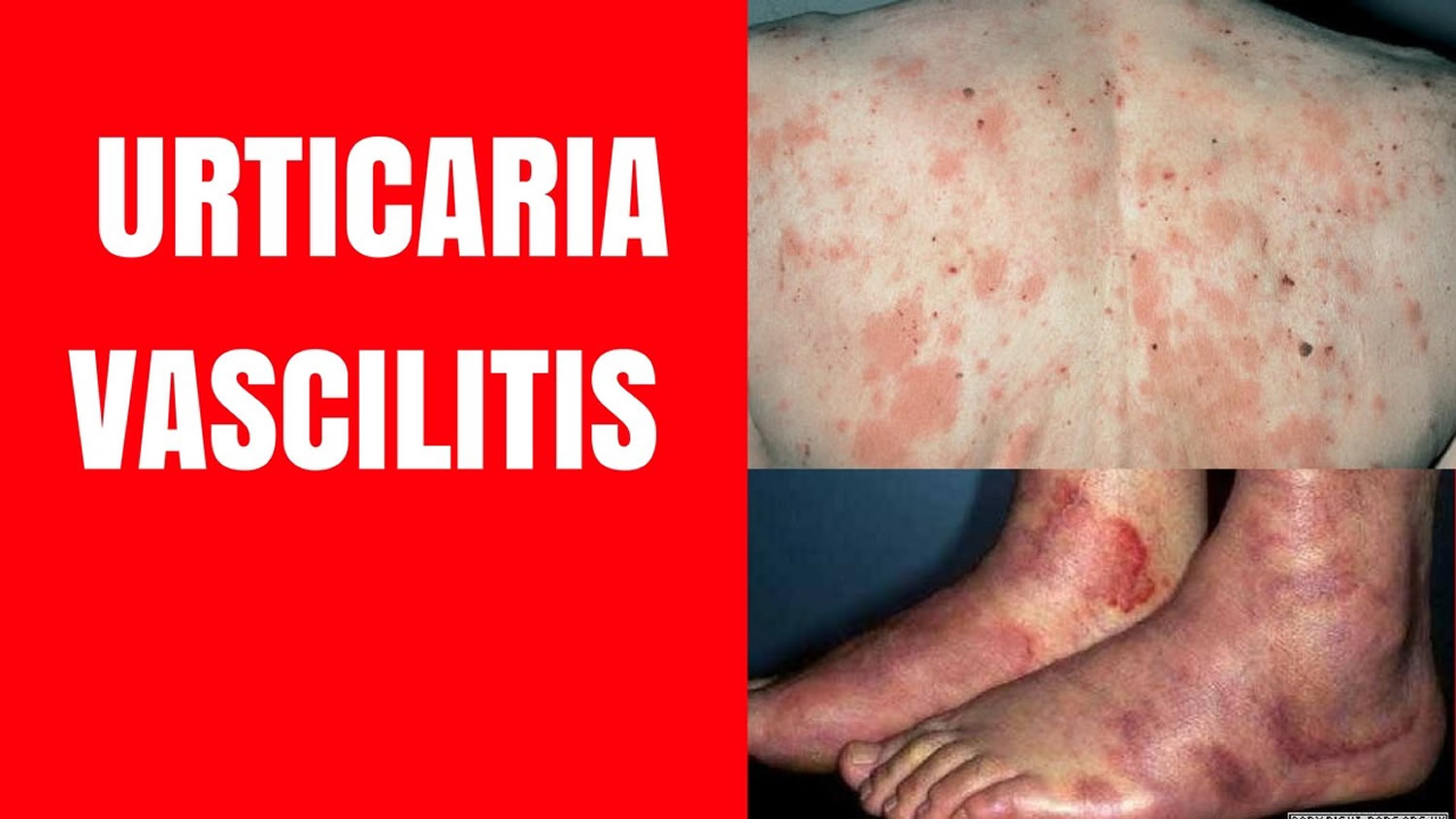
What does urticarial vasculitis look like?
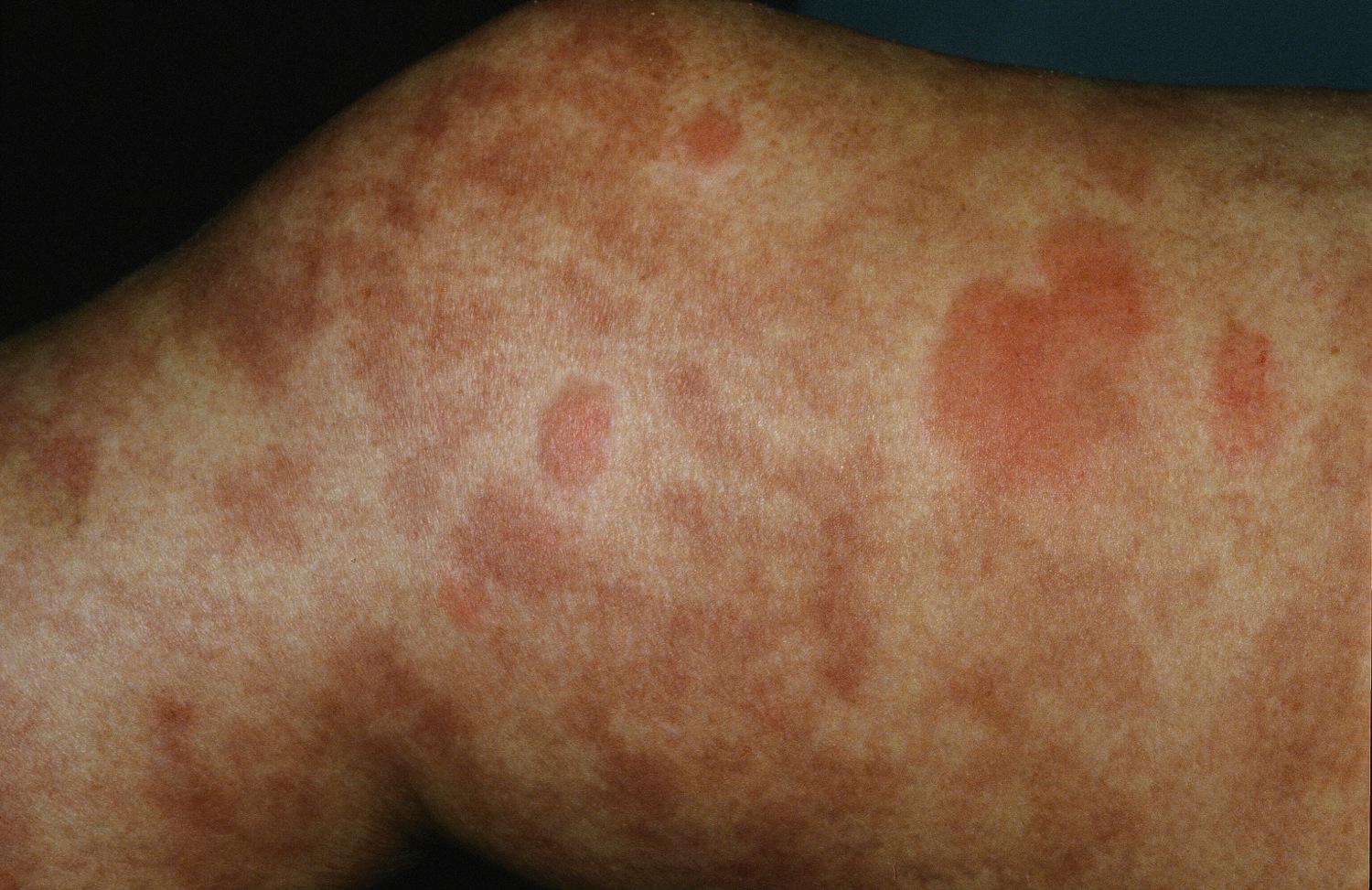
The first symptom of urticarial vasculitis is an urticarial eruption that is often painful or has a burning sensation. In some cases there may be pruritus. Lesions are red patches or plaques that may have a white centre, and petechiae may appear. They usually last for more than 24 hours in a fixed location, after which they will slowly resolve spontaneously. Ecchymoses or hyperpigmentation may occur in the healing process.
In addition to the skin lesions, patients with urticarial vasculitis may also develop systemic symptoms including photosensitivity, swollen lymph nodes, joint pain (50%), fever, abdominal pain (20%), difficulty breathing, and lung and kidney problems.
Figure 1. Urticarial vasculitis
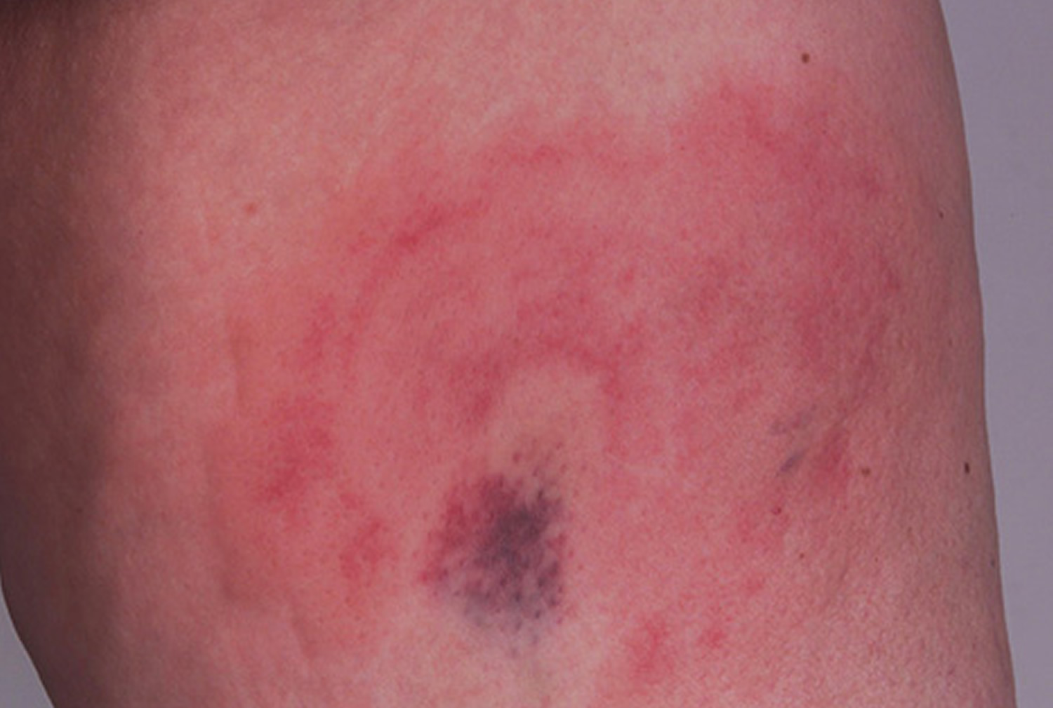
Figure 2. Urticarial vasculitis
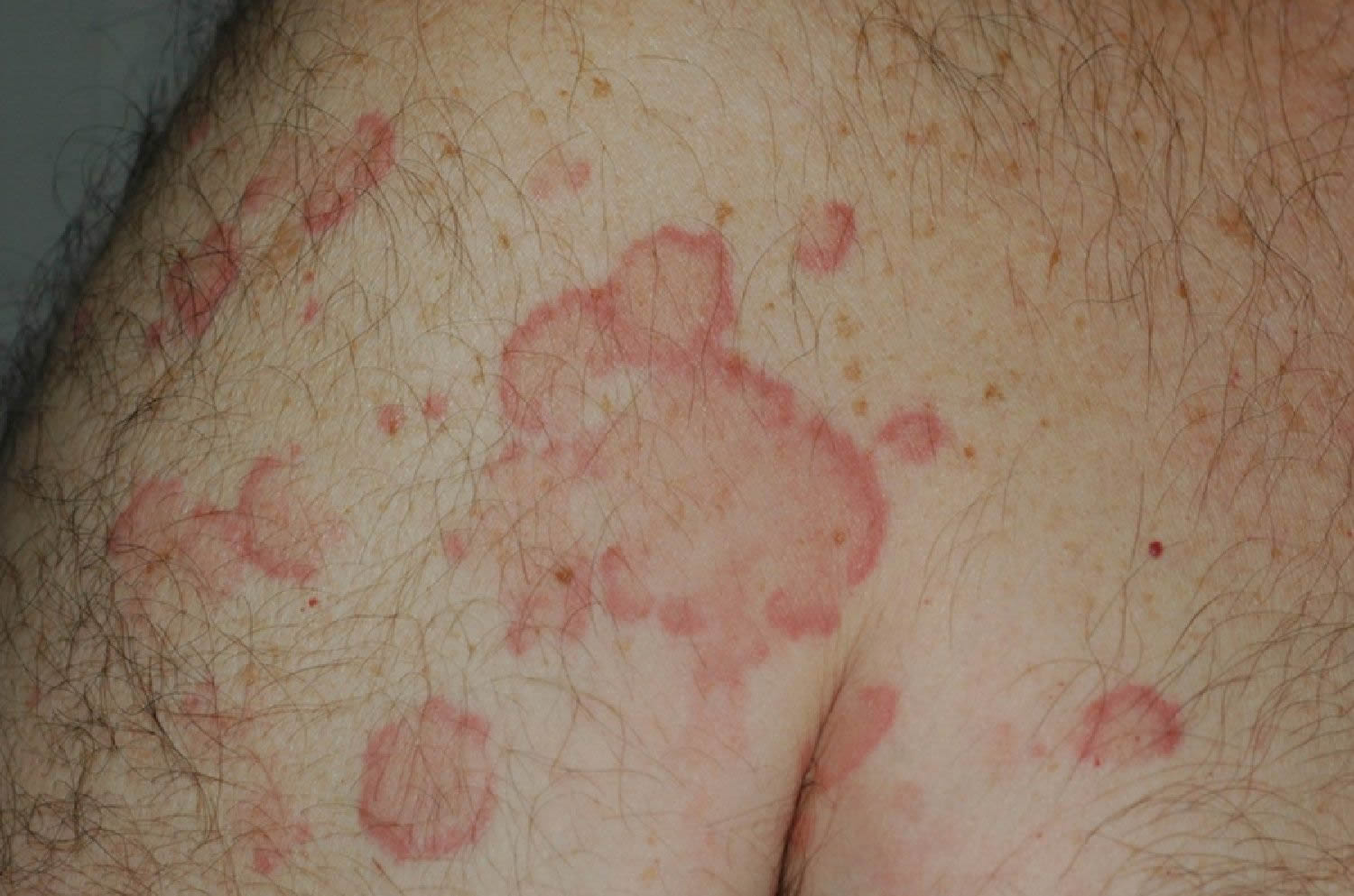
Figure 3. Urticarial vasculitis
What causes Urticarial Vasculitis?
The cause of most cases of urticarial vasculitis is unknown, but it has been associated with the following conditions:
- Inflammatory connective disorders such as systemic lupus erythematosus (SLE), rheumatoid arthritis and Sjögren syndrome.
- Immunoglobulin disorders such as immunoglobulin A and immunoglobulin M monoclonal gammopathies
- Some cancers, including leukemias and colon and pancreatic cancers
- Viral diseases such as hepatitis B, hepatitis C and infectious mononucleosis (glandular fever)
- Drug-induced conditions from ACE (angiotensin-converting-enzyme) inhibitors, penicillin, sulfonamides, fluoxetine and thiazides.
However, the majority of cases of urticarial vasculitis are idiopathic (i.e there is no known cause).
Urticarial vasculitis symptoms
Urticarial lesions lasting more than 24 hours at any given location are characteristic. The most common symptoms are hives that cause itching, pain and a burning feeling. Pain and burning are more common than with typical urticaria. Skin patches often are red-rimmed with white centers, and unlike common hives may have petechia or bleeding under the skin. Diascopy may blanch much of the erythema, but hyperpigmentation and/or purpura remain. When the urticarial changes resolve, the patches can be present for days and result in skin discoloration as they heal. Angioedema may also be associated. Some patients may also have fevers, joint and abdominal pain, shortness of breath and swollen lymph glands. Sometimes urticarial vasculitis even causes injury to vital organs including the gut, lungs and kidneys.
Urticarial vasculitis diagnosis
Diagnosis is based on characteristic patches in the skin. Sometimes a biopsy is ordered to show inflammation in the skin and damage of small blood vessels with white blood cells. Since it’s often associated with a number of different diseases, it’s often necessary to do other tests and exams to rule out underlying conditions like lupus erythematosus or cancer. Because this form of vasculitis can affect vital organs such as the kidneys, other tests, for example of kidney function and chest radiographs may be needed, especially when the blood levels of complement are low.
Systemic findings have included anemia, arthralgias, abdominal pain, glomerulonephritis, ocular inflammation, and pulmonary disease. Laboratory abnormalities may include elevated erythrocyte sedimentation rate (ESR), decreased complement levels, decreased or absent C1q, and C1q autoantibody. In one study, the C1q autoantibody was found in 100% of patients with hypocomplementemic urticarial vasculitis syndrome, 35% of patients with SLE, and almost no controls.
Tests
Workup may include drug history (e.g. dexfenfluramine), skin biopsy, complete blood count (CBC), antinuclear antibodies (ANA), Ro, La, rheumatoid factor, ESR, urine analysis, CH50, C3,C4, liver enzymes, hepatitis C serology, cryoglobulins, and serum protein electrophoresis.
Urticarial vasculitis treatment
Treatment depends on the extent of symptoms and organ involvement.
Patients with normocomplementaemic urticarial vasculitis usually have minimal or no systemic involvement and lesions often resolve on their own over time or with minimal treatment. In such cases, antihistamines or nonsteroidal drugs such as ibuprofen or naproxen may be helpful.
Treatment is based on systemic effects of the disease and extent of cutaneous involvement. To relieve cutaneous symptoms, antihistamines or non-steroidal anti-inflammatory drugs (NSAIDs) may be used.
Treatments that may be used in the long-term control of severe urticarial vasculitis that may be associated with systemic symptoms include:
- Dapsone
- Colchicine
- Hydroxychloroquine
- Indometacin/indomethacin (non-steroidal anti-inflammatory)
- Corticosteroids, e.g. oral prednisone
- Chemotherapies like azathioprine or cyclophosphamide
Prednisone may be used (e.g. 40-60 mg/day initially, then tapered). Azathioprine has been used (e.g. 50-100 mg/day). Treatment may be intermittent, although it is not uncommon for patients to need treatment for several years.
For cases of urticarial vasculitis that are resistant to treatment with corticosteroids other immunosuppressive agents such as azathioprine, cyclophosphamide or ciclosporin may be considered.


