Contents
What are eye floaters ?
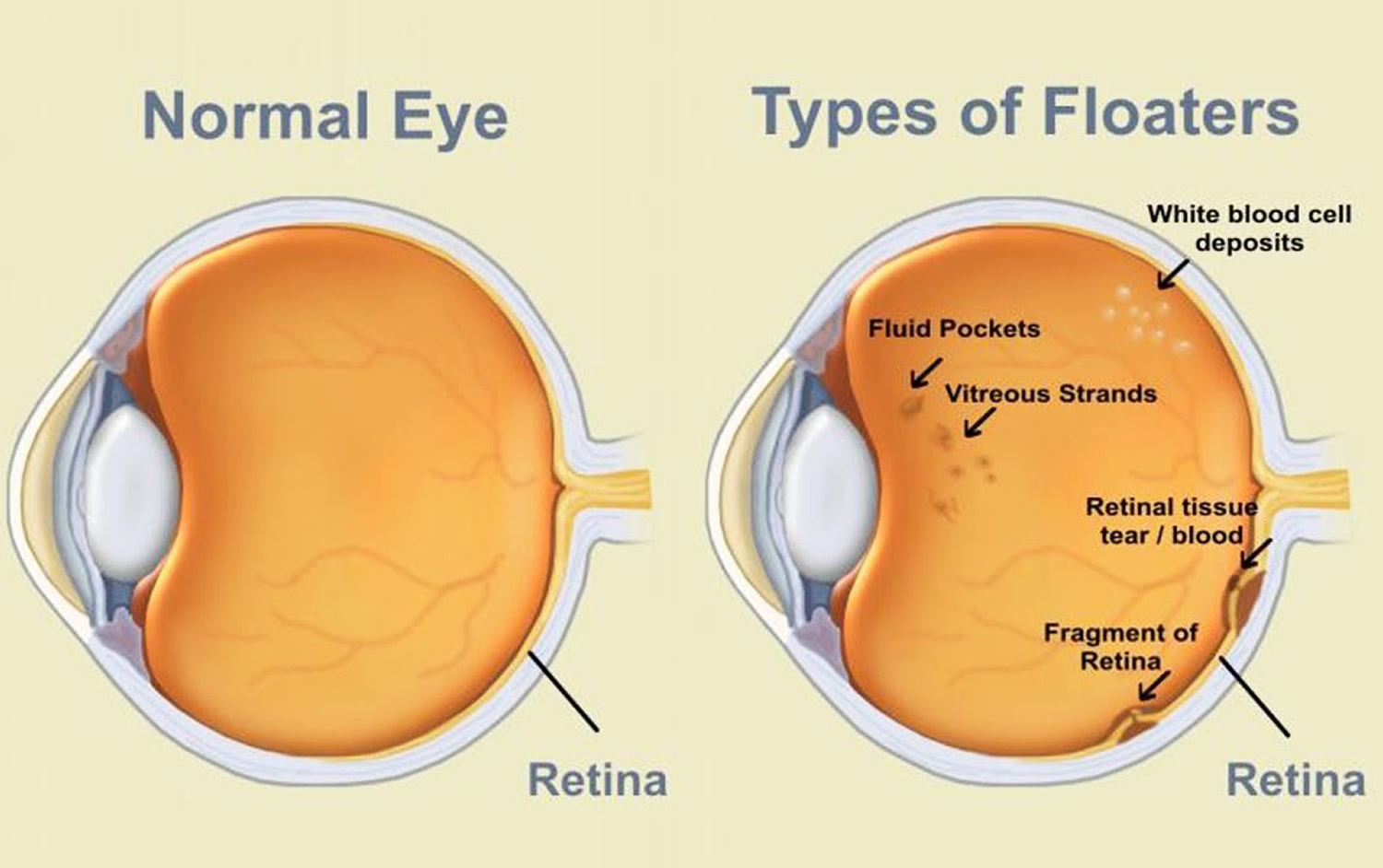
Eye floaters look like small dark specks, thread-like strands, shadowy shapes, dots, circles, squiggly lines or “cobwebs” that float in your field of vision 1. They move as your eyes move and seem to dart away when you try to look at them directly. They do not follow your eye movements precisely, and usually drift when your eyes stop moving 2. The floating specks you sometimes see in front of your eyes are not on the surface of your eyes, but inside them. These floaters are tiny clumps of gel or bits of cell debris that drift around in the fluid (the vitreous body or vitreous humor) that fills the back of your eye 3. What you see are the shadows these clumps cast on your retina. You usually notice floaters when looking at something plain, like a blank wall or a blue sky.
Most people have floaters and learn to ignore them; they are usually not noticed until they become numerous or more prominent. Floaters can become apparent when looking at something bright, such as white paper or a blue sky.
Most of the time floaters are harmless. However, they can be a symptom of a tear in the retina. The retina is the layer in the back of the eye that contain all the vision cells (cones and rods). The retina is a layer of tissue in the back of your eye that senses light and sends images to your brain. It provides the sharp, central vision needed for reading, driving, and seeing fine detail. If you notice a sudden increase in floaters or if you see floaters along with flashes of light in your side vision, this may be a symptom of a retinal tear or detachment 1. Go to an eye doctor or emergency room if you have these symptoms.
As you age, your vitreous body (vitreous humor) starts to thicken or shrink. Sometimes clumps or strands form in the vitreous. And sometimes a section of the vitreous pulls the fine fibers away from the retina all at once, rather than gradually, causing many new floaters to appear suddenly. This is called a vitreous detachment, which in most cases is not sight-threatening and requires no treatment.
You are more likely to get floaters if you:
- are nearsighted (you need glasses to see far away)
- have had surgery for cataracts
- have had inflammation (swelling) inside the eye.
If the vitreous pulls away from the back of the eye (the retina), it is called posterior vitreous detachment 1. Floaters usually happen with posterior vitreous detachment. They are not serious, and they tend to fade or go away over time. Severe floaters can be removed by surgery, but this is seldom necessary 1.
However, a sudden increase in floaters, possibly accompanied by light flashes or peripheral (side) vision loss, could indicate a retinal detachment. A retinal detachment occurs when any part of the retina, the eye’s light-sensitive tissue, is lifted or pulled from its normal position at the back wall of the eye.
- A retinal detachment is a serious condition and should always be considered an emergency. If left untreated, it can lead to permanent visual impairment within two or three days or even blindness in the eye.
Those who experience a sudden increase in floaters, flashes of light in peripheral vision, or a loss of peripheral vision should have an eye care professional examine their eyes as soon as possible.
Figure 1. Eye anatomy

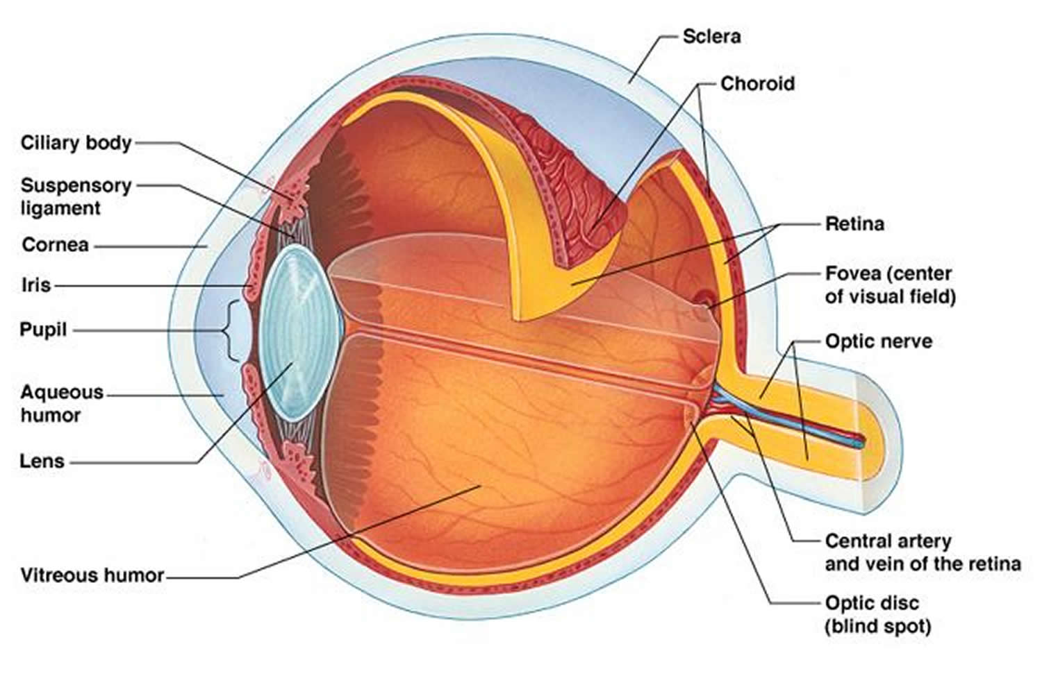
Figure 2. Types of eye floaters
What causes eye floaters
Eye floaters occur when the vitreous, a gel-like substance that fills about 80 percent of the eye and helps it maintain a round shape, slowly shrinks 2.
As the vitreous shrinks, it becomes somewhat stringy, and the strands can cast tiny shadows on the retina. These are floaters.
In most cases, floaters are part of the natural aging process and simply an annoyance 2. They can be distracting at first, but eventually tend to “settle” at the bottom of the eye, becoming less bothersome. They usually settle below the line of sight and do not go away completely 2.
However, there are other, more serious causes of floaters, including infection, inflammation (uveitis), hemorrhaging, retinal tears, and injury to the eye.
Who is at risk for floaters ?
Floaters are more likely to develop as we age and are more common in people who are very nearsighted, have diabetes, or who have had a cataract operation 2.
Symptoms and Detection of Eye Floaters
Eye floaters are little “cobwebs” or specks that float about in your field of vision. They are small, dark, shadowy shapes that can look like spots, thread-like strands, or squiggly lines. They move as your eyes move and seem to dart away when you try to look at them directly. They do not follow your eye movements precisely, and usually drift when your eyes stop moving.
Treatment of Eye Floaters
For people who have eye floaters that are simply annoying, no treatment is recommended 2.
On rare occasions, floaters can be so dense and numerous that they significantly affect vision. In these cases, a vitrectomy, a surgical procedure that removes floaters from the vitreous, may be needed 2.
A vitrectomy removes the vitreous gel, along with its floating debris, from the eye. The vitreous is replaced with a salt solution. Because the vitreous is mostly water, you will not notice any change between the salt solution and the original vitreous 2.
This operation carries significant risks to sight because of possible complications, which include retinal detachment, retinal tears, and cataract 2. Most eye surgeons are reluctant to recommend this surgery unless the floaters seriously interfere with vision 2.
What are eye flashes ?
Flashes can look like flashing lights or lightning streaks in your field of vision 1. Some people compare them to seeing “stars” after being hit on the head. You might see flashes on and off for weeks, or even months. Flashes happen when the vitreous rubs or pulls on your retina 1.
As people age, it is common to see flashes occasionally.
Flashes and migraines
Sometimes people have light flashes that look like jagged lines or heat waves. These can appear in one or both eyes and may last up to 20 minutes. This type of flash may be caused by a migraine. A migraine is a spasm of blood vessels in the brain.
When you get a headache after these flashes, it is called a “migraine headache.” But sometimes you only see the light flash without having a headache. This is called an “ophthalmic migraine” or “migraine without headache.”
When eye floaters and eye flashes are serious
Most eye floaters and eye flashes are not a problem. However, there are times when they can be signs of a serious condition. Here is when you should call an ophthalmologist right away 4:
- you notice a lot of new floaters
- you have a lot of flashes
- a shadow appears in your peripheral (side) vision
- a gray curtain covers part of your vision
These floaters and flashes could be symptoms of a torn or detached retina 4. This is when the retina pulls away from the back of your eye. This is a serious condition that needs to be treated.
What is Retinal Detachment
The retina is the light-sensitive layer of tissue that lines the inside of the eye and sends visual messages through the optic nerve to the brain. When the retina detaches, it is lifted or pulled from its normal position.
- A retinal detachment is a medical emergency. If not promptly treated, it can cause permanent vision loss. If you have any symptoms, see an eye care professional immediately. Treatment includes different types of surgery.
- If not promptly treated, retinal detachment can cause permanent vision loss.
In some cases there may be small areas of the retina that are torn. These areas, called retinal tears or retinal breaks, can lead to retinal detachment.
Retinal detachment can occur at any age, but it is more common in people over age 40 5. It affects men more than women and whites more than African Americans. A retinal detachment is also more likely to occur in people who:
- Are extremely nearsighted (myopia)
- Have had a retinal detachment in the other eye
- Have a family history of retinal detachment
- Have had cataract surgery
- Have other eye diseases or disorders
- Have had an eye injury
There are three different types of retinal detachment 6:
- Rhegmatogenous retinal detachment—A tear or break in the retina allows fluid to get under the retina and separate it from the retinal pigment epithelium, the pigmented cell layer that nourishes the retina. These types of retinal detachments are the most common.
- Tractional retinal detachment—In this type of detachment, scar tissue on the retina’s surface contracts and causes the retina to separate from the retinal pigment epithelium. This type of detachment is less common.
- Exudative retinal detachment—Frequently caused by retinal diseases, including inflammatory disorders and injury/trauma to the eye. In this type, fluid leaks into the area underneath the retina, but there are no tears or breaks in the retina.
Causes and Risk Factors for retinal detachment
A retinal detachment can occur at any age, but it is more common in people over age 40. It affects men more than women, and Whites more than African Americans.
A retinal detachment is also more likely to occur in people who:
- Are extremely nearsighted (myopia)
- Have had a retinal detachment in the other eye
- Have a family history of retinal detachment
- Have had cataract surgery
- Have other eye diseases or disorders, such as retinoschisis, uveitis, degenerative myopia, or lattice degeneration
- Have had an eye injury
What are the symptoms of retinal detachment ?
Symptoms include a sudden or gradual increase in either the number of floaters, which are little “cobwebs” or specks that float about in your field of vision, and/or light flashes in the eye. Another symptom is the appearance of a curtain over the field of vision. A retinal detachment is a medical emergency. Anyone experiencing the symptoms of a retinal detachment should see an eye care professional immediately.
Treatment for retinal detachment
Small holes and tears are treated with laser surgery or a freeze treatment called cryopexy. These procedures are usually performed in the doctor’s office. During laser surgery tiny burns are made around the hole to “weld” the retina back into place. Cryopexy freezes the area around the hole and helps reattach the retina.
Retinal detachments are treated with surgery that may require the patient to stay in the hospital. In some cases a scleral buckle, a tiny synthetic band, is attached to the outside of the eyeball to gently push the wall of the eye against the detached retina. If necessary, a vitrectomy may also be performed. During a vitrectomy, the doctor makes a tiny incision in the sclera (white of the eye). Next, a small instrument is placed into the eye to remove the vitreous, a gel-like substance that fills the center of the eye and helps the eye maintain a round shape. Gas is often injected to into the eye to replace the vitreous and reattach the retina; the gas pushes the retina back against the wall of the eye. During the healing process, the eye makes fluid that gradually replaces the gas and fills the eye. With all of these procedures, either laser or cryopexy is used to “weld” the retina back in place.
With modern therapy, over 90 percent of those with a retinal detachment can be successfully treated, although sometimes a second treatment is needed 6. However, the visual outcome is not always predictable 6. The final visual result may not be known for up to several months following surgery 6. Even under the best of circumstances, and even after multiple attempts at repair, treatment sometimes fails and vision may eventually be lost 6. Visual results are best if the retinal detachment is repaired before the macula (the center region of the retina responsible for fine, detailed vision) detaches. That is why it is important to contact an eye care professional immediately if you see a sudden or gradual increase in the number of floaters and/or light flashes, or a dark curtain over the field of vision.
Symptoms include an increase in the number of floaters, which are little “cobwebs” or specks that float about in your field of vision, and/or light flashes in the eye. It may also seem like there is a “curtain” over your field of vision.
A retinal detachment is a medical emergency. If not promptly treated, it can cause permanent vision loss. If you have any symptoms, see an eye care professional immediately. Treatment includes different types of surgery.
- What Are Floaters and Flashes ? American Academy of Ophthalmology. https://www.aao.org/eye-health/diseases/what-are-floaters-flashes[↩][↩][↩][↩][↩][↩]
- Facts About Floaters. National Eye Institute, The National Institutes of Health. https://nei.nih.gov/health/floaters/floaters[↩][↩][↩][↩][↩][↩][↩][↩][↩][↩]
- Eye floaters. Medline Plus. https://medlineplus.gov/ency/article/002085.htm[↩]
- Floaters and Flashes Treatment. American Academy of Ophthalmology. https://www.aao.org/eye-health/diseases/floaters-flashes-treatment[↩][↩]
- Retinal Detachment. Medline Plus. https://medlineplus.gov/retinaldetachment.html[↩]
- Facts About Retinal Detachment. National Eye Institute, The National Institutes of Health. https://nei.nih.gov/health/retinaldetach/retinaldetach[↩][↩][↩][↩][↩]


