Contents
Trigeminal nerve
The large trigeminal nerve or 5th cranial nerve has three branches: ophthalmic (V1), maxillary (V2), and mandibular (V3) divisions. Trigeminal nerve is a mixed nerve providing sensations of the face for touch, temperature, and pain from the upper, middle, and lower portions of the face, as well as the oral cavity, to the brain. The ophthalmic, or upper, branch supplies sensation to most of the scalp, forehead, and front of the head. The maxillary, or middle, branch stimulates the cheek, upper jaw, top lip, teeth and gums, and to the side of the nose. The mandibular, or lower, branch supplies nerves to the lower jaw, teeth and gums, and bottom lip. More than one nerve branch can be affected by the disorder.
The trigeminal nerve exits from the anterolateral surface of the pons as a large sensory root and a small motor root (Figure 1). These roots continue forward out of the posterior cranial fossa and into the middle cranial fossa by passing over the medial tip of the petrous part of the temporal bone (Figure 2).
In the middle cranial fossa the sensory root expands into the trigeminal ganglion (Figures 1 to 3), which contains cell bodies for the sensory neurons in the trigeminal nerve and is comparable to a spinal ganglion. The ganglion is in a depression (the trigeminal depression) on the anterior surface of the petrous part of the temporal bone, in a dural cave (the trigeminal cave). The motor root is below and completely separate from the sensory root at this point.
Arising from the anterior border of the trigeminal ganglion are the three terminal divisions of the trigeminal nerve, which in descending order are:
- The ophthalmic nerve (ophthalmic division V1) .
- The maxillary nerve (maxillary division V2), and
- The mandibular nerve (mandibular division V3).
Figure 1. Trigeminal nerve
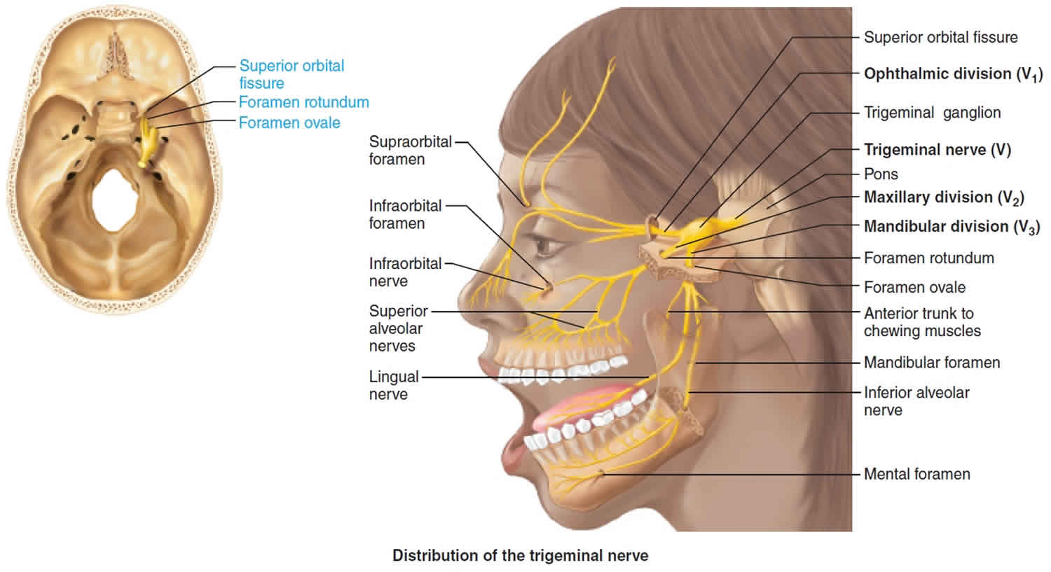
Figure 2. Trigeminal nerve exiting the cranial cavity
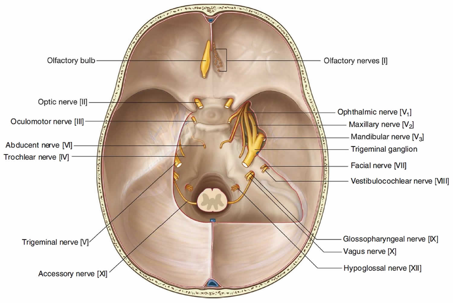
Figure 3. Trigeminal nerve distribution
Trigeminal nerve branches
Ophthalmic nerve
The ophthalmic nerve [V1] passes forward in the dura of the lateral wall of the cavernous sinus, leaves the cranial cavity, and enters the orbit through the superior orbital fissure (Figure 1). The ophthalmic nerve [V1] carries sensory branches from the eyes, conjunctiva, and orbital contents, including the lacrimal gland. The ophthalmic nerve [V1] also receives sensory branches from the nasal cavity, frontal sinus, ethmoidal cells, falx cerebri, dura in the anterior cranial fossa and superior parts of the tentorium cerebelli, upper eyelid, dorsum of the nose, and the anterior part of the scalp.
Maxillary nerve
The maxillary nerve [V2] passes forward in the dura mater of the lateral wall of the cavernous sinus just inferior to the ophthalmic nerve [V1], leaves the cranial cavity through the foramen rotundum and enters the pterygopalatine fossa. The maxillary nerve [V2] receives sensory branches from the dura in the middle cranial fossa, the nasopharynx, the palate, the nasal cavity, teeth of the upper jaw, maxillary sinus, and skin covering the side of the nose, the lower eyelid, the cheek, and the upper lip.
Mandibular nerve
The mandibular nerve [V3] leaves the inferior margin of the trigeminal ganglion and leaves the skull through the foramen ovale. The motor root of the trigeminal nerve also passes through the foramen ovale and unites with the sensory component of the mandibular nerve [V3] outside the skull. Thus the mandibular nerve [V3] is the only division of the trigeminal nerve that contains a motor component.
Outside the skull the motor fibers innervate the four muscles of mastication (temporalis, masseter, and medial and lateral pterygoids), as well as the tensor tympani muscle, the tensor veli palatini muscle, the anterior belly of the digastric muscle, and the mylohyoid muscle.
The mandibular nerve [V3] also receives sensory branches from the skin of the lower face, cheek, lower lip, anterior part of the external ear, part of the external acoustic meatus and the temporal region, the anterior two thirds of the tongue, the teeth of the lower jaw, the mastoid air cells, the mucous membranes of the cheek, the mandible, and dura in the middle cranial fossa.
Trigeminal nerve function
Trigeminal nerve provides sensory function via:
- Ophthalmic division [V1]: General somatic sensation from the eyes cornea, conjunctiva, orbital contents, nasal cavity mucosa, frontal sinus, ethmoidal cells, upper eyelid, lacrimal gland, dorsum of nose, skin of anterior part of scalp, dura in anterior cranial fossa, superior part of tentorium cerebelli;
- Maxillary nerve [V2]: General somatic sensation from skin of cheek, upper lip, and lower eyelid, nasal cavity mucosa, palate, upper teeth, maxillary sinus, skin covering the side of the nose, lower eyelid, cheek, upper lip, the dura in middle cranial fossa and nasopharynx.
- Mandibular division [V3]: General somatic sensation from skin of chin, cheek, lower lip, mucous membranes of cheek, anterior part of external ear, part of external acoustic meatus, temporal fossa, mastoid air cells, temporal region of scalp, anterior two-thirds of tongue, lower teeth, mandible and dura in middle cranial fossa.
Trigeminal nerve also innervates the muscles of mastication: temporalis, masseter, medial and lateral pterygoids, tensor tympani, tensor velipalatini, anterior belly of digastric and mylohyoid muscles. Afferent proprioceptor fibers return from these muscles.
Trigeminal nerve pain
Trigeminal nerve pain also trigeminal neuralgia or tic douloureux, is a chronic pain condition that affects the trigeminal nerve. Trigeminal neuralgia is a form of neuropathic pain (pain associated with nerve injury or nerve lesion. The typical or “classic” form of trigeminal neuralgia called “Type 1” or trigeminal neuralgia 1 causes extreme, sporadic, sudden burning or shock-like facial pain that lasts anywhere from a few seconds to as long as two minutes per episode. These attacks can occur in quick succession, in volleys lasting as long as two hours. The “atypical” form of trigeminal neuralgia called “Type 2” or trigeminal neuralgia 2, is characterized by constant aching, burning, stabbing pain of somewhat lower intensity than Type 1. Both forms of trigeminal nerve pain may occur in the same person, sometimes at the same time. The intensity of trigeminal nerve pain can be physically and mentally incapacitating. Rarely, both sides of the face may be affected at different times in an individual, or even more rarely at the same time called bilateral trigeminal neuralgia.
Trigeminal neuralgia affects women more often than men, and it’s more likely to occur in people who are older than 50, although it can occur at any age, including infancy. The possibility of trigeminal neuralgia being caused by multiple sclerosis increases when it occurs in young adults. The incidence of new cases is approximately 12 per 100,000 people per year; the disorder is more common in women than in men.
Because of the variety of treatment options available, having trigeminal neuralgia doesn’t necessarily mean you’re doomed to a life of pain. Doctors usually can effectively manage trigeminal neuralgia with medications, injections or surgery.
Trigeminal nerve pain causes
Trigeminal neuralgia is associated with a variety of conditions. Trigeminal neuralgia can be caused by a blood vessel pressing on the trigeminal nerve as it exits the brain stem. This compression causes the wearing away or damage to the protective coating around the nerve (the myelin sheath). Trigeminal neuralgia symptoms can also occur in people with multiple sclerosis, a disease that causes deterioration of the trigeminal nerve’s myelin sheath. Rarely, symptoms of trigeminal neuralgia may be caused by nerve compression from a tumor, or a tangle of arteries and veins called an arteriovenous malformation. Injury to the trigeminal nerve (perhaps the result of sinus surgery, oral surgery, stroke, or facial trauma) may also produce neuropathic facial pain.
Trigeminal neuralgia may be caused by:
- Multiple sclerosis (MS) or other diseases that damage the protective covering myelin of the nerves
- Pressure on the trigeminal nerve from a swollen blood vessel or tumor
- Injury to the trigeminal nerve, such as from trauma to the face or from oral or sinus surgery
Often, no exact cause is found. Trigeminal neuralgia usually affects adults above age 50 years, but it can occur at any age. Women are affected more often than men. When trigeminal neuralgia affects people younger than 40, it is often due to multiple sclerosis (MS) or a tumor.
Trigeminal nerve pain symptoms
Trigeminal neuralgia symptoms may include one or more of these patterns:
- Episodes of severe, shooting or jabbing pain that may feel like an electric shock spasms that usually last from several seconds to less than 2 minutes, but can become constant.
- Pain is usually only on one side of the face, often around the eye, cheek, and lower part of the face.
- Spontaneous attacks of pain or attacks triggered by things such as touching the face, chewing, speaking brushing teeth or sounds
- Bouts of pain lasting from a few seconds to several minutes
- Episodes of several attacks lasting days, weeks, months or longer — some people have periods when they experience no pain
- Constant aching, burning feeling that may occur before it evolves into the spasm-like pain of trigeminal neuralgia
- Pain in areas supplied by the trigeminal nerve, including the cheek, jaw, teeth, gums, lips, or less often the eye and forehead
- Pain affecting one side of the face at a time, though may rarely affect both sides of the face
- Pain focused in one spot or spread in a wider pattern
- Attacks that become more frequent and intense over time
- There is usually no loss of sensation or movement of the affected part of the face.
Painful attacks of trigeminal neuralgia can be triggered by common, everyday activities, such as:
- Talking
- Smiling
- Brushing teeth
- Chewing
- Drinking
- Eating
- Exposure to hot or cold temperature
- Touching the face
- Shaving
- Wind
- Applying make-up
- Washing your face
The right side of the face is mostly affected. In some cases, trigeminal neuralgia goes away on its own.
Trigeminal nerve pain diagnosis
Your doctor will diagnose trigeminal neuralgia mainly based on your description of the pain, including:
- Type. Pain related to trigeminal neuralgia is sudden, shock-like and brief.
- Location. The parts of your face that are affected by pain will tell your doctor if the trigeminal nerve is involved.
- Triggers. Trigeminal neuralgia-related pain usually is brought on by light stimulation of your cheeks, such as from eating, talking or even encountering a cool breeze.
Your doctor may conduct many tests to diagnose trigeminal neuralgia and determine underlying causes for your condition, including:
- A neurological examination. Touching and examining parts of your face can help your doctor determine exactly where the pain is occurring and — if you appear to have trigeminal neuralgia — which branches of the trigeminal nerve may be affected. Reflex tests also can help your doctor determine if your symptoms are caused by a compressed nerve or another condition.
- Magnetic resonance imaging (MRI). Your doctor may order an MRI scan of your head to determine if multiple sclerosis or a tumor is causing trigeminal neuralgia. In some cases, your doctor may inject a dye into a blood vessel to view the arteries and veins and highlight blood flow (magnetic resonance angiogram).
Your facial pain may be caused by many different conditions, so an accurate diagnosis is important. Your doctor may order additional tests to rule out other conditions.
Trigeminal nerve pain treatment
Trigeminal neuralgia treatment usually starts with medications, and some people don’t need any additional treatment. However, over time, some people with the condition may stop responding to medications, or they may experience unpleasant side effects. For those people, injections or surgery provide other trigeminal neuralgia treatment options.
If your condition is due to another cause, such as multiple sclerosis, your doctor will treat the underlying condition.
Medications
To treat trigeminal neuralgia, your doctor usually will prescribe medications to lessen or block the pain signals sent to your brain.
- Anticonvulsants. Doctors usually prescribe carbamazepine (Tegretol, Carbatrol, others) for trigeminal neuralgia, and it’s been shown to be effective in treating the condition. Other anticonvulsant drugs that may be used to treat trigeminal neuralgia include oxcarbazepine (Trileptal), lamotrigine (Lamictal) and phenytoin (Dilantin, Phenytek). Other drugs, including clonazepam (Klonopin) and gabapentin (Neurontin, Gralise, others), also may be used. If the anticonvulsant you’re using begins to lose effectiveness, your doctor may increase the dose or switch to another type. Side effects of anticonvulsants may include dizziness, confusion, drowsiness and nausea. Also, carbamazepine can trigger a serious drug reaction in some people, mainly those of Asian descent, so genetic testing may be recommended before you start carbamazepine.
- Antispasmodic agents. Muscle-relaxing agents such as baclofen (Gablofen, Lioresal) may be used alone or in combination with carbamazepine. Side effects may include confusion, nausea and drowsiness.
- Botox injections. Small studies have shown that onabotulinumtoxinA (Botox) injections may reduce pain from trigeminal neuralgia in people who are no longer helped by medications. However, more research needs to be done before this treatment is widely used for this condition.
Surgery
Surgical options for trigeminal neuralgia include:
- Microvascular decompression. This procedure involves relocating or removing blood vessels that are in contact with the trigeminal root to stop the nerve from malfunctioning. During microvascular decompression, your doctor makes an incision behind the ear on the side of your pain. Then, through a small hole in your skull, your surgeon moves any arteries that are in contact with the trigeminal nerve away from the nerve, and places a soft cushion between the nerve and the arteries. If a vein is compressing the nerve, your surgeon may remove it. Doctors may also cut part of the trigeminal nerve (neurectomy) during this procedure if arteries aren’t pressing on the nerve. Microvascular decompression can successfully eliminate or reduce pain most of the time, but pain can recur in some people. Microvascular decompression has some risks, including decreased hearing, facial weakness, facial numbness, a stroke or other complications. Most people who have this procedure have no facial numbness afterward.
- Brain stereotactic radiosurgery (Gamma knife). In this procedure, a surgeon directs a focused dose of radiation to the root of your trigeminal nerve. This procedure uses radiation to damage the trigeminal nerve and reduce or eliminate pain. Relief occurs gradually and may take up to a month. Brain stereotactic radiosurgery is successful in eliminating pain for the majority of people. If pain recurs, the procedure can be repeated. Facial numbness can be a side effect.
Other procedures may be used to treat trigeminal neuralgia, such as a rhizotomy. In a rhizotomy, your surgeon destroys nerve fibers to reduce pain, and this causes some facial numbness. Types of rhizotomy include:
- Glycerol injection. During this procedure, your doctor inserts a needle through your face and into an opening in the base of your skull. Your doctor guides the needle into the trigeminal cistern, a small sac of spinal fluid that surrounds the trigeminal nerve ganglion — where the trigeminal nerve divides into three branches — and part of its root. Then, your doctor will inject a small amount of sterile glycerol, which damages the trigeminal nerve and blocks pain signals. This procedure often relieves pain. However, some people have a later recurrence of pain, and many experience facial numbness or tingling.
- Balloon compression. In balloon compression, your doctor inserts a hollow needle through your face and guides it to a part of your trigeminal nerve that goes through the base of your skull. Then, your doctor threads a thin, flexible tube (catheter) with a balloon on the end through the needle. Your doctor inflates the balloon with enough pressure to damage the trigeminal nerve and block pain signals. Balloon compression successfully controls pain in most people, at least for a period of time. Most people undergoing this procedure experience at least some transient facial numbness.
- Radiofrequency thermal lesioning. This procedure selectively destroys nerve fibers associated with pain. While you’re sedated, your surgeon inserts a hollow needle through your face and guides it to a part of the trigeminal nerve that goes through an opening at the base of your skull. Once the needle is positioned, your surgeon will briefly wake you from sedation. Your surgeon inserts an electrode through the needle and sends a mild electrical current through the tip of the electrode. You’ll be asked to indicate when and where you feel tingling. When your neurosurgeon locates the part of the nerve involved in your pain, you’re returned to sedation. Then the electrode is heated until it damages the nerve fibers, creating an area of injury (lesion). If your pain isn’t eliminated, your doctor may create additional lesions. Radiofrequency thermal lesioning usually results in some temporary facial numbness after the procedure. Pain may return after three to four years.
- A neurectomy (also called partial nerve section), which involves cutting part of the nerve, may be performed near the entrance point of the nerve at the brain stem during an attempted microvascular decompression if no vessel is found to be pressing on the trigeminal nerve. Neurectomies also may be performed by cutting superficial branches of the trigeminal nerve in the face. When done during microvascular decompression, a neurectomy will cause more long-lasting numbness in the area of the face that is supplied by the nerve or nerve branch that is cut. However, when the operation is performed in the face, the nerve may grow back and in time sensation may return. With neurectomy, there is risk of creating anesthesia dolorosa.
Surgical treatment for trigeminal neuralgia 2 is usually more problematic than for trigeminal neuralgia 1, particularly where vascular compression is not detected in brain imaging prior to a proposed procedure. Many neurosurgeons advise against the use of microvascular decompression or rhizotomy in individuals for whom trigeminal neuralgia2 symptoms predominate over trigeminal neuralgia 1, unless vascular compression has been confirmed. Microvascular decompression for trigeminal neuralgia2 is also less successful than for trigeminal neuralgia 1.
Alternative medicine
Alternative treatments for trigeminal neuralgia generally haven’t been as well-studied as medications or surgical procedures, so there’s often little evidence to support their use.
However, some people have found improvement with treatments, such as low-impact exercise, yoga, creative visualization, aroma therapy, acupuncture, biofeedback, upper cervical chiropractic, and vitamin or nutritional therapy. Be sure to check with your doctor before trying an alternative treatment because it may interact with your other treatments. Some people report modest pain relief after injections of botulinum toxin to block activity of sensory nerves.
Coping and support
Living with trigeminal neuralgia can be difficult. The disorder may affect your interaction with friends and family, your productivity at work, and the overall quality of your life.
You may find encouragement and understanding in a support group. Group members often know about the latest treatments and tend to share their own experiences. If you’re interested, your doctor may be able to recommend a group in your area.