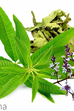
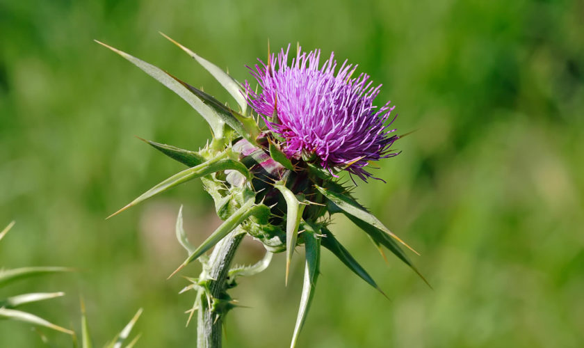
What is Sandalwood
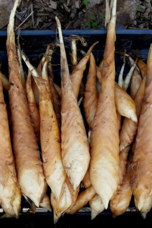
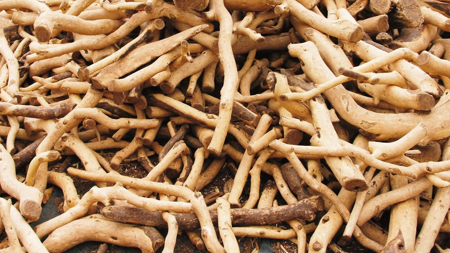
Sandalwood is the general name for woody perennials of the Santalum genus (Santalaceae), which are exploited for their fragrant heartwood. Sandalwoods are slow growing hemiparasitic trees distributed throughout the tropical and temperate regions of India, Indonesia, Australia and the Pacific Islands 1. The oil extracted from the sandalwood tree stems and roots are highly sought after by the fragrance and perfume industry. Santalum album, also known as tropical or Indian sandalwood, is the most valuable of the commercially used species due to the high heartwood oil content (6–10% by dry weight) and desirable odor characteristics. Approximately 90% of Sandalwood essential oil is composed of the sesquiterpene alcohols α-, β-, and epi-β-santalol and α-exo-bergamotol. The α- and β-santalols are the most important contributors to sandalwood oil fragrance 2. Lanceol and α-bisabolol are also found in modest concentrations 3. While the demand for sandalwood oil is increasing, disease, grazing animals and unsustainable exploitation of sandalwood trees has led to the demise of many natural populations. Plantations provide a more sustainable alternative to wild harvesting; however, slow growth rates, high potential for disease and substantial variation in oil yield hamper productivity. Alternatively, chemical approaches to synthesize the santalols have been attempted 4, key to these approaches is the elucidation of the biosynthesis of the santalols, bergamotols, and other sesquiterpene compounds characteristic of sandalwood oil. But multiple low-recovery steps make chemical synthesis uneconomical at an industrial scale.
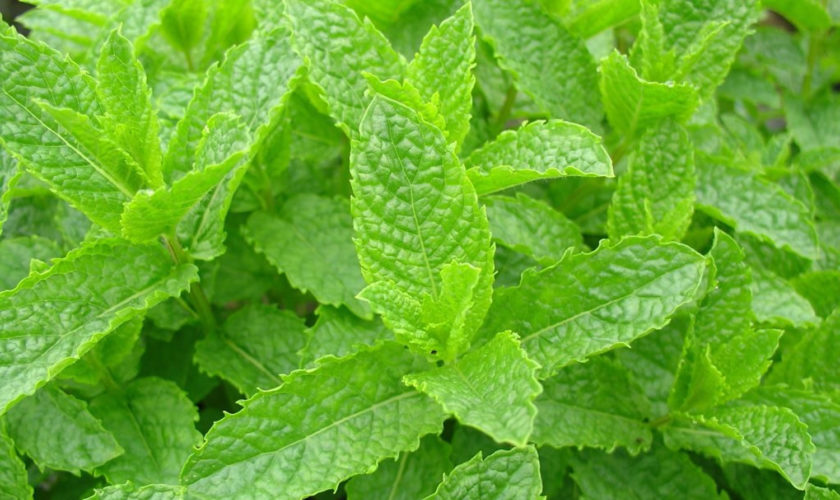
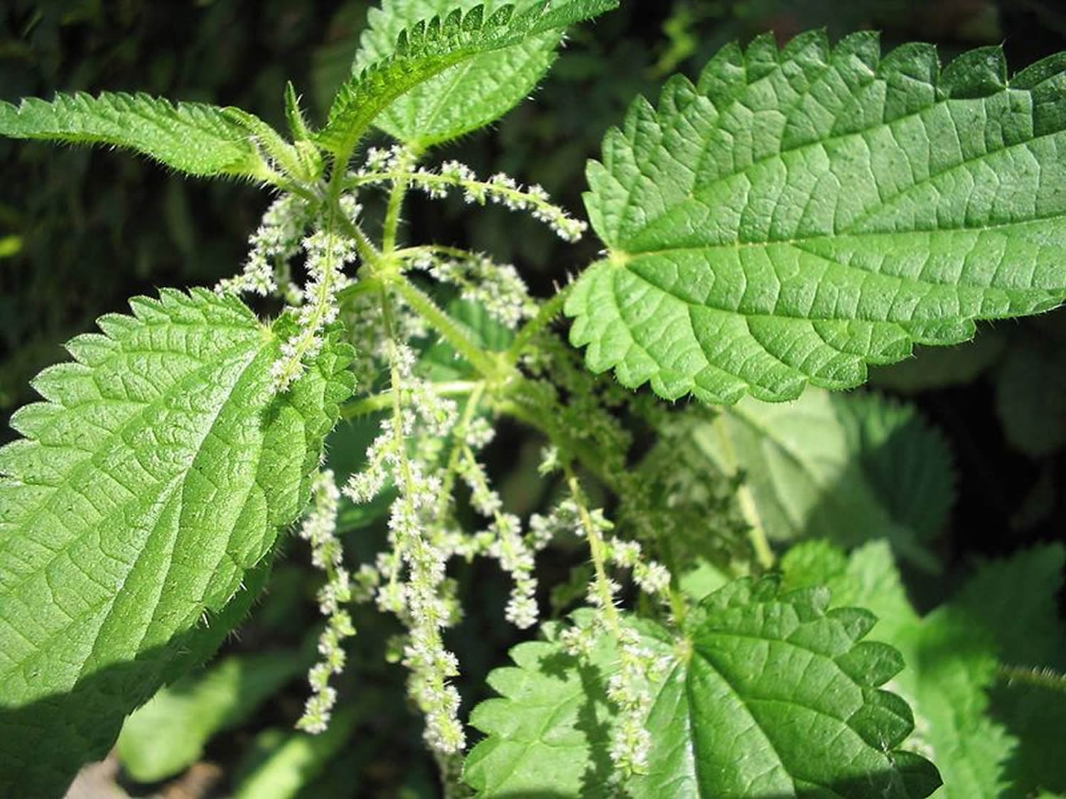
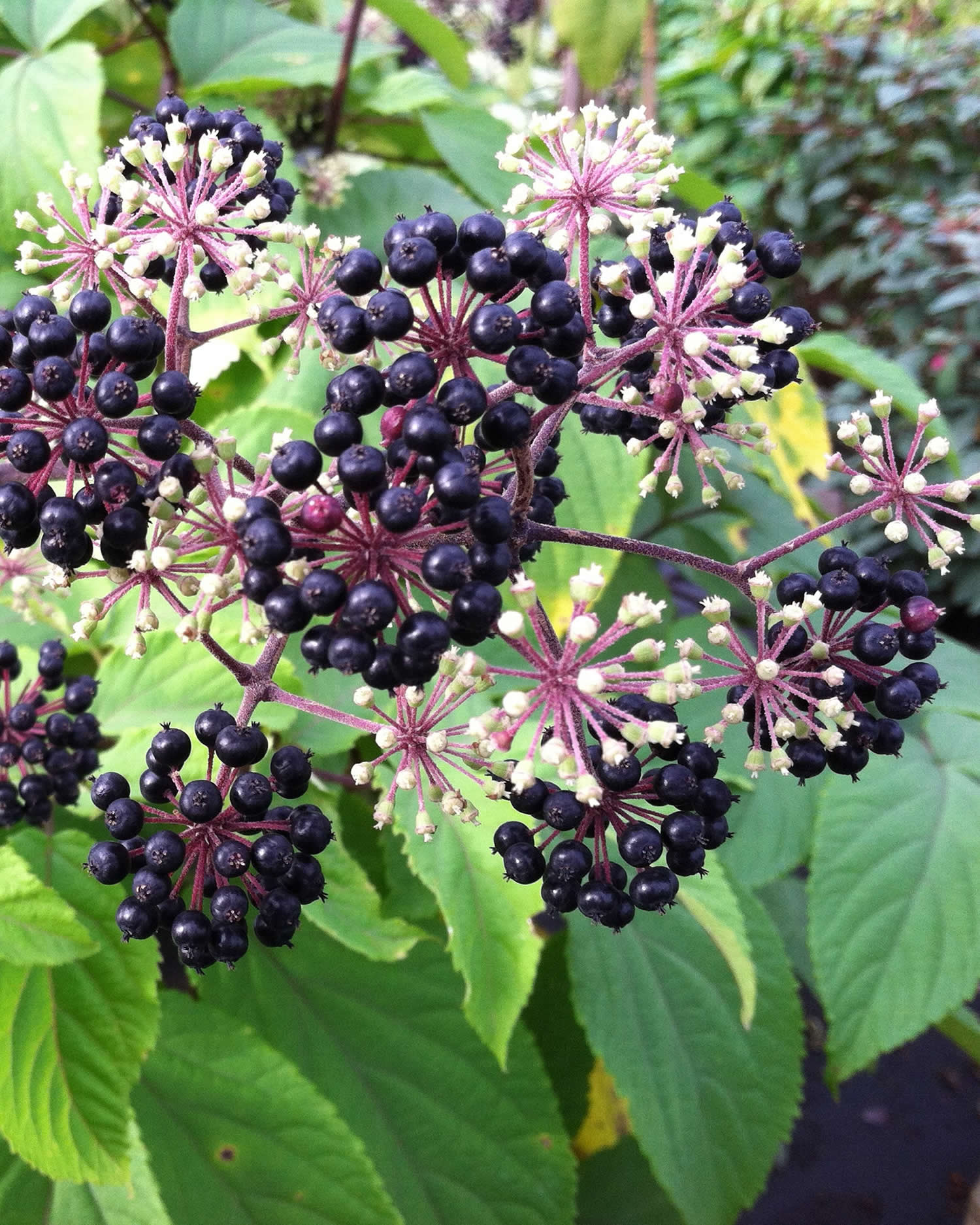
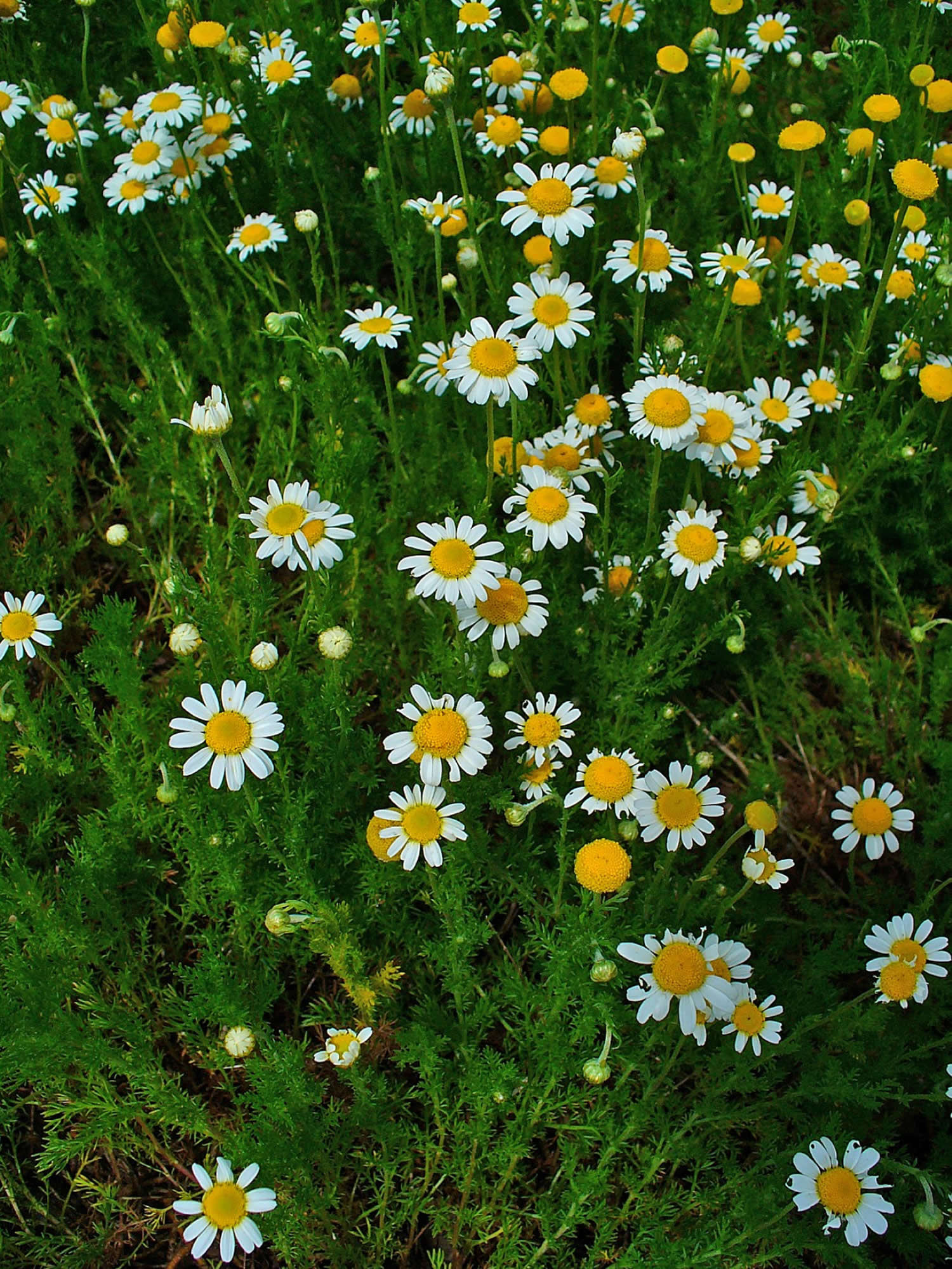
Figure 1. Sandalwood
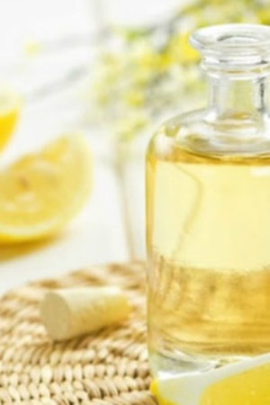
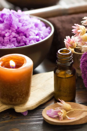
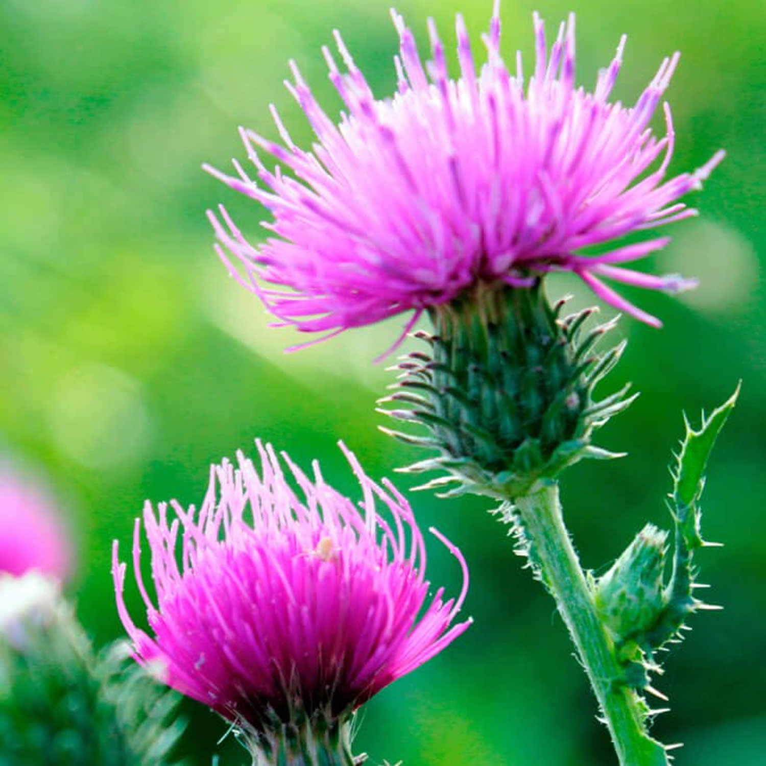
Sandalwood essential oil
Indian sandalwood (Santalum album L.) is an economically important plant species because of its ability to produce highly valued perfume oils. Sandalwood oil is an essential oil obtained from the steam distillation of chips and billets cut from the mature heartwood of various species of sandalwood trees – the most common are the Indian sandalwood (Santalum album) and Australian sandalwood (Santalum spicatum) followed by the species found in Hawaii (Santalum ellipticum), New Caledonia (Santalum austrocaledonicum), and French Polynesia (Santalum insulare) 5. The chemical composition of sandalwood essential oil has been studied in detail. At least 300 chemical constituents have been identified. The essential oil obtained from the well matured sandalwood tree is represented by a mixture of sesquiterpene olefins and alcohols such as: (Z)-α-santalol, (Z)-β-santalol, (Z)-epi-β-santalol, (Z)-α-trans-bergamotol, α-bisabolol, (Z)-lanceol, sesquisabinene hydrate and (E,E)-farnesol, along with small amounts (up to 2%) of corresponding precursor sesquiterpene hydrocarbons 6. Four sesquiterpenols, α-, β-, and epi-β-santalol and α-exo-bergamotol, make up approximately 90% of the oil of Indian sandalwood, where (Z)-α-santalol (alpha-santalol) and (Z)-β-santalol (beta-santalol) constitute the most abundant 7 and the major active components 8.
Table 1. Chemical composition of New Caledonia Sandalwood (Santalum austrocaledonicum) essential oil
| Compound | Percent (%) |
|---|---|
| 1-Furfurylpyrrole | 0.11 |
| 7-Epi-sesquithujene | 0.08 |
| α-Santalene | 0.67 |
| Trans-α-bergamotene | 0.11 |
| Epi-β-santalene | 0.43 |
| β-Acoradiene | 0.13 |
| γ-Curcumene | 0.27 |
| α-Curcumene | 0.28 |
| Helifolen-12-al | 0.07 |
| β-Bisabolene | 0.21 |
| β-Curcumene | 0.54 |
| β-Sesquiphellandrene | 0.14 |
| 8,14-Cedranoxide | 0.09 |
| (E)-Nerolidol | 0.53 |
| Dendrolasin | 0.38 |
| Sesquiterpenoids | 2.66 |
| β-Bisabolol | 1.60 |
| (Z)-α-Santalol | 25.34 |
| α-Santalal | 0.95 |
| (Z)-Trans-α-Bergamotol | 4.35 |
| 8-Cedren-13-ol | 1.83 |
| (Z)-Epi-β-Santalol | 4.07 |
| (Z,Z)-Farnesol | 1.56 |
| (E)-β-Santalol | 10.97 |
| (Z)-Nuciferol | 18.34 |
| (E)-α-Santalol | 0.48 |
| (Z)-α-Cis-Bergamotol | 0.76 |
| (E)-Nuciferol | 10.46 |
| (Z)-Lanceol | 7.34 |
| (Z)-α-Trans-bergamotol acetate | 1.33 |
| (E)-α-Trans-bergamotol acetate | 0.59 |
| (E)-Lanceol acetate | 0.83 |
| (E)-β-Santalol acetate | 0.15 |
| 1,3-Hydroxy-bisabola-2,10-diene | 0.57 |
| Total | 98.22 |
Sandalwood essential oil is widely used for several purposes such as in perfumery, cosmetics, aromatherapy, as an antidepressant, anti-inflammatory, antifungal, astringent, sedative, insecticide, antiseptic and in sacred unguents 10, 11. Sandalwood essential oil is also used in the food industry as a flavoring ingredient with a daily consumption of 0.0074 mg/kg 12. Sandalwood oil and its major constituent have low acute oral and dermal toxicity in laboratory animals 12. Although the available information on toxicity of sandalwood oil is limited, it has a long history of oral use without any reported adverse effects and is considered safe at present use levels 12.
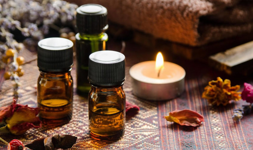
Sandalwood essential oil uses
Sandalwood essential oil is used in perfumes, cosmetics, sacred unguents, and as a mild food flavoring. Due to its highly coveted fragrance, the essential oil produced from Sandalwood is often used in aromatherapy and is added to soaps and cosmetics. It is also used in Ayurvedic medicine for the treatment of both somatic and mental disorders, including common colds, bronchitis, fever, urinary tract infections, and inflammation 13. A study investigating the effects of inhalation of East Indian sandalwood oil and its main compound, α-santalol, on human physiological parameters found that the compounds elevated pulse rate, skin conductance, and systolic blood pressure 13.
Sandalwood essential oil has also been studied as a chemopreventive agent for skin papillomas in mice 14 and in vitro in human epidermoid carcinoma cells 15. Matsuo and Mimaki studied the cytotoxicity of various α-santalol derivatives and found that some of them present tumor-specific cytotoxicity 16. This group also found that α-santalol induces DNA fragmentation as a result of apoptosis 16. Even though previous studies have provided valuable data on the pharmacological properties of individual constituents of sandalwood essential oil, there is no information on the activity of whole sandalwood essential oil in human breast cells, or on its DNA-damaging potential. Due to the increased popularity of essential oils for massage and aromatherapy, people are frequently exposed to sandalwood essential oil through various routes of administration including the skin. Essential oils can easily be absorbed through the human skin due to their lipid solubility and extremely low molecular size and the lipophilic nature of the skin itself 17.
Folk medicine uses
Sandalwood has been used in traditional medicine and Ayurveda to treat many conditions such as:
- Lymphatic system – Venous and lymphatic stasis such as varicose veins and swollen lymph nodes.
- Nervous system – Sandalwood oil has a relaxing effect on the nerves and may be used for hot, agitated emotional states leading to conditions such as headaches, insomnia and nervous tension.
- Respiratory system – Can treat respiratory tract infections, especially when its soothing, demulcent effects are required.
- Genitourinary – Has been used for years for genitourinary tract infections (e.g. Cystitis and gonorrhoea). “Sandalwood is an astringent and helps to resolve mucous congestion. Sandalwood oil helps to restore the mucous membrane and minimise the risk of infection”
- Integumentary system– “Applied to the skin, sandalwood oil is soothing, cooling and moisturising and primarily used for dry skin conditions caused by loss of moisture and skin inflammations. It may be used to relieve eczema and psoriasis and for the treatment of oily skin and acne”
- Anti-viral(herpes simplex/cold sores)
- Anti-cancer e.g. skin cancer, breast cancer
- Anti-microbial
- Anti-fungal
Summary
Our take is that sandalwood and sandalwood essential oil uses lack proper clinical scientific evidence to be of any use apart for its use in the food flavoring and perfumery industry. More studies especially well designed human clinical trials are needed to substantiate these pseudo-science or anecdotal therapeutic effects or benefits.
- Geographic and phenotypic variation in heartwood and essential-oil characters in natural populations of Santalum austrocaledonicum in Vanuatu. Page T, Southwell I, Russell M, Tate H, Tungon J, Sam C, Dickinson G, Robson K, Leakey RR. Chem Biodivers. 2010 Aug; 7(8):1990-2006. https://www.ncbi.nlm.nih.gov/pubmed/20730962/[↩]
- Baldovini N, Delasalle C, Joulain D (2011) Phytochemistry of the heartwood from fragrant Santalum species: a review. Flavour Fragrance J 26: 7–26[↩]
- Jones CG, Plummer JA, Barbour EL (2007) Non-destructive sampling of Indian sandalwood (Santalum album L.) for oil content and composition. J Essent Oil Res 19: 157–164[↩]
- Brocke C, Eh M, Finke A (2008) Recent developments in the chemistry of sandalwood odorants. Chem Biodivers 5: 1000–1010. https://www.ncbi.nlm.nih.gov/pubmed/18618410[↩]
- Phylogeny and biogeography of the sandalwoods (Santalum, Santalaceae): repeated dispersals throughout the Pacific. Harbaugh DT, Baldwin BG. Am J Bot. 2007 Jun; 94(6):1028-40. http://www.amjbot.org/content/94/6/1028.long[↩]
- Preparative separation of α- and β-santalenes and (Z)-α- and (Z)-β-santalols using silver nitrate-impregnated silica gel medium pressure liquid chromatography and analysis of sandalwood oil. Daramwar PP, Srivastava PL, Priyadarshini B, Thulasiram HV. Analyst. 2012 Oct 7; 137(19):4564-70. https://www.ncbi.nlm.nih.gov/pubmed/22900258/[↩]
- Structure-odor relationships of α-santalol derivatives with modified side chains. Hasegawa T, Izumi H, Tajima Y, Yamada H. Molecules. 2012 Feb 22; 17(2):2259-70.[↩]
- Phylogeny and biogeography of the sandalwoods (Santalum, Santalaceae): repeated dispersals throughout the Pacific. Harbaugh DT, Baldwin BG. Am J Bot. 2007 Jun; 94(6):1028-40.[↩]
- Ortiz C, Morales L, Sastre M, Haskins WE, Matta J. Cytotoxicity and Genotoxicity Assessment of Sandalwood Essential Oil in Human Breast Cell Lines MCF-7 and MCF-10A. Evidence-based Complementary and Alternative Medicine : eCAM. 2016;2016:3696232. doi:10.1155/2016/3696232. https://www.ncbi.nlm.nih.gov/pmc/articles/PMC4879231/[↩]
- Jirovetz L. et al. Comparative study on the antimicrobial activities of different sandalwood essential oils of various origin. Flavour Frag. J. 21, 465–468 (2006).[↩]
- Chemopreventive effects of various concentrations of alpha-santalol on skin cancer development in CD-1 mice. Dwivedi C, Maydew ER, Hora JJ, Ramaeker DM, Guan X. Eur J Cancer Prev. 2005 Oct; 14(5):473-6.[↩]
- Safety assessment of sandalwood oil (Santalum album L.). Burdock GA, Carabin IG. Food Chem Toxicol. 2008 Feb; 46(2):421-32. https://www.ncbi.nlm.nih.gov/pubmed/17980948/[↩][↩][↩]
- East Indian Sandalwood and alpha-santalol odor increase physiological and self-rated arousal in humans. Planta Med. 2006 Jul;72(9):792-800. Epub 2006 Jun 19. https://www.ncbi.nlm.nih.gov/pubmed/16783696[↩][↩]
- Sandalwood oil prevent skin tumour development in CD1 mice. Dwivedi C, Zhang Y. Eur J Cancer Prev. 1999 Oct; 8(5):449-55.[↩]
- Skin cancer chemopreventive agent, {alpha}-santalol, induces apoptotic death of human epidermoid carcinoma A431 cells via caspase activation together with dissipation of mitochondrial membrane potential and cytochrome c release. Kaur M, Agarwal C, Singh RP, Guan X, Dwivedi C, Agarwal R. Carcinogenesis. 2005 Feb; 26(2):369-80. https://www.ncbi.nlm.nih.gov/pubmed/15528219/[↩]
- α-Santalol derivatives from Santalum album and their cytotoxic activities. Matsuo Y, Mimaki Y. Phytochemistry. 2012 May; 77():304-11. https://www.ncbi.nlm.nih.gov/pubmed/22410352/[↩][↩]
- Keville K., Green M. Aromatherapy: A Complete Guide to the Healing Art. Freedom, Calif, USA: The Crossing Press; 1995. The sense of smell; pp. 9–10.[↩]
