Natural Remedies

Boswellia

Boswellia-serrata

What is boswellia serrata

Boswellia serrata (also called Frankincense or olibanum) has been used in the traditional herbal formulas to treat inflammatory arthritis in China and India 1. Boswellia serrata contains boswellic acids which have been shown to exhibit anti-inflammatory and antiarthritic properties.

The gum resin extracted from Boswellia serrata has gained considerable attention as a potent anti-inflammatory, anti-arthritic and analgesic agent 2. 3-O-Acetyl-11-keto-beta-boswellic acid (AKBA) is the most active compound of Boswellia extract and is a potent inhibitor of 5-lipoxygenase (5-LOX), a key enzyme in the biosynthesis of leukotrienes from arachidonic acid in the cellular inflammatory cascade 3. A number of independent clinical studies support the anti-inflammatory and anti-arthritic properties of Boswellia extracts 4, 5.

In the last decade preparations from the oleogum resin of Boswellia serrata and other Boswellia species, have become more and more popular in some European countries for the treatment of a variety of chronic inflammatory diseases including rheumatoid arthritis, chronic bowel diseases, bronchial asthma, peritumoural brain oedema and others 6. However, studies indicate that upon oral administration, Boswellia extracts exhibit poor intestinal absorption of 3-O-Acetyl-11-keto-beta-boswellic acid (AKBA) and poor bioavailability which limits its anti-inflammatory efficacy 7, 8.

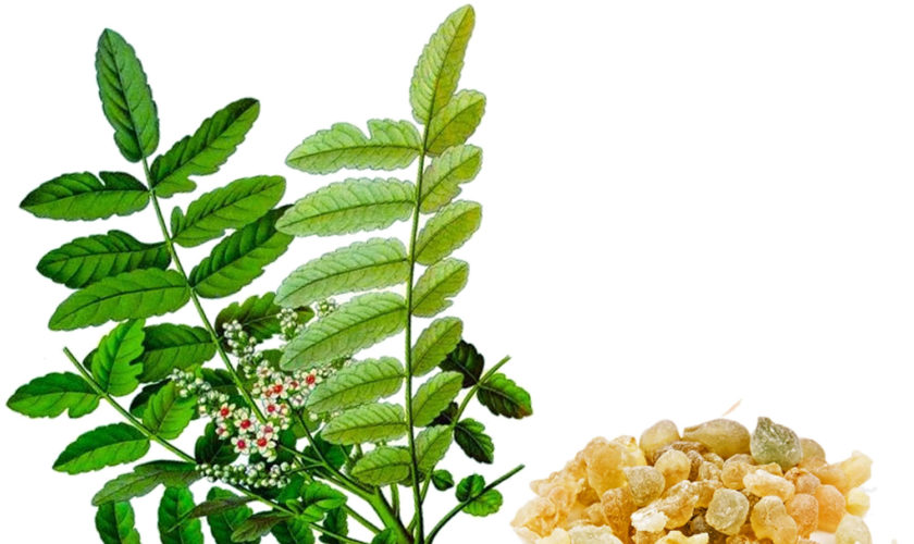
Boswellia are moderate-sized flowering plants, including both trees and shrubs, and are native to tropical regions of Africa and Asia. The distributions of the species are primarily associated with the tropics. The greatest diversity of species presently is in Africa and India 9.

Frank (“pure”) incense is the oleogum resin produced in the bark of different Boswellia species belonging to the family of Burseraceae. There are four main species of Boswellia which produce true frankincense. Boswellia sacra (synonyms B. carteri and B. bhaw-dajiana), Boswellia frereana, Boswellia papyrifera, and Boswellia serrata 10 and each type of resin is available in various grades. The grades depend on the time of harvesting, and the resin is hand sorted for quality. The term guggals collectively refers to gum resins.

Table 1. Boswellia serrata (Salai guggal) traditional Indian Ayurvedic Medicine Uses

Organs and functional systemsEffects
Nervous systemAnalgesic
Mental tonic
Stimulation
Eye tonic
Cardiovascular systemCardiotonic
Gastrointestinal tractRegulating colour of stool
Carminative, stomachic
Improving digestion, antidiarrhoeic
Improving taste
Anthelmintic
Urogenital systemDiuretic
Aphrodisiac
Improving menstruation
FeverAntipyretic
SkinIncreases perspiration
Wound cleaning
Whole organismAnti-inflammatory
Antiseptic
Reducing fat
Haemostypic
Connecting tissue
Decreasing Kapha diseases
(in Ayurvedic nomenclature)
[Source 6]

Composition of Boswellia Extract Oleogum Resins

More than 200 different compounds were identified in the boswellia extract oleogum resin of different Boswellia species. Main components are volatile oil, pure resin and mucus. The content of these differs from species to species, between different harvestings and different locations. An approximate composition of some oleogum resins is listed in Table 2. The resins of Boswellia species contain pentacyclic and tetracyclic triterpenes. Among the pentacyclic triterpenes, some boswellic acids are mainly responsible for many of the pharmacological effects. Further compounds are tetracyclic triterpenic acids among which tirucallic acids were also shown to be biologically active.

Table 2. Composition of boswellia extract oleogum resin of two different Boswellia species

Boswellia carteri Birdw.Boswellia serrata Roxb.
Volatile oil5 – 9 %7.5 – 9 % to 15 %
Pure resin≈ 66 %55 – 57 %
Mucus≈ 12 – 20 %≈ 23 %
[Source 11]

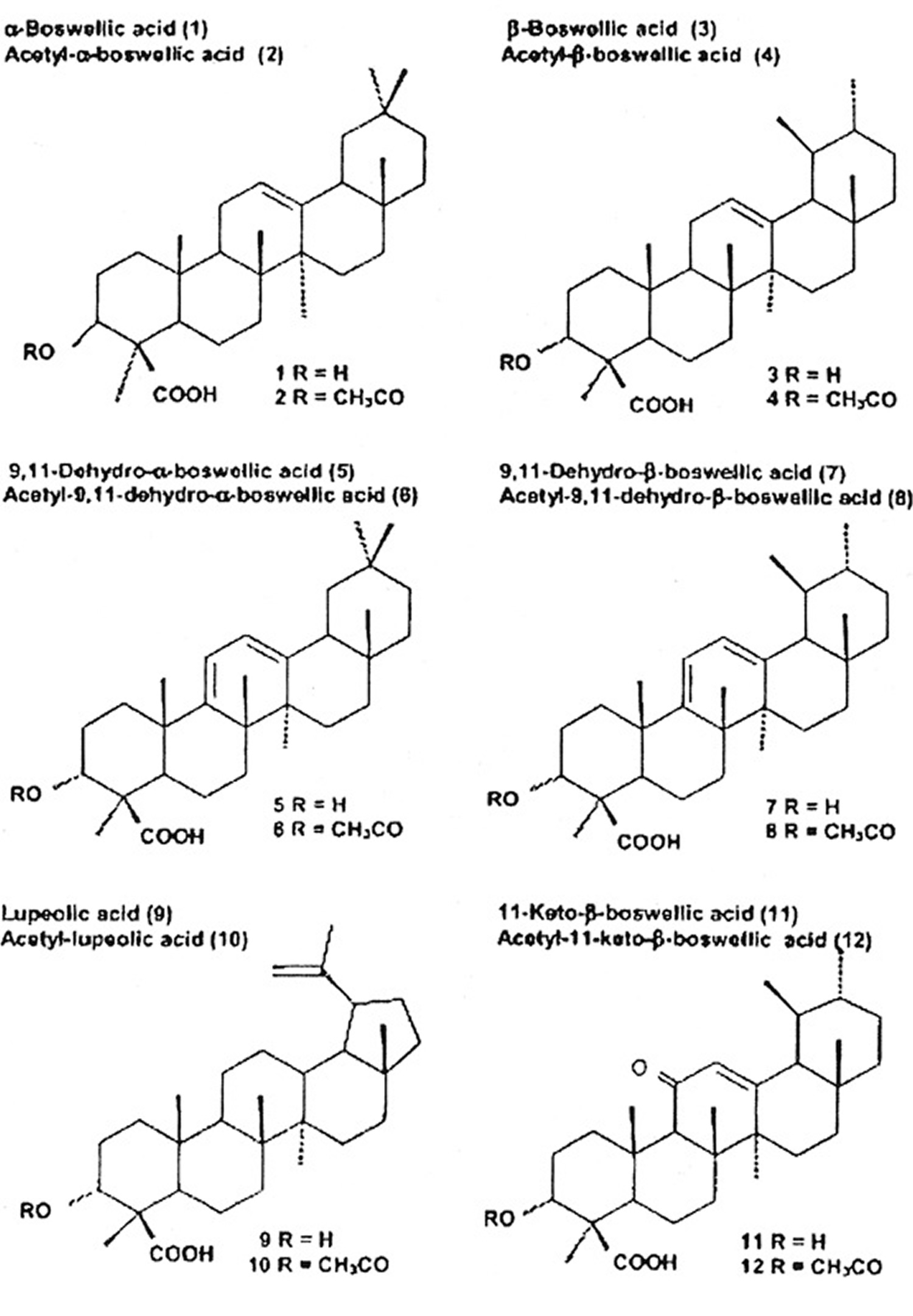
Among the pentacyclic triterpenes, a variety of boswellic acids and other compounds were identified. Some of them are closely related to the pharmacological effects of boswellia serrata. Figure 1 shows their chemical structure and the content in the oleogum resins of boswellia serrata and African species. In 2003, Büchele et al. 12 identified 12 different pentacyclic triterpenes in different samples of Boswellia, i. e., frankincense from India and Africa using an extract. The authors reported marked differences in different species. A striking difference was observed in the content of the main active boswellic acids 3-O-Acetyl-11-keto-beta-boswellic acid (AKBA) and 11-keto-β-boswellic acid (KBA). It is evident that the Indian sample contained quite similar amounts of 3-O-Acetyl-11-keto-beta-boswellic acid (AKBA) and 11-keto-β-boswellic acid (KBA) whereas the African samples contained less 11-keto-β-boswellic acid (KBA) than 3-O-Acetyl-11-keto-beta-boswellic acid (AKBA). A new pentacyclic triterpene from boswellia serrata, i. e., 3α-acetyl-20(29)-lupene-24-oic acid was recently identified by Beisner et al. 13. Employing a commercial extract from boswellia serrata (H 15 AyurmedicaTM), ∼2.6 mg/100 mg KBA and ∼2.8 mg/100 mg AKBA were detected on average in 11 different lots. Ganzera et al. 14 studied 4 different commercial products containing the oleogum resin of boswellia serrata together with up to 10 other plant extracts. Considering the manufacturer’s dosing recommendations, the daily intake of total boswellic acids varies up to 6-fold (18.49 to 109.62 mg per day). Hamm et al. 15 tested volatile and semi-volatile terpenes from 6 different olibanum samples, i. e., B. carterii, B. sacra, B. serrata, B. papyfera and B. frereana. The chemical composition was different in all species and allowed identification of the taxonomic origin of frankincense samples purchased from various markets.

Among the tetracyclic triterpenes, three tirucallic acids were identified, i. e., 3-oxotirucallic acid, 3-hydroxytirucallic acid and 3-acetoxytirucallic acid, which were also shown to interact with the 5-LO-system 16.

Figure 1. Triterpenic acids present in Frankincense

triterpenic acids present in frankincense

Boswellia benefits

There are several severe chronic diseases mostly related to autoimmune disorders. Among these are rheumatic diseases, inflammatory bowel diseases, bronchial asthma and others.

In fact, several clinical studies were performed on the effect of boswellia serrata. However, the results published so far must be regarded as inconclusive and early preliminary in character because some were performed in test tubes and in animals order to test theoretical hypothesis that may also be of clinical relevance. Therefore, further studies involving human subjects confirming these findings are necessary.

Rheumatoid Arthritis and Osteoarthritis

In traditional Indian Ayurvedic medicine, Salai guggal is used to treat rheumatoid arthritis. In order to support this traditional use scientifically, Singh and Atal 17 from the Regional Research Laboratory in Jammu, India described for the first time anti-inflammatory properties of Salai guggal ex Boswellia serrata in experimental animals. Further studies with the rat adjuvant arthritis model showed protective effects of Salai guggal and boswellic acids. Classical animal models for testing antiarthritic actions of drugs are formaldehyde- and adjuvant-produced arthritis and the cotton pellet-induced granuloma test. In the study of Singh and Atal 17, arthritis was induced by injecting 0.1 ml of formaldehyde (2 % v/v in normal saline) in the subplantar region on the 1st and 3rd day of experiment. Paw volume was measured before formaldehyde injection and during drug treatment. Drugs were administered orally daily. In a dose range of 50 – 200 mg/kg orally, the alcoholic extract of the oleogum resin of boswellia serrata resulted in marked inhibition of swelling in these rats.

Adjuvant arthritis was caused by injecting 0.05 ml of a (0.5 % w/v) suspension of killed Mycobacterium tuberculosis homogenised in liquid paraffin into the left hind foot. Oral administration of the test drug was started on the day before the injection of Mycobacterium and continued until day 14. Paw volume was measured on alternate days, and percent inhibition was calculated on day 14. In this model, boswellia extract at 100 mg/kg caused a reduction in swelling by 45 %.

In the adjuvant arthritis model, Kesava-Reddy et al. 18 studied the effect of an extract of oleogum resin of boswellia serrata [prepared according to Singh and Atal 17] on urinary excretion of connective tissue metabolites, including hydroxyproline, hexosamine and uronic acid. Compared to controls, the arthritic animals showed increased excretion of these metabolites in the urine. The elevated levels of urinary hydroxyproline (free, total, non-dialysable and dialysable), hexosamine and uronic acid in the arthritic animals were found to be slightly decreased in the acute phase and significantly decreased in the chronic phase of the disease following administration of the drug suggesting a beneficial action.

In another study, Sharma et al. 19 investigated the effect of a boswellia extract on bovine serum albumin (BSA)-induced arthritis in rabbits. Oral administration of the boswellia extract (25, 50 and 100 mg/kg/day) significantly reduced the population of leukocytes in a BSA-injected knee and changed the electrophoretic pattern of the synovial fluid proteins. The local injection of the extract (5, 10 and 20 mg) into the knee 15 min prior to BSA challenge also significantly reduced the infiltration of leukocytes into the knee joint, reduced the infiltration of leukocytes into the pleural cavity and inhibited the migration of PMN in vitro. The leukocyte-inhibitory activity of boswellic acids was not due to a cytotoxic effect and could later be explained by inhibition of leukotriene synthesis and therefore by a failure of the chemotactic action. The antiarthritic action of an acetone extract of Boswellia carterii Birdw. in adjuvant-induced arthritis of Lewin rats has been recently confirmed by Fan et al. 20.

Taken together, these studies suggest an anti-inflammatory action of boswellia extracts in experimental arthritis.

In 2006 a double-blind randomised controlled trial 21 designed to assess the efficacy and safety of 5-Loxin [5-Loxin® is a novel B. serrata extract enriched to 30% AKBA (US Patent publication no.: 2004/0073060A1)] for osteoarthritis of the knee. The 5-Loxin treatment is a gum extract of the ancient herb Boswellia serrata (frankincense), which is enriched with other chemicals to create a compound that inhibits the enzyme 5-lipoxygenase, which is known to be a key enzyme in the inflammatory process.

The trial was carried out in India and 75 people with mild-to-moderate osteoarthritis of the knee were randomised to receive either 100mg (low dose) or 250mg (high dose) of 5-Loxin per day, or identical placebo capsules. Some 236 outpatients were originally selected based on symptoms and signs of osteoarthritis. To be included in the trial, people had to have been suffering knee pain for longer than three months, be between 40 and 80 years old, and score between 4 and 7 on a 10-point visual analogue pain scale once they had stopped taking their usual medication (daily anti-inflammatory drugs or paracetamol) for one week. The researchers excluded all those with inflammatory conditions, such as rheumatoid arthritis, gout, knee injury, need for steroid injection in the past three months, obese people, those with high alcohol intake, or those with medical conditions that may put them at risk from the treatments (e.g. liver or kidney conditions).

The patients completed a baseline questionnaire about their medical history and nutritional status. They were followed up at seven, 30, 60 and 90 days. Pain, stiffness and physical function scores were assessed at each visit, and blood tests for inflammatory markers and urine samples were taken. Based on pain scores, the patients could receive “rescue” anti-inflammatory drugs if they were needed. If these were taken, the patients were advised to discontinue taking the “rescue” treatment three days before each assessment. No other treatments were taken.

At baseline and 90 days, the patients had fluid taken from the knee joint to look at the concentration of matrix metalloproteinase-3 (MMP3) – an enzyme which can break down bone cartilage. Adverse effects were self-reported by the patients. The primary outcome for the study was the difference in pain, stiffness and physical function with 5-Loxin compared with the placebo.

What were the results of the study ?

Seventy patients completed the study. There was significant improvement in pain score with both low- and high-dose 5-Loxin compared with the placebo. Compared with the placebo, low-dose 5-Loxin improved pain score by 49%, 24% and 40% on the three different scales used. However, the 43% improvement in stiffness and 29% improvement in function were not significant when compared with the placebo. In the high-dose group, percentage improvements were greater compared with the placebo across all measures of pain, stiffness and function, and these were significant. Both dose groups showed significant improvement in pain at one week compared with the placebo.

There was no change in MMP3 concentration in the fluid from the knee in the placebo group. However, 5-Loxin significantly reduced MMP3 concentration (by 31% at the low dose and 46% at the high dose). The reduction in MMP3 with high-dose 5-Loxin was also significantly greater than with the low dose. There was no difference in adverse effects seen in the treatment or control groups.

The researchers conclude that 5-Loxin is safe and significantly reduces pain and improves physical functioning in people with osteoarthritis.

What interpretations can we make of this study ?

This is reportedly the first trial of the drug 5-Loxin, and it provides promising results. However, results should be considered preliminary. There are a number of limitations to the interpretation of this study:

  • This trial was relatively small, and this may have affected the ability of randomisation to balance the groups for important characteristics. For example, on average those in the high-dose 5-Loxin group were about 6kg lighter, and had a BMI about 3.5 units lower, than those in the other groups.
  • The method of how participants were randomly assigned into groups was not described, and some methods of randomisation are not as robust as others.
  • The placebo pills were described as being “similar” in appearance, colour and taste to the 5-Loxin pills, but it is not clear if they were similar enough to prevent participants guessing which treatment they were receiving.
  • The trial was relatively short. Longer trials will be needed to confirm long-term safety.

In order to confirm efficacy and safety results, further research is needed in larger numbers of people for longer periods of time. Research is also required for people with different characteristics (strict inclusion and exclusion criteria were used here), and for those with osteoarthritis of joints other than the knee. Efficacy compared with the wide range of other medical and surgical treatments used in the treatment of osteoarthritis will also be needed. The possible role of this treatment in arthritic conditions aside from osteoarthritis is not known from this research.

In another double blind randomized clinical study done subsequent to the above 5-Loxin study 22 using a new boswellia extract formulation called Aflapin [which contains Boswellia serrata extract enriched to 20% AKBA and B. serrata non-volatile oil (PCT/IN2009/000505)] in knee osteoarthritis. Sixty eligible osteoarthritis subjects selected through screening were included in the study. The subjects received either 100 mg (n=30) of Aflapin® or placebo (n=30) daily for 30 days. Each subject was evaluated for pain and physical functions by using the standard tools (visual analog scale, Lequesne’s Functional Index, and Western Ontario and McMaster Universities Osteoarthritis Index) at the baseline (day 0), and at days 5, 15 and 30. A series of biochemical tests in serum, urine and hematological parameters established the safety of Aflapin. The observations suggest that Aflapin conferred clinically and statistically significant improvements in pain scores and physical function scores in osteoarthritis subjects. Aflapin provided significant improvements in pain score and functional ability in as early as 5 days of treatment. In conclusion, the researchers state that Aflapin is more efficacious as an anti-inflammatory agent compared to the existing Boswellia products, 5-Loxin® and traditional 65% Boswellia extract in alleviating pain, joint stiffness and improving physical functions in degenerative joint disease (osteoarthritis) subjects 22. In comparison with the placebo, at the end of the study, the Aflapin supplemented group showed statistically significant improvements in all pain scores.

Boswellia side effects

During the course of the 30-day Aflapin study 22, no major adverse events were reported. However, nausea and headache were reported as minor adverse events by two subjects during the study; one each from placebo and Aflapin supplemented groups.

One subject from placebo groups was dropped out from the study due to un-availability for the follow up evaluations.

During the course of the 90-day 5-Loxin study 21 period, some minor adverse events were noted: diarrhoea, nausea, abdominal pain, mild fever (up to 37.5°C [99.5°F]) and general weakness. The patients who reported these minor events were distributed evenly throughout the placebo and active treatment groups.

Five patients (one from the low-dose [100 mg 5-Loxin®] group, and two each from placebo and high-dose [250 mg 5-Loxin®] group) were excluded from the study because they were suffering from a nonfatal viral infection during the course of study.

  1. Chevrier MR, Ryan AE, Lee DY-W, Zhongze M, Wu-Yan Z, Via CS. Boswellia carterii Extract Inhibits TH1 Cytokines and Promotes TH2 Cytokines In Vitro. Clinical and Diagnostic Laboratory Immunology. 2005;12(5):575-580. doi:10.1128/CDLI.12.5.575-580.2005. https://www.ncbi.nlm.nih.gov/pmc/articles/PMC1112084/[]
  2. Boswellia: an evidence-based systematic review by the Natural Standard Research Collaboration. Basch E, Boon H, Davies-Heerema T, Foppo I, Hashmi S, Hasskarl J, Sollars D, Ulbricht C. J Herb Pharmacother. 2004; 4(3):63-83. https://www.ncbi.nlm.nih.gov/pubmed/15829470/[]
  3. Acetyl-11-keto-beta-boswellic acid (AKBA): structure requirements for binding and 5-lipoxygenase inhibitory activity. Sailer ER, Subramanian LR, Rall B, Hoernlein RF, Ammon HP, Safayhi H. Br J Pharmacol. 1996 Feb; 117(4):615-8. https://www.ncbi.nlm.nih.gov/pmc/articles/PMC1909340/[]
  4. Use of complementary and alternative medicine in Germany – a survey of patients with inflammatory bowel disease. Joos S, Rosemann T, Szecsenyi J, Hahn EG, Willich SN, Brinkhaus B. BMC Complement Altern Med. 2006 May 22; 6():19. https://www.ncbi.nlm.nih.gov/pmc/articles/PMC1539021/[]
  5. Efficacy and tolerability of Boswellia serrata extract in treatment of osteoarthritis of knee–a randomized double blind placebo controlled trial. Kimmatkar N, Thawani V, Hingorani L, Khiyani R. Phytomedicine. 2003 Jan; 10(1):3-7. https://www.ncbi.nlm.nih.gov/pubmed/12622457/[]
  6. Boswellic Acids in Chronic Inflammatory Diseases. Planta Med 2006; 72(12): 1100-1116. https://www.thieme-connect.com/products/ejournals/html/10.1055/s-2006-947227[][]
  7. Metabolism of boswellic acids in vitro and in vivo. Krüger P, Daneshfar R, Eckert GP, Klein J, Volmer DA, Bahr U, Müller WE, Karas M, Schubert-Zsilavecz M, Abdel-Tawab M. Drug Metab Dispos. 2008 Jun; 36(6):1135-42. http://dmd.aspetjournals.org/content/36/6/1135.long[]
  8. Permeation of Boswellia extract in the Caco-2 model and possible interactions of its constituents KBA and AKBA with OATP1B3 and MRP2. Krüger P, Kanzer J, Hummel J, Fricker G, Schubert-Zsilavecz M, Abdel-Tawab M. Eur J Pharm Sci. 2009 Feb 15; 36(2-3):275-84. https://www.ncbi.nlm.nih.gov/pubmed/19010411/[]
  9. Weeks, A., Daly, D.C. and B.B. Simpson. 2005. The phylogenetic history and biogeography of the frankincense and myrrh family (Burseraceae) based on nuclear and chloroplast sequence data. Molecular Phylogenetics and Evolution, 35: 85-101.[]
  10. Frankincense and myrrh. Tucker, A.O. Econ Bot (1986) 40: 425. https://doi.org/10.1007/BF02859654[]
  11. Kreck C, Saller R. Indischer Weihrauch und seine Zubereitungen einschließlich H15 als traditionelles und modernes Therapeutikum. Internist Prax. 1998; 38 857-72.[]
  12. Büchele B, Zugmaier W, Simmet T h. Analysis of pentacyclic triterpenic acids from frankincense gum resins and related phytopharmaceuticals by high-performance liquid chromatography. Identification of lupeolic acid, a novel pentacyclic triterpene. J Chromatogr B. 2003; 791 21-30.[]
  13. Beisner K, Büchele B, Werz U, Simmet T h. Structural analysis of 3-α-acetyl-20(29)-lupene-24-oic acid, a novel pentacyclic triterpene isolated from the gum resin of Boswellia serrata, by NMR spectroscopy. Magn Reson Chem. 2003; 41 629-32.[]
  14. Ganzera M, Khan I A. A reversed phase high performance liquid chromatography method for the analysis of boswellic acids in Boswellia serrata . Planta Med. 2001; 67 778-80[]
  15. Hamm S, Bleton J, Connan J, Tchapla A. A chemical investigation by headspace SPME and GC-MS of volatile and semi-volatile terpenes in various olibanum samples. Phytochemistry. 2005; 66 1499-514[]
  16. Boden S E, Schweizer S, Bertsche T h, Dufer M, Drews G, Safayhi H. Stimulation of leukotriene synthesis in intact polymorphonuclear cells by the 5-lipoxygenase inhibitor 3-oxotirucallic acid. Mol Pharmacol. 2001; 60 267-73[]
  17. Singh G B, Atal C K. Pharmacology of an extract of salai guggal ex Boswellia serrata, a new non-steroidal anti-inflammatory agent. Agents Actions. 1986; 18 407-12[][][]
  18. Kesava Reddy G, Dhar S C, Singh G B. Urinary excretion of connective tissue metabolites under the influence of a new non-steroidal anti-inflammatory agent in adjuvant induced arthritis. Agents Actions. 1987; 22 99-105.[]
  19. Sharma M L, Bani S, Singh G B. Anti-arthritic activity of boswellic acids in bovine serum albumin (BSA)-induced arthritis. Int J Immunopharmacol. 1989; 11 647-52[]
  20. Fan A Y, Lao L, Zhang R X, Wang L B, Lee D Y, Ma Z Z. et al . Effects of an acetone extract of Boswellia carterii Birdw. (Burseraceae) gum resin on rats with persistent inflammation. J Altern Complement Med. 2005; 11 323-31[]
  21. Sengupta K, Alluri KV, Satish AR, et al. A double blind, randomized, placebo controlled study of the efficacy and safety of 5-Loxin® for treatment of osteoarthritis of the knee. Arthritis Research & Therapy. 2008;10(4):R85. doi:10.1186/ar2461. https://www.ncbi.nlm.nih.gov/pmc/articles/PMC2575633/[][]
  22. Vishal AA, Mishra A, Raychaudhuri SP. A Double Blind, Randomized, Placebo Controlled Clinical Study Evaluates the Early Efficacy of Aflapin® in Subjects with Osteoarthritis of Knee. International Journal of Medical Sciences. 2011;8(7):615-622. https://www.ncbi.nlm.nih.gov/pmc/articles/PMC3198257/[][][]
read more

Vitex chasteberry review

vitex

What is vitex chasteberry

Chasteberry (Vitex agnus-castus) or monk pepper is the fruit of the chaste tree, a genus of flowering plants in the family Lamiaceae, which is native to Central Asia and the Mediterranean region 1. Chasteberry has been used for more than 2,500 years to treat various conditions. In ancient Egypt, Greece, and Rome, it was used for a variety of gynecologic conditions and reproductive disorders. The plant was believed to promote chastity (hence its name). In medieval Europe, chasteberry was popular among celibate Monks for its purported ability to reduce unwanted sexual libido.

Over the past 50 years, chasteberry has been used widely in Europe for gynecologic conditions such as premenstrual syndrome (PMS), cyclical breast discomfort, menstrual cycle irregularities, and dysfunctional uterine bleeding. The German Commission E approves the use of chasteberry for irregularities of the menstrual cycle, cyclical breast discomfort, and premenstrual syndrome (PMS) 2 and it is widely prescribed by family physicians and gynecologists in Germany 3.

Today, chasteberry is used as a dietary supplement for menstrual problems, menopause symptoms, infertility, and other conditions.

Active Chemical Compounds in Chasteberry (Vitex agnus-castus)

The berry of the chaste tree contains a number of active constituents: flavonoids (i.e., casticin, kaempferol, orientin, quercetagetin, and isovitexin), iridoid glycosides (i.e., agnuside and aucubin), and essential oils (i.e., limonene, cineol, pinene, and sabinene) 4, 5, 6, 7, 8. Chasteberry shows central dopaminergic activity in vitro 9 and in vivo.11 This dopaminergic effect inhibits basal- and thyrotropin-releasing hormone–stimulated prolactin release 10, 11.

Chasteberry’s therapeutic effects are attributed to its indirect effects on various hormones, especially prolactin and progesterone. This hormonal effect appears to be dose-dependent: low doses of extract have resulted in decreased estrogen levels and increased progesterone and prolactin levels, possibly caused by an inhibition of the release of follicle-stimulating hormone (FSH) and stimulation of luteinizing hormone (LH) levels 12. However, in some studies of persons receiving higher doses, FSH and LH levels remained unchanged 10, while prolactin release was decreased 4. These effects may explain why lower doses of the herb might stimulate breast milk production, whereas higher doses seem to have the opposite effect.

Vitex benefits

There’s not a lot of good strong research on the effectiveness of chasteberry for any condition.

  • A few preliminary studies found that chasteberry may improve some symptoms of premenstrual syndrome but the evidence isn’t firm (see below on PMS and PMDD) 13, 14.
  • Researchers have studied chasteberry for breast pain and infertility, but there isn’t enough reliable scientific evidence to know if it helps.
  • There’s no evidence that chasteberry helps with menopausal symptoms 15, 16.

Premenstrual Dysphoric Disorder (PMDD) and Premenstrual Syndrome (PMS)

What is Premenstrual Syndrome (PMS) ?

Premenstrual syndrome (PMS) refers to a wide range of symptoms. The symptoms start during the second half of the menstrual cycle (14 or more days after the first day of your last menstrual period). These usually go away 1 to 2 days after the menstrual period starts. The exact cause of premenstrual syndrome (PMS) is not known. Changes in brain hormone levels may play a role. However, this has not been proven. Women with premenstrual syndrome (PMS) may also respond differently to these hormones.

Premenstrual syndrome (PMS) may be related to social, cultural, biological, and psychological factors.

About 75% of women of childbearing age have some premenstrual syndrome (PMS) problems. About 2% to 10% of women in this age group have premenstrual dysphoric disorder (PMDD).

Most women experience premenstrual syndrome (PMS) symptoms during their childbearing years. premenstrual syndrome (PMS) occurs more often in women:

  • Between their late 20s and late 40s
  • Who have had at least one child
  • With a personal or family history of major depression
  • With a history of postpartum depression or an affective mood disorder

The symptoms often get worse in a woman’s late 30s and 40s as menopause approaches.

The most common symptoms of premenstrual syndrome (PMS) include:

  • Bloating or feeling gassy
  • Breast tenderness
  • Clumsiness
  • Constipation or diarrhea
  • Food cravings
  • Headache
  • Less tolerance for noises and lights

Other symptoms include:

  • Confusion, trouble concentrating, or forgetfulness
  • Fatigue and feeling slow or sluggish
  • Feelings of sadness or hopelessness
  • Feelings of tension, anxiety, or edginess
  • Irritable, hostile, or aggressive behavior, with outbursts of anger toward self or others
  • Loss of sex drive (may increase in some women)
  • Mood swings
  • Poor judgment
  • Poor self-image, feelings of guilt, or increased fears
  • Sleep problems (sleeping too much or too little).

Treatment for premenstrual syndrome (PMS)

A healthy lifestyle is the first step to managing premenstrual syndrome (PMS). For many women, lifestyle approaches are often enough to control symptoms. To manage PMS:

  • Drink plenty of fluids like water or juice. Do not drink soft drinks, alcohol, or other beverages with caffeine. This will help reduce bloating, fluid retention, and other symptoms.
  • Eat a balanced diet. Include extra whole grains, vegetables, and fruit in your diet. Limit your intake of salt and sugar.
  • Your provider may recommend that you take nutritional supplements. Vitamin B6, calcium, and magnesium are commonly used. Tryptophan, which is found in dairy products, may also be helpful.
  • Get regular aerobic exercise throughout the month. This helps in reducing the severity of PMS symptoms. Exercise more often and harder during the weeks when you have PMS.
  • Try changing your nighttime sleep habits before taking drugs for insomnia.

Symptoms such as headache, backache, menstrual cramping, and breast tenderness may be treated with:

  • Aspirin
  • Ibuprofen
  • Other NSAIDs

Birth control pills may decrease or increase premenstrual syndrome (PMS) symptoms.

In severe cases, medicines to treat depression may be helpful. Antidepressants known as selective serotonin reuptake inhibitors (SSRIs) are often tried first. These have been shown to be very helpful. You may also want to seek the advice of a counselor or therapist.

Other medicines that you may use include:

  • Anti-anxiety drugs for severe anxiety
  • Diuretics, which may help with severe fluid retention, which causes bloating, breast tenderness, and weight gain

What is Premenstrual Dysphoric Disorder (PMDD) ?

Premenstrual Dysphoric Disorder (PMDD) is a severe form of premenstrual syndrome (PMS). In premenstrual dysphoric disorder (PMDD) a woman often has severe depression symptoms, irritability, tension and more severe physical symptoms before menstruation.

Many women with premenstrual dysphoric disorder (PMDD) have:

  • Anxiety
  • Severe depression
  • Seasonal affective disorder (SAD)

Other factors that may play a role include:

  • Alcohol or substance abuse
  • Thyroid disorders
  • Being overweight
  • Having a mother with a history of the disorder
  • Lack of exercise.

The symptoms of premenstrual dysphoric disorder (PMDD) are similar to those of premenstrual syndrome (PMS). However, they are very often more severe and debilitating. They also include at least one mood or emotion-related symptom. Symptoms occur during the week just before menstrual bleeding. They most often get better within a few days after the period starts.

Here is a list of common premenstrual dysphoric disorder (PMDD) symptoms:

  • Lack of interest in daily activities and relationships
  • Fatigue or low energy
  • Sadness or hopelessness, possibly thoughts of suicide
  • Anxiety
  • Out of control feeling
  • Food cravings or binge eating
  • Mood swings with bouts of crying
  • Panic attacks
  • Irritability or anger that affects other people
  • Bloating, breast tenderness, headaches, and joint or muscle pain
  • Problems sleeping
  • Trouble concentrating.

Treatment for premenstrual dysphoric disorder (PMDD)

A healthy lifestyle is the first step to managing premenstrual dysphoric disorder (PMDD).

  • Eat healthy foods with whole grains, vegetables, fruit, and little or no salt, sugar, alcohol, and caffeine.
  • Get regular aerobic exercise throughout the month to reduce the severity of PMS symptoms.
  • If you have problems sleeping, try changing your sleep habits before taking medicines for insomnia.

Antidepressants may be helpful. The first option is most often an antidepressant known as a selective serotonin-reuptake inhibitor (SSRI). You can take SSRIs in the second part of your cycle up until your period starts. You may also take it the whole month. Ask your provider.

Cognitive behavioral therapy may be used either with or instead of antidepressants. During cognitive behavioral therapy, you have about 10 visits with a mental health professional over several weeks.

Other treatments that may help include:

  • Birth control pills typically help reduce PMS symptoms. Continuous dosing types are most effective, especially those that contain a hormone called drospirenone.
  • Diuretics may be useful for women who have significant short-term weight gain from fluid retention.
  • Other medicines (such as Depo-Lupron) suppress the ovaries and ovulation.
  • Pain relievers such as aspirin or ibuprofen may be prescribed for headache, backache, menstrual cramps, and breast tenderness.

A recent systematic review 14 on randomized controlled trials using Chasteberry (Vitex agnus-castus) to treat premenstrual syndrome (PMS) and premenstrual dysphoric disorder (PMDD). A total of eight randomized controlled trials were included in this review. Most studies focused on premenstrual syndrome (PMS), and the diagnostic criteria of PMS and PMDD changed over the years. Three different preparations of Chasteberry (Vitex agnus-castus) were tested and there was significant variability in the measurement of treatment outcomes between the studies. Nevertheless, all eight studies were positive for Chasteberry (Vitex agnus-castus) in the treatment of PMS or PMDD and Chasteberry (Vitex agnus-castus) was overall well tolerated. Main limitations were differences in definition of diagnostic criteria, the instruments used as main outcome measures, and different preparations of Chasteberry (Vitex agnus-castus) extracts limit the comparison of results between studies. In conclusion, the randomized controlled trials using Chasteberry (Vitex agnus-castus) for treatment of PMS/PMDD suggested that the Chasteberry (Vitex agnus-castus) extract is a safe and efficacious alternative to be considered for the treatment of PMS/PMDD symptoms 14.

Premenstrual syndrome (PMS) and Cyclical Breast Discomfort

In clinical trials for the treatment of PMS, chasteberry reduced some symptoms, especially breast pain or tenderness, edema, constipation, irritability, depressed mood, anger, and headache 17, 18.

Numerous double-blind randomized controlled trials have evaluated different chasteberry preparations for treating the symptoms of PMS. Two trials were of lesser quality 19, 18 and found little difference or small improvement of breast pain. A recent higher-quality, prospective double-blind randomized controlled trial 20 showed significant improvement in self-assessed symptoms associated with PMS and in physician-assessed clinical global impressions scores measuring severity of condition, global improvement, and overall benefit versus risk (number need to treat = 4). The study, which specifically addressed breast fullness but not pain, involved 170 women with a diagnosis of PMS who received a standardized extract of chasteberry fruit (Ze 440, standardized to casticin) or placebo for three menstrual cycles. More than one half of the women experienced a 50 percent or greater reduction in symptoms 20. Patient acceptance was high, and side effects were few and mild. Another placebo-controlled, double-blind trial 21 of 104 women examined the effects of two forms of chasteberry (liquid and tablet) for at least three menstrual cycles. The women in the treatment group showed improvement of cyclical breast discomfort 21.

A previous study 22 found comparable efficacy for chasteberry and vitamin B6, both of which decreased symptoms by almost 50 percent. This study was placebo controlled, but not double blinded, and was of suboptimal sample size. Three open trials 23, 24, 25 involving between 400 and 1,600 patients also showed significant improvement of various PMS symptoms but were not controlled and were of lesser quality.

Menstrual Disorders, Amenorrhea and Fertility

Patients use chasteberry for a variety of menstrual irregularities and fertility disorders 26; in Germany, physicians prescribe the herb for luteal phase disorders.

Results of a small randomized clinical trial 27 involving 96 women with fertility disorders (38 women with secondary amenorrhea, 31 with luteal insufficiency, and 27 with idiopathic infertility) suggested that patients receiving chasteberry achieved pregnancy more readily than did women in the placebo group. The subjects received chasteberry or placebo twice daily for three months. Hormone levels did not differ, but in women with amenorrhea or luteal insufficiency, pregnancy occurred in the active treatment group more than twice as often as in the group receiving placebo. However, the total number of patients conceiving was small (15 women), the treatment was only administered for three months, and the product used (Mastodynon, not currently available in the United States) contains five additional herbs that are not approved by the German Commission E reports 28.

In another small randomized clinical trial 29 involving 52 patients with luteal phase defects, women in the active treatment group were found to have reduced prolactin release, normalized luteal phases, improved luteal phase progesterone synthesis, and increased luteal phase estradiol. This study appears to support the use of chasteberry for luteal phase disorders, but the actual effect on fertility was not mentioned. A recent double-blind placebo-controlled pilot study 27 of 30 women showed an increasing trend in midluteal phase progesterone level and an increased number of pregnancies in the 15 women who took a nutritional supplement containing chasteberry for five months.

Lactation and Libido

There is insufficient evidence to support chasteberry’s traditional use to enhance breast milk production (i.e., as a galactagogue). Some evidence suggests that low doses might increase milk production in women who are lactating 4, 7. A small study 30 involving 20 healthy men showed increased prolactin levels in those receiving a low dose of chasteberry (120 mg per day) but a decrease of prolactin secretion with higher doses (480 mg per day).

Chasteberry also has been used to modify libido, most often to reduce sexual desire,28 but sometimes to improve decreased libido 4. However, no clinical data exist to support these indications.

Dosage and Preparations of Chasteberry

The dosages and preparations of chasteberry used in different clinical trials vary widely. In many studies, 4 mg per day of an extract standardized to 6 percent of the constituent agnuside (a chemical compound found in Vitex agnus-castus) has been used. In the United States, this formulation is available as Femaprin from Nature’s Way.

Dosage of the fruit extract is 20 to 40 mg per day 2, 20, although higher doses (up to 1,800 mg per day) also have been used 4. Fluid extract (40 drops daily) 4 and tincture (35 to 45 drops, three times daily) also have been used 5.

Vitex side effects

The most frequent adverse events are nausea, headache, dry mouth, dizziness, tiredness, gastrointestinal disturbances, menstrual disorders, acne, pruritus and erythematous rash 31, 32. No drug interactions were reported.

  • Use of Chasteberry (Vitex agnus-castus) should be avoided during pregnancy or lactation. In the case of lactation, theoretical and expert opinion conflict as to whether chaste tree increases or decreases lactation 33, 34, 35, 36.
  • Theoretically, Chasteberry (Vitex agnus-castus) might also interfere with dopaminergic antagonists. People taking dopamine-related medications, such as certain antipsychotic drugs and Parkinson’s disease medications should avoid using chasteberry.
  • Women on birth control pills or hormone replacement therapy, or who have a hormone-sensitive condition (such as breast cancer) should not use chasteberry.

Although further rigorous studies are needed to assess the safety of Chasteberry (Vitex agnus-castus), the data available seem to indicate that Chasteberry (Vitex agnus-castus) is a safe herbal medicine.

  1. Chasteberry. https://nccih.nih.gov/health/chasteberry[]
  2. Blumenthal M. German Federal Institute for Drugs and Medical Devices. Commission E. Herbal Medicine: expanded Commission E monographs. 1st ed. Newton, Mass.: Integrative Medicine Communications, 2000.[][]
  3. Schilcher H. Phytotherapy and classical medicine. Journal of Herbs, Spices, and Medicinal Plants. 1994;2:71–80.[]
  4. Mills S, Bone K. Principles and practice of phytotherapy: modern herbal medicine. Edinburgh: Churchill Livingstone, 2000.[][][][][][]
  5. Upton R. Chaste tree fruit: Vitex agnus-castus: standards of analysis, quality control, and therapeutics. American Herbal Pharmacopoeia and therapeutic compendium. Santa Cruz, Calif,: American Herbal Pharmacopoeia, 2001.[][]
  6. Schulz V. Rational phytotherapy: a physician’s guide to herbal medicine. 3d ed. Berlin: Springer, 1998.[]
  7. Brown D. Vitex agnus castus clinical monograph. Quarterly Review of Natural Medicine. 1994;2:111–21.[][]
  8. Jellin JM, Gregory P, Batz F, Hitchens K, et al, eds. Pharmacist’s letter/prescriber’s letter. Natural Medicines Comprehensive Database. 3d ed. Stockton, Calif.: Therapeutic Research Faculty, 2000.[]
  9. Meier B, Berger D, Hoberg E, Sticher O, Schaffner W. Pharmacological activities of Vitex agnus-castus extracts in vitro. Phytomedicine. 2000;7:373–81.[]
  10. Jarry H, Leonhardt S, Gorkow C, Wuttke W. In vitro prolactin but not LH and FSH release is inhibited by compounds in extracts of Agnus castus: direct evidence for a dopaminergic principle by the dopamine receptor assay. Exp Clin Endocrinol. 1994;102:448–54.[][]
  11. Wuttke W. Dopaminergic action of extracts of Agnus Castus. Forschende Komplementarmedizen. 1996;3:329–30.[]
  12. Merz PG, Gorkow C, Schrodter A, Rietbrock S, Sieder C, Loew D, et al. The effects of a special Agnus castus extract (BP1095E1) on prolactin secretion in healthy male subjects. Exp Clin Endocrinol Diabetes. 1996;104:447–53[]
  13. Dante G, Facchinetti F. Herbal treatments for alleviating premenstrual symptoms: a systematic review. Journal of Psychosomatic Obstetrics & Gynecology. 2011;32(1):42-51. https://www.ncbi.nlm.nih.gov/pubmed/21171936[]
  14. Vitex agnus castus for premenstrual syndrome and premenstrual dysphoric disorder: a systematic review. Cerqueira, R.O., Frey, B.N., Leclerc, E. et al. Arch Womens Ment Health (2017). https://doi.org/10.1007/s00737-017-0791-0[][][]
  15. Laakmann E, Grajecki D, Doege K, et al. Efficacy of Cimicifuga racemosa, Hypericum perforatum and Agnus castus in the treatment of climacteric complaints: a systematic review. Gynecological Endocrinology. 2012;28(9):703-709. https://www.ncbi.nlm.nih.gov/pubmed/22385322[]
  16. van Die MD, Burger HG, Bone KM, et al. Hypericum perforatum with Vitex agnus-castus in menopausal symptoms: a randomized, controlled trial. Menopause. 2009;16(1):156-163. https://www.ncbi.nlm.nih.gov/pubmed/18791483[]
  17. Blumenthal M. German Federal Institute for Drugs and Medical Devices. Commission E. The complete German Commission E monographs: therapeutic guide to herbal medicines. Austin, Tex.: American Botanical Council, 1998.[]
  18. Halaska M, Raus K, Beles P, Martan A, Paithner KG. Treatment of cyclical mastodynia using an extract of Vitex agnus castus: results of a double-blind comparison with a placebo [in Czech]. Ceska Gynekol. 1998;63:388–92.[][]
  19. Turner S, Mills S. A double-blind clinical trail on a herbal remedy for premenstrual syndrome: a case study. Complement Ther Med. 1993;1:73–7.[]
  20. Schellenberg R. Treatment for the premenstrual syndrome with agnus castus fruit extract: prospective, randomised, placebo controlled study. BMJ. 2001;322:134–7.[][][]
  21. Wuttke W, Splitt G, Gorkow C, et al. Treatment of cyclical mastalgia: results of a randomised, placebo-controlled, double-blind study [in German]. Geburtshilfe Frauenheilkd. 1997;57:569–74.[][]
  22. Lauritzen C, Reuter HD, Repges R, Bohnert KJ, Schmidt U. Treatment of premenstrual tension syndrome with Vitex agnus castus. Controlled, double-blind study versus pyridoxine. Phytomedicine. 1997;4:183–9.[]
  23. Loch EG, Selle H, Boblitz N. Treatment of premenstrual syndrome with a phytopharmaceutical formulation containing Vitex agnus castus. J Womens Health Gend Based Med. 2000;9:315–20.[]
  24. Peteres-Welter C, Albrecht M. Menstrual abnormalities and PMS. Vitex agnus-castus in a study of application. Therapiewoche Gynäkol. 1994;7:49–52.[]
  25. Dittmar F, Böhnert K, Peeters M, et al. Premenstrual syndrome: treatment with a phytopharmaceutical. Therapiewoche Gynäkol. 1992;5:60–8.[]
  26. Tyler VE. Herbs of choice: the therapeutic use of phytomedicinals. New York: Pharmaceutical Products Press, 1994.[]
  27. Gerhard I, Patek A, Monga B, Blank A, Gorkow C. Mastodynon (R) bei weiblicher Sterilitat. Forsch Komplementarmed. 1998;5:272–8.[]
  28. Blumenthal M. German Federal Institute for Drugs and Medical Devices. Commission E. Herbal Medicine: expanded Commission E monographs. 1st ed. Newton, Mass.: Integrative Medicine Communications, 2000[]
  29. Milewicz A, Gejdel E, Sworen H, Sienkiewicz K, Jedrzejak J, Teucher T, et al. Vitex agnus castus extract in the treatment of luteal phase defects due to latent hyperprolactinemia. Results of a randomized placebo-controlled double-blind study [in German]. Arzneimittelforschung. 1993;43:752–6.[]
  30. Merz PG, Gorkow C, Schrodter A, Rietbrock S, Sieder C, Loew D, et al. The effects of a special Agnus castus extract (BP1095E1) on prolactin secretion in healthy male subjects. Exp Clin Endocrinol Diabetes. 1996;104:447–53.[]
  31. Vitex agnus castus: a systematic review of adverse events. Drug Safety 2005;28(4):319-32. https://www.ncbi.nlm.nih.gov/pubmedhealth/PMH0022475/[]
  32. Rani A, Sharma A. The genus Vitex: A review. Pharmacognosy Reviews. 2013;7(14):188-198. doi:10.4103/0973-7847.120522. https://www.ncbi.nlm.nih.gov/pmc/articles/PMC3841997/[]
  33. Dugoua JJ, Seely D, Perri D, Koren G, Mills E. Safety and efficacy of chastetree (Vitex agnus-castus) during pregnancy and lactation. Can J Clin Pharmacol. 2008;15:e74–9. https://www.ncbi.nlm.nih.gov/pubmed/18204102[]
  34. Assunta H. Vitex agnus castus chaste tree berry/Monk’s pepper: What’s in a name? Aust Center Complement Med. 2006;25:888.[]
  35. Roemheld-Hamm B. Chasteberry. Am Fam Physician. 2005;72:821–4. https://www.ncbi.nlm.nih.gov/pubmed/16156340[]
  36. Daniele C, Thompson Coon J, Pittler MH, Ernst E. Vitex agnus castus: A systematic review of adverse events. Drug Safety. 2005;28:319–32. https://www.ncbi.nlm.nih.gov/pubmedhealth/PMH0022475/[]
read more

L glutamine

L-Glutamine

What is L-Glutamine

L-glutamine is in a class of medications called amino acids. It works by helping to prevent damage to red blood cells. L-glutamine is a non-essential branched-chain amino acid that is present abundantly throughout the body and is involved in many metabolic processes 1. L-glutamine is an important non-toxic nitrogen carrier in the body and an essential component of diet, especially dairy products, fish and green leafy vegetables 2. L-glutamine participates in a variety of physiological functions, and is a major fuel source of enterocytes (intestine or gut cells) and is a substrate for gluconeogenesis (glucose formation) in the kidney, lymphocytes (white blood cells) and monocytes (white blood cells). L-glutamine is also a nutrient in muscle protein metabolism in response to infection, inflammation and muscle trauma 3. Because of glutamine’s importance as a nitrogen carrier and respiratory fuel for enterocytes of the gut and other rapidly proliferating cells, including lymphocytes and fibroblasts, glutamine can be considered as a conditionally essential amino acid 4. Although there are no known drug interactions with glutamine, physiological antagonism may occur with lactulose when given to treat high ammonia levels in liver failures. In some patients, glutamate may lead to brain excitation, and in patients with seizure, may make the drug less effective.

L-glutamine is used by doctors as prescription medicine to reduce the frequency of painful episodes (crises) in adults and children 5 years of age and older with sickle cell anemia (an inherited blood disorder in which the red blood cells are abnormally shaped [shaped like a sickle] and cannot bring enough oxygen to all parts of the body) 5. And L-glutamine is often prescribed to treat short bowel syndrome (short gut syndrome). Glutamine is used together with human growth hormone and a specialized diet to treat short bowel syndrome. This medicine is available only with your doctor’s prescription.

  • L-glutamine has Pregnancy Category C: Animal studies have shown an adverse effect and there are no adequate studies in pregnant women OR no animal studies have been conducted and there are no adequate studies in pregnant women.
  • Breast Feeding: There are no adequate studies in women for determining infant risk when using this medication during breastfeeding. Weigh the potential benefits against the potential risks before taking this medication while breastfeeding.

In addition to L-glutamine usage in sickle cell anemia and short bowel syndrome, L-glutamine supplement may have an important role in the prevention of gastrointestinal, neurologic and, possibly, cardiac complications of cancer therapy 2. The influence of glutamine on body homeostasis is protean (able to do many different things, versatile). States of physiologic stress, including those resulting from the treatment of malignant disease, are characterized by a relative deficiency of glutamine. These complications often negatively affect the quality of life and may also lead to changes in therapy, which potentially alter efficacy. L-glutamine may also improve the therapeutic index of both chemotherapy and radiation, increasing cytotoxicity while concurrently protecting against toxicity. However, the current evidence is not sufficient to recommend its regular use. Further studies of glutamine supplementation in these areas is warranted and multicentric, placebo-controlled phase III studies are needed to evaluate the role of L-glutamine for the prevention of mucositis, neurotoxicity and cardiotoxicity, and for the prevention of hepatic venoocclusive disease in patients undergoing hematopoietic cell transplantation before any definitive recommendation can be made.

Figure 1. L-Glutamine

L-Glutamine


What does L-glutamine do?

Glutamine is a major component of tissue of the skeletal muscle, which is the main site for the synthesis and storage of L-glutamine. When the supply of glutamine in plasma is inadequate to meet the demand, glutamine synthesis occurs in skeletal muscle and liver. Glutamine is transported to the neurons and, by the enzyme glutaminase, is converted to glutamate – the potential excitotoxin. L-glutamine accounts for 30–35% of the amino acid nitrogen in the plasma. It contains two ammonia groups, one from its precursor, glutamate, and the other from free ammonia in the bloodstream. Glutamine plays an important role to prevent fluctuations in the levels of ammonia in blood by acting as a “nitrogen shuttle.” It does so by acting as a buffer, accepting and then releasing excess ammonia when needed to form other amino acids, amino sugars, nucleotides and urea. This capacity to accept and donate nitrogen makes glutamine the major vehicle for nitrogen transfer among tissues. Glutamine is one of the three amino acids involved in glutathione synthesis. Glutathione synthesis, an important intracellular antioxidant and hepatic detoxifier, is comprised of glutamic acid, cysteine and glycine 6. Glutamine is one of the most important substrates for ammoniagenesis in the gut and the kidney due to its important role in the regulation of acid–base homeostasis 7. It decomposes readily to yield ammonia and glutamate or via intramolecular catalysis to pyroglutamate. Deamidation of glutamine via the enzyme glutaminase produces glutamate, a precursor of gamma-amino butyric acid. The transfer of the amide nitrogen from glutamine via the amido transferase reaction is involved in the biosynthesis of purines and pyrimidines and in the production of hexosamines. Glutamine via glutamate is converted to α-ketoglutarate, an integral component of the citric acid cycle. It is a component of the antioxidant glutathione synthesis and of the polyglutamated folic acid. The cyclization of glutamate produces proline, an amino acid important for the synthesis of collagen and connective tissue. However, excess glutamine in a protein is of pathological importance, and a number of neurodegenerative diseases have been found to be due to a CAG expansion that causes expansion of glutamine repeats in affected proteins (CAA and CAG codons are responsible for the insertion of glutamine from its transfer RNA with its anti-codon triplet into the genetically determined position of the coded polypeptide chain). This leads to abnormal protein folding 8 and neuronal diseases 9.

Glutamine is involved in many metabolic processes in the body. Glutamine is converted to glucose when more glucose is required by the body as an energy source. Glutamine also plays a part in maintaining proper blood glucose levels and the right pH range. It is also used by white blood cells and is important for immune function. Glutamine assists in maintaining the proper acid/alkaline balance in the body, and is the basis of the building blocks for the synthesis of RNA and deoxyribonucleic acid (DNA). Glutamine regulates the expression of certain genes, including those that govern certain protective enzymes, and helps regulate the biosynthesis of DNA and RNA. Construction of DNA is dependent on adequate amounts of glutamine. It also increases the body’s ability to secrete human growth hormone, which assists in metabolizing body fat and helps to support new muscle tissue growth. The glutamic acid–glutamine interconversion is of central importance to the regulation of the levels of toxic ammonia in the body, and thus among all the amino acids of blood plasma, glutamine has the highest concentration.

L-glutamine benefits

Glutamine is one of the most common plasma amino acids, and its concentration often decreases post-operatively 10, during sepsis 11 and after multiple trauma 12 or major burns 13, similar to a fall in the concentrations of many other amino acids, electrolytes, minerals and trace elements; therefore, it seems prudent to give glutamine supplementation in all these conditions 14.

Short Bowel Syndrome

Short bowel syndrome is a group of problems related to poor absorption of nutrients. Short bowel syndrome typically occurs in people who have:

  • had at least half of their small intestine removed and sometimes all or part of their large intestine removed
  • significant damage of the small intestine
  • poor motility, or movement, inside the intestines

Short bowel syndrome may be mild, moderate, or severe, depending on how well the small intestine is working.

People with short bowel syndrome cannot absorb enough water, vitamins, minerals, protein, fat, calories, and other nutrients from food. What nutrients the small intestine has trouble absorbing depends on which section of the small intestine has been damaged or removed.

What causes Short Bowel Syndrome ?

The main cause of short bowel syndrome is surgery to remove a portion of the small intestine. This surgery can treat intestinal diseases, injuries, or birth defects.

Some children are born with an abnormally short small intestine or with part of their bowel missing, which can cause short bowel syndrome. In infants, short bowel syndrome most commonly occurs following surgery to treat necrotizing enterocolitis, a condition in which part of the tissue in the intestines is destroyed 15.

Short bowel syndrome may also occur following surgery to treat conditions such as:

  • cancer and damage to the intestines caused by cancer treatment
  • Crohn’s disease, a disorder that causes inflammation, or swelling, and irritation of any part of the digestive tract
  • gastroschisis, which occurs when the intestines stick out of the body through one side of the umbilical cord
  • internal hernia, which occurs when the small intestine is displaced into pockets in the abdominal lining
  • intestinal atresia, which occurs when a part of the intestines doesn’t form completely
  • intestinal injury from loss of blood flow due to a blocked blood vessel
  • intestinal injury from trauma
  • intussusception, in which one section of either the large or small intestine folds into itself, much like a collapsible telescope
  • meconium ileus, which occurs when the meconium, a newborn’s first stool, is thicker and stickier than normal and blocks the ileum
  • midgut volvulus, which occurs when blood supply to the middle of the small intestine is completely cut off
  • omphalocele, which occurs when the intestines, liver, or other organs stick out through the navel, or belly button

Even if a person does not have surgery, disease or injury can damage the small intestine.

How common is Short Bowel Syndrome ?

Short bowel syndrome is a rare condition. Each year, short bowel syndrome affects about three out of every million people 15.

What are the signs and symptoms of Short Bowel Syndrome ?

The main symptom of short bowel syndrome is diarrhea—loose, watery stools. Diarrhea can lead to dehydration, malnutrition, and weight loss. Dehydration means the body lacks enough fluid and electrolytes—chemicals in salts, including sodium, potassium, and chloride—to work properly. Malnutrition is a condition that develops when the body does not get the right amount of vitamins, minerals, and nutrients it needs to maintain healthy tissues and organ function. Loose stools contain more fluid and electrolytes than solid stools. These problems can be severe and can be life threatening without proper treatment.

Other signs and symptoms may include:

  • bloating
  • cramping
  • fatigue, or feeling tired
  • foul-smelling stool
  • heartburn
  • too much gas
  • vomiting
  • weakness

People with short bowel syndrome are also more likely to develop food allergies and sensitivities, such as lactose intolerance. Lactose intolerance is a condition in which people have digestive symptoms—such as bloating, diarrhea, and gas—after eating or drinking milk or milk products.

What are the complications of Short Bowel Syndrome ?

The complications of short bowel syndrome may include

  • malnutrition
  • peptic ulcers—sores on the lining of the stomach or duodenum caused by too much gastric acid
  • kidney stones—solid pieces of material that form in the kidneys
  • small intestinal bacterial overgrowth—a condition in which abnormally large numbers of bacteria grow in the small intestine

How is Short Bowel Syndrome treated ?

A health care provider will recommend treatment for short bowel syndrome based on a patient’s nutritional needs. Treatment may include

  • nutritional support
  • medications
  • surgery
  • intestinal transplant

Nutritional Support

The main treatment for short bowel syndrome is nutritional support, which may include the following:

  • Oral rehydration. Adults should drink water, sports drinks, sodas without caffeine, and salty broths. Children should drink oral rehydration solutions—special drinks that contain salts and minerals to prevent dehydration—such as Pedialyte, Naturalyte, Infalyte, and CeraLyte, which are sold in most grocery stores and drugstores.
  • Parenteral nutrition. This treatment delivers fluids, electrolytes, and liquid vitamins and minerals into the bloodstream through an intravenous (IV) tube—a tube placed into a vein. Health care providers give parenteral nutrition to people who cannot or should not get their nutrition or enough fluids through eating.
  • Enteral nutrition. This treatment delivers liquid food to the stomach or small intestine through a feeding tube—a small, soft, plastic tube placed through the nose or mouth into the stomach. Gallstones—small, pebble like substances that develop in the gallbladder—are a complication of enteral nutrition.
  • Vitamin and mineral supplements. A person may need to take vitamin and mineral supplements during or after parenteral or enteral nutrition.
  • Special diet. A health care provider can recommend a specific diet plan for the patient that may include:
    • small, frequent feedings
    • avoiding foods that can cause diarrhea, such as foods high in sugar, protein, and fiber
    • avoiding high-fat foods.

Bone marrow transplant

Bone marrow transplant is a sophisticated procedure consisting of the administration of high-dose chemoradiotherapy followed by intravenous infusion of hemopoietic stem cells to re-establish marrow function when the bone marrow is damaged or defective. Bone marrow transplant is used in the treatment of solid tumors, hematological diseases and autoimmune disorders. Glutamine has protein-anabolic effects and has shown a clear reduction of complications in patients undergoing bone marrow transplant who exhibit post-transplant body protein wasting, gut mucosal injury leading to mucositis of gastrointestinal tract, acute graft versus host disease and immunodeficiency. Studies indicate that enteral and parenteral glutamine supplementation is well tolerated and potentially efficacious after high-dose chemotherapy or bone marrow transplant for cancer treatment. Although not all studies demonstrate benefits, sufficient data has been published to suggest that this nutrient should be considered as adjunctive metabolic support of some individuals undergoing marrow transplant 16. However, bone marrow transplant is a rapidly evolving clinical procedure with regard to the conditioning and supportive protocols used. Thus, additional randomized, double-blind, controlled clinical trials are indicated to define the efficacy of glutamine with current bone marrow transplant regimens 17.

Glutamine and Cancer

Numerous studies on glutamine metabolism in cancer indicate that many tumors are avid glutamine consumers in vivo and in vitro. As a consequence of progressive tumor growth, host glutamine depletion develops and becomes a hallmark. This glutamine depletion occurs in part because the tumor behaves as a “glutamine trap” and also because of cytokine-mediated alterations in glutamine metabolism in host tissues. Animal and human studies that have investigated the use of glutamine-supplemented nutrition in the host with cancer suggest that pharmacologic doses of dietary glutamine may be beneficial. Understanding the control of glutamine metabolism in the tumor-bearing host not only improves the knowledge of metabolic regulation in the patient with cancer but also leads to improved nutritional support regimens targeted to benefit the host.

Glutamine supplementation in chemotherapy

The results of glutamine supplementation and oncology in animals and humans are conflicting 18. In vitro (test tube) studies reveal an increase in cellular growth with glutamine supplementation 19. While in vivo (animal) studies show the opposite effect, i.e. reduction in tumor growth 20. Glutamine uptake in patients with colon cancer, regardless of tumor size and cell type, is comparable to uptake in patients with healthy intestinal tissue 21, also enteral diet containing glutamine increase muscle glutamine in rats by 60% without increasing tumor growth or tumor glutamine use 22. Glutamine supplementation in rats receiving methotrexate chemotherapy causes reduction in methotrexate-induced side-effects, including mucositis, and improved survival is observed 23. Mucosal ulceration in rats subjected to abdominal radiation is also prevented 24.

GLUTAMINE: ROLE IN INCREASING SELECTIVITY OF CHEMOTHERAPEUTIC AGENTS

Chemotherapy doses are limited by toxicity to normal tissues. Intravenous glutamine protects liver cells from oxidant injury by increasing intracellular glutathione synthesis content 25. The effects of oral glutamine on tumor and host glutathione synthesis metabolism and response to methotrexate have been studied in rat models of sarcoma as well as in human patients with inflammatory breast cancer. Feeding the glutamine-enriched diets to rats receiving methotrexate decreases tumor glutathione synthesis while increasing or maintaining host glutathione synthesis stores 26. Diminished glutathione synthesis levels in tumor cells increases susceptibility to chemotherapy. Significantly decreased glutathione synthesis content in tumor cells in the glutamine-supplemented group correlates with enhanced tumor volume loss 27. These data suggest that oral glutamine supplementation will enhance the selectivity of antitumor drugs by protecting normal tissues from and possibly sensitizing tumor cells to radiation-induced and chemotherapy treatment-related injury 28.

GLYCYL-GLUTAMINE-DIPEPTIDE IN THE PARENTERAL NUTRITION OF PATIENTS WITH ACUTE LEUKEMIA UNDERGOING INTENSIVE CHEMOTHERAPY

The effects of parenteral glycyl-glutamine supplementation in patients with acute leukemia receiving intensive conventional chemotherapy was evaluated in a randomized, double-blind, controlled study that compared a standard glutamine-free parenteral nutrition with a glycyl-glutamine-supplemented parenteral nutrition containing 20 g of glutamine. There was significant faster neutrophil recovery in the group that received glutamine supplementation along with high-dose cytarabine chemotherapy as compared with those patients receiving cytarabine regimen alone. There was no significant difference in the recovery of CD4+ or CD8+ lymphocytes or monocyte activation between the two groups. The authors concluded that there is a possible role of glutamine in the stimulation of lymphocyte proliferation 29.

Other Uses of L-glutamine

Possibly Effective for:

  • Burns. Administering glutamine through a feeding tube or intravenously (by IV) seems to reduce infections, shorten hospital stays, and improve wound healing in people with burns.
  • Critical illness (trauma). There is some evidence that glutamine keeps bacteria from moving out of the intestine and infecting other parts of the body after major injuries. However, not all evidence is consistent. It is not clear if glutamine reduces the risk of death in critically ill people. Some studies suggest that it might reduce the risk of death, while others do not.
  • Treating weight loss and intestinal problems in people with HIV/AIDs disease. Taking glutamine by mouth seems to help HIV/AIDS patients absorb food better and gain weight. Doses of 40 grams per day seem to produce the best effect.
  • Soreness and swelling inside the mouth, caused by chemotherapy treatments. Some evidence suggests that glutamine reduces soreness and swelling inside the mouth caused by chemotherapy. However, glutamine does not seem to have this effect for all chemotherapy patients. It is not clear which patients are likely to benefit. Some researchers suspect that chemotherapy patients who do not have enough glutamine to start with are most likely to be helped.
  • Surgery. Giving glutamine intravenously (by IV) along with intravenous nutrition seems to improve immune function and reduce complications related to infections after major surgery. Also, giving glutamine intravenously (by IV) along with intravenous nutrition after a bone marrow transplant seems to reduce the risk of infection and improve recovery compared to intravenous nutrition alone. However, not all people who undergo major surgery or who receive bone marrow transplants seem to benefit from glutamine.

Possibly Ineffective for 30:

  • Athletic performance. Taking glutamine by mouth does not seem to improve athletic performance.
  • Crohn’s disease. Taking glutamine by mouth does not seem to improve symptoms of Crohn’s disease.
  • Inherited disease that causes stones in the kidneys or bladder (Cystinuria). Taking glutamine by mouth does not seem to improve an inherited condition that causes stones to form in the kidneys or bladder.
  • Muscular dystrophy. Research shows that taking glutamine by mouth does not improve muscle strength in children with muscular dystrophy.

Insufficient Evidence for 30:

  • Diarrhea caused by drugs used to treat HIV. Early research shows that taking glutamine by mouth reduces the severity of diarrhea in people with HIV who are taking the drug nelfinavir.
  • Diarrhea caused by chemotherapy treatments. There is some evidence that glutamine might help to prevent diarrhea after chemotherapy, but not all research findings agree.
  • Reducing damage to the immune system during cancer treatment. There is some evidence that glutamine reduces damage to the immune system caused by chemotherapy. However, not all research findings agree.
  • Diarrhea. There are inconsistent findings about the effects of glutamine when used to treat diarrhea in children and infants. One early study suggests that taking glutamine by mouth reduces the duration of diarrhea in children. However, taking glutamine by mouth along with conventional rehydration solutions does not appear to have an advantage over rehydration solutions alone.
  • Low birth weight. There are inconsistent findings about the effects of glutamine in infants with low to very low birth weight. Some research suggests that using glutamine in feeding tubes decreases infections in some low birth weight infants. However, most research suggests that it does not benefit low birth weight infants.
  • Muscle and joint pains caused by the drug paclitaxel (Taxol, used to treat cancer). There is some evidence that glutamine might help to reduce muscle and joint pains caused by paclitaxel.
  • Inflammation of the pancreas (pancreatitis). An early study shows that giving glutamine intravenously (by IV) along with intravenous nutrition improves immune function but does not reduce the risk for complications or the amount of time spent in the hospital in people with pancreatitis.
  • Nutrition problems after major gut surgery (short bowel syndrome). Researchers have studied whether glutamine combined with growth hormone is effective in treating short bowel syndrome. This combination seems to help some patients become less dependent on tube feeding. However, glutamine alone does not seem to be effective.
  • Depression.
  • Moodiness.
  • Irritability.
  • Anxiety.
  • Attention deficit-hyperactivity disorder (ADHD).
  • Insomnia.
  • Stomach ulcers.
  • Ulcerative colitis.
  • Sickle cell anemia.
  • Treating alcoholism.
  • Other conditions.

More evidence is needed to rate glutamine for these uses.

When to take L-glutamine supplement

L-glutamine comes as a powder to be mixed with a liquid or soft wet food and taken by mouth twice a day. Take L-glutamine at around the same times every day. Follow the directions on your prescription label carefully, and ask your doctor or pharmacist to explain any part you do not understand. Take L-glutamine exactly as directed. Do not take more or less of it or take it more often than prescribed by your doctor.

You will need to mix the medication powder with 8 ounces (240 ml) of a liquid such as water, milk, or apple juice, or 4 to 6 ounces (120 to 180 ml) of a soft wet food such as applesauce or yogurt right before you take it. The liquid or food must be cold or room temperature. The powder does not need to be completely dissolved in the liquid or food before you take the mixture.

What special precautions should I follow when taking L-glutamine ?

Before taking L-glutamine:

  • tell your doctor and pharmacist if you are allergic to L-glutamine, any other medications.
  • tell your doctor and pharmacist what prescription and nonprescription medications, vitamins, nutritional supplements, and herbal products you are taking or plan to take. Your doctor may need to change the doses of your medications or monitor you carefully for side effects.
  • tell your doctor if you are pregnant, plan to become pregnant, or are breastfeeding. If you become pregnant while taking L-glutamine, call your doctor.

What special dietary instructions should I follow when taking L-glutamine ?

Unless your doctor tells you otherwise, continue your normal diet.

What should I do if I forget a dose ?

Take the missed dose as soon as you remember it. However, if it is almost time for the next dose, skip the missed dose and continue your regular dosing schedule. Do not take a double dose to make up for a missed one.

L-glutamine dosage

Your doctor will tell you how much L-glutamine medicine to use. Take this medicine exactly as directed by your doctor. Do not take more of it, do not take it more often, and do not take it for a longer time than your doctor ordered.

For patients using the oral powder for solution:

  • Mix a packet of this medicine with water just before using it.
  • Take it with a meal or snack every 2 to 3 hours while you are awake. Be sure to drink all of the mixture.
  • Do not use the medicine during the night unless your doctor tells you to.

For patients using the oral powder:

  • Mix the oral powder with 4 to 6 ounces (oz) of food (eg, applesauce, yogurt) or 8 oz of cold or room temperature beverage (eg, water, milk, or apple juice).
  • Complete dissolution of the mixture is not required.
  • Be sure to drink or swallow all of the mixture.

Dosing

The dose of this medicine will be different for different patients. Follow your doctor’s orders or the directions on the label. The following information includes only the average doses of this medicine. If your dose is different, do not change it unless your doctor tells you to do so.

The amount of medicine that you take depends on the strength of the medicine. Also, the number of doses you take each day, the time allowed between doses, and the length of time you take the medicine depend on the medical problem for which you are using the medicine.

  • For oral dosage form (powder for oral solution):

    • For short bowel syndrome:

      • Adults—30 grams (g) per day in divided doses (5 g taken 6 times a day) for up to 16 weeks. Taken with meals or snacks, 2 to 3 hours apart while awake. Be sure to drink all of the mixture. Do not use this medicine during the night unless your doctor tells you to.
      • Children—Use and dose must be determined by your doctor.
  • For oral dosage form (oral powder):

    • For sickle cell disease:

      • Adults and children 5 years of age and older and weighs greater than 65 kilograms (kg)—15 grams (g) per dose (3 packets per dose) 2 times a day or 30 g per day (6 packets per day).
      • Adults and children 5 years of age and older and weighs 30 to 65 kg—10 g per dose (2 packets per dose) 2 times a day or 20 g per day (4 packets per day).
      • Children 5 years of age and older and weighs less than 30 kg—5 g per dose (1 packet per dose) 2 times a day or 10 g per day (2 packets per day).
      • Children younger than 5 years of age—Use and dose must be determined by your doctor.
      • Mix the oral powder with 4 to 6 ounces (oz) of food (including apple sauce or yogurt) or with 8 oz of cold or room temperature beverage (including water, milk, or apple juice) as instructed by your doctor or pharmacist. Be sure to drink or swallow all of the mixture. Complete dissolution of the mixture is not required.

Missed dose: Take a dose as soon as you remember. If it is almost time for your next dose, wait until then and take a regular dose. Do not take extra medicine to make up for a missed dose.

Storage

Store the medicine in a closed container at room temperature, away from heat, moisture, and direct light. Keep from freezing.

Keep out of the reach of children.

Do not keep outdated medicine or medicine no longer needed.

Ask your healthcare professional how you should dispose of any medicine you do not use.

L-glutamine side effects and safety

Special Precautions & Warnings 31:

Children: Glutamine is POSSBILY SAFE when taken by mouth appropriately. Children aged 3 to 18 years should not be given doses that are larger than 0.7 grams per kg of weight daily. Not enough information is known about the safety of higher doses in children.

Pregnancy and breast-feeding: Not enough is known about the use of glutamine during pregnancy and breast-feeding. Stay on the safe side and avoid use.

Cirrhosis: Glutamine could make this condition worse. People with this condition should avoid glutamine supplements.

Severe liver disease with difficulty thinking or confusion (hepatic encephalopathy): Glutamine could make this condition worse. Do not use it.

Monosodium glutamate (MSG) sensitivity (also known as “Chinese restaurant syndrome”): If you are sensitive to MSG, you might also be sensitive to glutamine, because the body converts glutamine to glutamate.

Mania, a mental disorder: Glutamine might cause some mental changes in people with mania. Avoid use.

Seizures: There is some concern that glutamine might increase the likelihood of seizures in some people. Avoid use.

Drug Interactions

Although certain medicines should not be used together at all, in other cases two different medicines may be used together even if an interaction might occur. In these cases, your doctor may want to change the dose, or other precautions may be necessary. Tell your healthcare professional if you are taking any other prescription or nonprescription (over-the-counter [OTC]) medicine.

Other Interactions

Certain medicines should not be used at or around the time of eating food or eating certain types of food since interactions may occur. Using alcohol or tobacco with certain medicines may also cause interactions to occur. Discuss with your healthcare professional the use of your medicine with food, alcohol, or tobacco.

L-glutamine common side effects.

Tell your doctor if any of these symptoms are severe or do not go away:

  • constipation
  • nausea
  • headache
  • abdominal pain
  • cough
  • back, leg, feet, hands, or arm pain

L-glutamine may cause other side effects. See your doctor if you have any unusual problems while taking this medication.

Less common side effects

  • Blood in urine
  • changes in skin color
  • chills
  • cold hands and feet
  • confusion
  • cough
  • difficulty swallowing
  • dizziness
  • fainting
  • fast heartbeat
  • fever
  • frequent and painful urination
  • headache
  • hives, itching, skin rash
  • light headedness
  • lower back or side pain
  • pain, redness, or swelling in the arm or leg
  • puffiness or swelling of the eyelids or around the eyes, face, lips, or tongue
  • rapid, shallow breathing
  • stomach pain
  • sudden decrease in amount of urine
  • tightness in the chest
  • unusual tiredness or weakness
  • wheezing

Some side effects may occur that usually do not need medical attention. These side effects may go away during treatment as your body adjusts to the medicine. Also, your health care professional may be able to tell you about ways to prevent or reduce some of these side effects. Check with your health care professional if any of the following side effects continue or are bothersome or if you have any questions about them.

Other Less common side effects

    1. Abnormal or decreased touch sensation
    2. back pain
    3. bacterial infection
    4. bleeding after defecation
    5. bleeding, blistering, burning, coldness, discoloration of skin, feeling of pressure, hives, infection, inflammation, itching, lumps, numbness, pain, rash, redness, scarring, soreness, stinging, swelling, tenderness, tingling, ulceration, or warmth at site
    6. bloated full feeling
    7. body aches or pain
    8. breast pain, female
    9. chest pain
    10. change in the color, amount, or odor of vaginal discharge
    11. congestion
    12. constipation
    13. Crohn’s disease, aggravated
    14. dark urine
    15. decreased urination
    16. diarrhea
    17. difficulty having a bowel movement (stool)
    18. difficulty in moving
    19. discoloration of fingernails or toenails
    20. discouragement
    21. dry mouth
    22. dryness or soreness of throat
    23. ear or hearing symptoms
    24. excess air or gas in stomach or intestines
    25. feeling sad or empty
    26. feeling unusually cold shivering
    27. flatulence
    28. full or bloated feeling
    29. general feeling of discomfort or illness
    30. increase in heart rate
    31. indigestion
    32. irritability
    33. joint pain;
    34. lack of appetite
    35. light-colored stools
    36. loss of appetite
    37. loss of interest or pleasure
    38. muscle aches and pains
    39. muscle pain or stiffness
    40. nausea
    41. pain in joints
    42. pain or burning while urinating
    43. pains in stomach, side, or abdomen, possibly radiating to the back
    44. passing gas
    45. pressure in the stomach
    46. rash
    47. rectal bleeding
    48. runny nose
    49. shivering
    50. sleeplessness
    51. sneezing
    52. sore throat
    53. stomach bloating, burning, cramping, or pain
    54. stuffy nose
    55. sunken eyes
    56. sweating
    57. swelling of abdominal or stomach area
    58. swelling of face
    59. swelling of hands, ankles, feet, or lower legs
    60. swollen joints
    61. tender, swollen glands in neck;
    62. thirst
    63. trouble concentrating
    64. trouble sleeping
    65. trouble in swallowing
    66. unable to sleep
    67. uncomfortable swelling around anus
    68. unpleasant breath odor
    69. unusual tiredness or weakness
    70. voice changes
    71. vomiting
    72. vomiting of blood
    73. weight loss
    74. wrinkled skin
    75. yellow eyes or skin

Other side effects not listed may also occur in some patients. If you notice any other effects, check with your healthcare professional.

Call your doctor for medical advice about side effects.

  1. L-glutamine. https://pubchem.ncbi.nlm.nih.gov/compound/L-glutamine[]
  2. Gaurav K, Goel RK, Shukla M, Pandey M. Glutamine: A novel approach to chemotherapy-induced toxicity. Indian Journal of Medical and Paediatric Oncology : Official Journal of Indian Society of Medical & Paediatric Oncology. 2012;33(1):13-20. doi:10.4103/0971-5851.96962. https://www.ncbi.nlm.nih.gov/pmc/articles/PMC3385273/[][]
  3. Intracellular free amino acid concentration in human muscle tissue. Bergström J, Fürst P, Norée LO, Vinnars E. J Appl Physiol. 1974 Jun; 36(6):693-7. https://www.ncbi.nlm.nih.gov/pubmed/4829908/[]
  4. Is glutamine a conditionally essential amino acid? Lacey JM, Wilmore DW. Nutr Rev. 1990 Aug; 48(8):297-309. https://www.ncbi.nlm.nih.gov/pubmed/2080048/[]
  5. L-glutamine. Medline Plus. https://medlineplus.gov/druginfo/meds/a617035.html[]
  6. Souba WW. Glutamine Physiology, Biochemistry, and Nutrition in Critical Illness. Austin, TX: R.G. Landes Co; 1992.[]
  7. Halperin HL, Kamel HS, Ethier JH. Biochemistry and physiology of ammonium excretion. In: Seldin DW, Giebisch L, editors. The kidney: Physiology and pathology. New York: Raven Press; 1992. pp. 2645–80[]
  8. Polyglutamine aggregates alter protein folding homeostasis in Caenorhabditis elegans. Satyal SH, Schmidt E, Kitagawa K, Sondheimer N, Lindquist S, Kramer JM, Morimoto RI. Proc Natl Acad Sci U S A. 2000 May 23; 97(11):5750-5. https://www.ncbi.nlm.nih.gov/pmc/articles/PMC18505/[]
  9. Bacterial and yeast chaperones reduce both aggregate formation and cell death in mammalian cell models of Huntington’s disease. Carmichael J, Chatellier J, Woolfson A, Milstein C, Fersht AR, Rubinsztein DC. Proc Natl Acad Sci U S A. 2000 Aug 15; 97(17):9701-5. https://www.ncbi.nlm.nih.gov/pmc/articles/PMC16928/[]
  10. Blomqvist BI, Hammarqvist F, von der Decken A, Wernerman J. Glutamine and alpha-ketoglutarate prevent the decrease in muscle free glutamine concentration and influence protein synthesis after total hip replacement. Metabolism. 1995;44:1215–22. https://www.ncbi.nlm.nih.gov/pubmed/7666798[]
  11. Planas M, Schwartz S, Arbós MA, Farriol M. Plasma glutamine levels in septic patients. J Parenter Enteral Nutr. 1993;17:299–300. https://www.ncbi.nlm.nih.gov/pubmed/8505843[]
  12. Wernerman J, Hammarkvist F, Ali MR, Vinnars E. Glutamine and ornithine-alpha-ketoglutarate but not branched-chain amino acids reduce the loss of muscle glutamine after surgical trauma. Metabolism. 1989;38:63–6. https://www.ncbi.nlm.nih.gov/pubmed/2503684[]
  13. Parry-Billings M, Evans J, Calder PC, Newsholme EA. Does glutamine contribute to immunosuppression after major burns? Lancet. 1990;336:523–5. https://www.ncbi.nlm.nih.gov/pubmed/1975037[]
  14. Ziegler TR. Glutamine and Acute Illness. Br J Nutr. 2002;87(Suppl 1):S9–15. https://www.ncbi.nlm.nih.gov/pubmed/11895159[]
  15. Thompson JS, Rochling FA, Weseman RA, Mercer DF. Current management of short bowel syndrome. Current Problems in Surgery. 2012;49(2):52–115.[][]
  16. Goringe AP, Brown S, O’Callaghan U, Rees J, Jebb S, Elia M, et al. Glutamine and vitamin E in the treatment of hepatic veno-occlusive disease following high-dose chemotherapy. Bone Marrow Transplant. 1998;22:2879–84. https://www.ncbi.nlm.nih.gov/pubmed/9603409[]
  17. Muscaritoli M, Grieco G, Capria S, Iori AP, Rossi Fanelli F. Nutritional and metabolic support in patients undergoing bone marrow transplantation. Am J Clin Nutr. 2002;75:183–90. http://ajcn.nutrition.org/content/75/2/183.long[]
  18. Miller AL. Therapeutic considerations of L-glutamine: A review of the literature. Alternat Med Rev. 1999;4:239–48. https://www.ncbi.nlm.nih.gov/pubmed/10468648[]
  19. Kang YJ, Feng Y, Hatcher EL. Glutathione stimulates A549 cell proliferation in glutamine-deficient culture: The effect of glutamine supplementation. J Cell Physiol. 1994;161:589–96. https://www.ncbi.nlm.nih.gov/pubmed/7962140[]
  20. Bartlett DL, Charland S, Torosian MH. Effect of glutamine on tumor and host growth. Ann Surg Oncol. 1995;2:71–6. https://www.ncbi.nlm.nih.gov/pubmed/7530589[]
  21. Van der Hulst RR, von Meyenfeldt MF, Deutz NE, Soeters PB. Glutamine extraction by the gut is reduced in depleted [corrected] patients with gastrointestinal cancer. Annals of Surgery. 1997;225(1):112-121. https://www.ncbi.nlm.nih.gov/pmc/articles/PMC1190613/[]
  22. Klimberg VS, Souba WW, Dolson DJ, Salloum RM, Hautamaki RD, Plumley DA, et al. Prophylactic glutamine protects the intestinal mucosa from radiation injury. Cancer. 1990;66:62–8. https://www.ncbi.nlm.nih.gov/pubmed/2354410[]
  23. Fox AD, Kripke SA, De Paula J, Berman JM, Settle RG, Rombeau JL. Effect of a glutamine-supplemented enteral diet on methotrexate-induced enterocolitis. JPEN J Parenter Enteral Nutr. 1988;12:325–31. https://www.ncbi.nlm.nih.gov/pubmed/3138440[]
  24. Klimberg VS, Souba WW, Salloum RM, Plumley DA, Cohen FS, Dolson DJ, et al. Glutamine-enriched diets support muscle glutamine metabolism without stimulating tumor growth. J Surg Res. 1990;48:319–23. https://www.ncbi.nlm.nih.gov/pubmed/2338817[]
  25. Smith CV, Mitchell JR. Pharmacological aspects of glutathione in drug metabolism. In: Dolphin D, Poulson R, Avramovic O, editors. Coenzymes and Cofactors. New York: John Wiley and Sons; 1989. pp. 1–44.[]
  26. Nirenberg A, Mosende C, Mehta BM, Gisolfi AL, Rosen G. High-dose methotrexate concentrations and corrective measures to avert toxicity. Cancer Treat Rep. 1977;61:779–83. https://www.ncbi.nlm.nih.gov/pubmed/302143[]
  27. Jensen JC, Nwokedi E, Baker ML, Bevans DW, 3rd, Baker ML, Pappas AA, et al. Prevention of chronic radiation enteropathy by dietary glutamine. Ann Surg Oncol. 1994;1:157–63. https://www.ncbi.nlm.nih.gov/pubmed/7834441[]
  28. Rouse K, Nwokedi E, Woodliff JE, Epstein J, Klimberg VS. Glutamine enhances selectivity of chemotherapy through changes in glutathione metabolism. Annals of Surgery. 1995;221(4):420-426. https://www.ncbi.nlm.nih.gov/pmc/articles/PMC1234593/[]
  29. Scheid C, Hermann K, Kremer G, Holsing A, Heck G, Fuchs M, et al. Randomized, double-blind, controlled study of glycyl-glutamine-dipeptide in the parenteral nutrition of patients with acute leukemia undergoing intensive chemotherapy. Nutrition. 2004;20:249–54. https://www.ncbi.nlm.nih.gov/pubmed/14990264[]
  30. GLUTAMINE. https://www.webmd.com/vitamins-supplements/ingredientmono-878-glutamine.aspx?activeingredientid=878[][]
  31. Glutamine (Oral route). https://www.ncbi.nlm.nih.gov/pubmedhealth/PMH0046028/[]
read more

Is kombucha good for you

kombucha

What is kombucha

Kombucha tea is a fermented drink made with tea, sugar, bacteria and yeast 1. Although it’s sometimes referred to as kombucha mushroom tea, kombucha is not a mushroom — it’s a colony of bacteria and yeast. Kombucha tea is made by adding the colony (bacteria and yeasts) to sugar and tea, and allowing the mix to ferment 2. The resulting liquid contains vinegar, B vitamins and a number of other chemical compounds. A floating cellulosic pellicle layer and the sour liquid broth are the 2 portions of kombucha tea (Figure 1). It tastes like sparkling apple cider and can be produced in the home by fermentation using mail order or locally available tea fungus. Though green tea can be used for kombucha preparation, black tea and white sugar are considered the finest substrates.

Traditional substrate for the kombucha fermentation is black or green tea extract sweetened with 5% to 8% sucrose. Different tea leaf varieties, amounts of sugar, fermentation time, and composition of tea fungus may account for differences in composition and therefore also the biological activities of kombucha tea.

Figure 1. Kombucha black tea having fermented broth and tea fungus

kombucha tea

Proponents claim kombucha tea helps prevent and manage serious health conditions, from blood pressure to cancer. These claims are not backed by scientific evidence 3. Limited evidence suggests kombucha tea may offer benefits similar to probiotic supplements, including promoting a healthy immune system and preventing constipation. All the biological activities have been investigated using animal experimental models. At present, however, valid medical studies of kombucha tea’s role in human health are very limited — and there are risks to consider.

There have been reports of adverse effects, such as stomach upset, infections and allergic reactions in kombucha tea drinkers. Kombucha tea is often brewed in homes under nonsterile conditions, making contamination likely. When improperly manufactured ceramic pots have been used for brewing, lead poisoning has occurred — the acids in the tea can leach lead from the ceramic glaze.

In short, there isn’t enough evidence that kombucha tea delivers on its health claims. At the same time, several cases of harm have been reported. Therefore, the prudent approach is to avoid kombucha tea until more definitive information is available.

Microorganisms of kombucha tea

Tea fungus or kombucha is the common name given to a symbiotic growth of acetic acid bacteria and osmophilic yeast species in a zoogleal mat which has to be cultured in sugared tea. According to Jarrell and others 4, kombucha is a consortium of yeasts and bacteria. The formal botanical name Medusomyces gisevii was given to it by Lindau (Hesseltine 1965). Tea fungus is not a mushroom. That name is wrongly given due to the ability of bacteria to synthesize a floating cellulose network which appears like surface mold on the undisturbed, unshaken medium.

Similarly to milk-derived kefir, the exact microbial composition of kombucha cannot be given because it varies. It depends on the source of the inoculum for the tea fermentation. One of the clearer accounts of the microbes found in kombucha starter is from Hesseltine 5. He isolated an Acetobacter sp. (NRRL B-2357) and 2 yeasts (NRRL YB-4810, NRRL YB-4882) from a kombucha sample received from Switzerland and used these microorganisms to produce kombucha tea.

The most abundant prokaryotes in this culture belong to the bacterial genera Acetobacter and Gluconobacter. The basic bacterium is Acetobacter xylinum 6. It produces a cellulosic floating network on the surface of the fermenting liquid. The network is the secondary metabolite of kombucha fermentation but also one of the unique features of the culture 7. Sievers and others 8 reported that the microflora embedded in the cellulose layer was a mixed culture of A. xylinum and a Zygosaccharomyces sp. The predominant acetic acid bacteria found in the tea fungus are A. xylium, A. pasteurianus, A. aceti, and Gluconobacter oxydans 9. Gluconacetobacter sp. A4 (G. sp. A4), which has strong ability to produce D-saccharic acid-1,4-lactone (DSL), was the key functional bacterial species isolated from a preserved kombucha by Yang and others 10. Strains of a new species in the genus Acetobacter, namely Acetobacter. intermedius sp. nov., were isolated from kombucha beverage and characterized by Boesch and others 11. Dutta and Gachhui 12 isolated the novel nitrogen-fixing Acetobacter nitrogenifigens sp. nov., and the nitrogen-fixing, cellulose-producing Gluconacetobacter kombuchae sp. nov., from kombucha tea. An investigation by Marsh and others 13 indicated that the dominant bacteria in 5 kombucha samples (2 from Canada and one each from Ireland, the United States, and the United Kingdom) belong to Gluconacetobacter (over 85% in most samples) and Lactobacillus (up to 30%) species. Acetobacter was determined in very small number (lower than 2%).

In addition to acetic acid bacteria there are many yeast species in kombucha. A broad spectrum of yeasts has been reported including species of Saccharomyces, Saccharomycodes, Schizosaccharomyces, Zygosaccharomyces, Brettanomyces/Dekkera, Candida, Torulospora, Koleckera, Pichia, Mycotorula, and Mycoderma. The yeasts of Saccharomyces species were identified as Saccharomyces sp. 14 and as Saccharomyces cerevisiae 15, Saccharomyces bisporus 7, Saccharomycoides ludwigii 16, Schizosaccharomyces pombe 17, Zygosaccharomyces sp. 13, Zygosaccharomyces rouxii 18, and Zygosaccharomyces bailii 19. The genus Brettanomyces was isolated by several workers. Herrera and Calderon-Villagomez 18 isolated Brettanomyces intermedius, Liu and others 9 and Teoh and others 17 isolated Brettanomyces bruxellensis, and Jayabalan and others 19 isolated B. claussenii. An examination of 2 commercial kombucha and 32 cultures from private households in Germany 20 showed variable compositions of yeasts. The predominant yeasts were Brettanomyces, Zygosaccharomyces, and Saccharomyces spp. Roussin 6 determined Zygosaccharomyces and S. cerevisiae as the typical yeasts in North American kombucha. Kurtzman and others 21 isolated an ascosporogenous yeast, Zygosaccharomyces kombuchaensis sp. n. (type strain NRRL YB-4811, CBS 8849), from kombucha. An investigation of the physiology of Z. kombuchaensis sp. n., related to the spoilage yeasts Zygosaccharomyces lentus, clearly showed that these 2 species were not same 22.

Candida sp. is included in a great number of kombucha beverages. Kozaki and others 14 isolated Candida famata, Candida guilliermondii, and Candida obutsa. In kombucha samples from Mexico, Herrera and Calderon-Villagomez 18 detected C. famata. Teoh and others 17 identified Candida stellata. From a local kombucha in Saudi Arabia, Ramadani and Abulreesh 16 isolated and identified 4 yeasts: Candida guilliermondi, Candida colleculosa, Candida kefyr, and Candida krusei. C. krusei were identified in kombucha from a district of Ankara 15.

Chemical composition of kombucha tea

Chemical analysis of kombucha showed the presence of various organic acids, such as acetic, gluconic, glucuronic, citric, L-lactic, malic, tartaric, malonic, oxalic, succinic, pyruvic, usnic; also sugars, such as sucrose, glucose, and fructose; the vitamins B1, B2, B6, B12, and C; 14 amino acids, biogenic amines, purines, pigments, lipids, proteins, some hydrolytic enzymes, ethanol, antibiotically active matter, carbon dioxide, phenol, as well as some tea polyphenols, minerals, anions, DSL, as well as insufficiently known products of yeast and bacterial metabolites 23.

Yeasts and bacteria in kombucha are involved in such metabolic activities that utilize substrates by different and in complementary ways. Yeasts hydrolyze sucrose into glucose and fructose by invertase and produce ethanol via glycolysis, with a preference for fructose as a substrate. Acetic acid bacteria make use of glucose to produce gluconic acid and ethanol to produce acetic acid. The pH value of kombucha beverage decreases due to the production of organic acids during fermentation 24.

The results presented in Table 1 indicate the predominant components of traditional kombucha beverage. These data suggest the heterogeneity of investigations performed on kombucha. The main differences in the investigated components are related to the duration of fermentation and the content of black tea. The researchers from different parts of the world used the same initial content of sucrose (10%). Researchers used different amounts of kombucha tea broth for the initial inoculation: 20% 25 and 10% 26. The fermentation process was performed in small volume reactors (glass jar or beaker), up to 1 L. The measured values of components propose that applied parameters (fermentation temperature, fermentation time, and initial content of sucrose and black tea), as well as the composition of kombucha culture have impact on the metabolic activity of kombucha, and therefore, on the end products of the metabolism.

Table 1. Predominant components in kombucha tea at the end of the fermentation on sugared black tea infusion

ComponentComponent content (g/L)Initial sucrose (%)Black teaFermentation temperature (°C)Fermentation time (d)Reference
Acetic acid8102 bags24 ± 360Chen and Liu 25
4.691012 g/L24 ± 318Jayabalan and others 26
Glucuronic acid0.003151.5 g/L2821Lončar and others 27
0.002671.5 g/L2821Lončar and others 28
0.0034101.5 g/L2821Lončar and others 29
1.711012 g/L24 ± 318Jayabalan and others 26
Gluconic acid39102 bags24 ± 360Chen and Liu 25
Glucose179.571.5 g/L2821Malbaša and others 30
24.5971.5 g/L2821Lončar and others 27
12102 bags24 ± 360Chen and Liu 25
Fructose76.971.5 g/L2821Malbaša and others 30
5.4071.5 g/L2821Lončar and others 27
55102 bags24 ± 360Chen and Liu 25
Remained sucrose192.871.5 g/L2821Malbaša and others 30
11102 bags24 ± 360Chen and Liu 25
2.0971.5 g/L2821Lončar and others 27

Acetic acid bacteria from kombucha produce acetic acid, as one of the main metabolites, when sucrose is used as a carbon source. Many authors determined the content of acetic acid in the beverage obtained after cultivation of kombucha on traditional substrate. Chen and Liu 25 followed extended kombucha fermentation and determined the highest rate of 11 g/L after 30 d. The trend of acetic acid content was slow, increased with time, and then gradually decreased to 8 g/L, at the end of fermentation (60 d; Table 1). The same pattern was established by Jayabalan and others26 who monitored the fermentation until the 18th day on green tea (12 g/L) sweetened with 10% sucrose. The highest content was 9.5 g/L on the 15th day. Molasses was used in place of sucrose by Malbaša and others 30. Kombucha fermentation on molasses produced only 50% of acetic acid in comparison with sucrose at the same stage of fermentation. This might be due to the poor growth of acetic acid bacteria on molasses.

Glucuronic and gluconic acids are also major organic acids that are produced as a result of the kombucha fermentation process on traditional substrate. Lončar and others 27 determined the glucuronic acid after kombucha fermentation on sweetened black tea. The highest amount was measured after 7, and 21 d (0.0034 g/L; Table 1). Jayabalan and others 26 established the maximum value of 2.33 g/L D-glucuronic acid after 12 d of fermentation. Chen and Liu 25 determined that gluconic acid was not produced until the 6th day of fermentation. The ending concentration amounted the about 39 g/L after 60 d (Table 1).

Yavari and others 31 cultivated kombucha on sour cherry juice sweetened with 0.6%, 0.8%, and 1% sucrose. Glucuronic acid was produced in very large amounts of 132.5 g/L which was determined on the 14th day of fermentation, in substrate with 0.8% sucrose. The fermentation process was conducted at 37 °C. Yavari and others 32 used response surface methodology to predict the value of glucuronic acid content in kombucha beverage obtained after fermentation on grape juice sweetened with 0.7% sucrose, and the highest value was achieved after 14 d of fermentation at 37 °C. Franco and others 33 established the presence of glucuronic (0.07 to 9.63 g/L) and gluconic (0.04 to 1.16 g/L) acids in a product obtained after kombucha cultivation on black tea sweetened with glucose (0.062% to 1.51%). Yang and others 10 also determined the presence of gluconic acid and 2-keto gluconic acid, after cultivation of Gluconacetobacter sp. A4 isolated from kombucha and a strain of lactic acid bacteria, on 5 g/L black tea sweetened with 10% glucose.

L-lactic acid is not a characteristic compound for traditional kombucha beverage, but it is detected and determined. Jayabalan and others 26 examined kombucha prepared with green tea to have a higher concentration of lactic acid than kombucha prepared from black tea and tea waste material. The maximum value of 0.54 g/L was established on the 3rd day. Malbaša and others 34 measured the content of L-lactic acid after kombucha fermentation on molasses and established that it is a metabolic product present in large amounts. The presence of L-lactic acid after kombucha fermentation on molasses can be correlated to the L-lactic content of molasses itself which can be produced as a result of degradation of invert sugar in molasses. Molasses also contains amino nitrogen and biotin, which affect the intensity of kombucha fermentation.

The composition of kombucha beverage indicates the presence of numerous compounds and it depends on cultivation substrate, time and temperature of fermentation process, as well as the microorganisms present in the culture, but also on the applied method of analysis.

How to make kombucha

The amounts of tea, sugar, and tea fungus differ in different places. The standard procedure is as follows:

  1. Tap water (1 L) is boiled and during boiling 50 g sucrose is stirred in.
  2. Then 5 g tea leaves is added and removed by filtration after 5 min.
  3. After cooling to room temperature (20 ºC) the tea is inoculated with 24 g tea fungus (the culture) and poured into a beaker (1 L) previously sterilized with boiling water.
  4. The growth of undesirable microorganisms is inhibited by the addition of 0.2 L previously fermented kombucha, thus lowering the pH.
  5. The beaker is covered with a paper towel to keep insects, especially Drosophila fruit flies away.
  6. The incubation is carried out at 20 ºC to 22 ºC. The optimal temperature is in the wide range of 18 ºC and 26 ºC.
  7. In the next few days, the newly formed daughter culture will start to float and form a clear thin gel-like membrane across the available surface. This is the newly formed tea fungus available as a new layer above the old tea fungus which was inoculated to begin the fermentation. At this time, the tea will start to smell fermented and there will be gas bubbles appearing from the carbonic acid produced during the fermentation. The mother culture will remain at its original volume as it sinks to the bottom of the tea broth where it remains under the newly forming daughter culture.
  8. After 10 to 14 d, a new tea fungus will have developed on the surface of the tea as a disc of 2-cm thickness covering the whole diameter of the beaker. The newly formed tea fungus is removed with a spoon and kept in a small volume of fermented tea. The remaining beverage is filtered and stored in capped bottles at 4 ºC 35.
  9. The taste of the kombucha changes during fermentation from a pleasantly fruity sour-like sparkling flavor after a few days to a mild vinegar-like taste after a long incubation period. The 50 g sucrose/L provide the optimal concentrations of ethanol and lactic acid, and this sugar concentration has been used in traditional recipes for the preparation of “teakwass” (another name for kombucha) for a long time 35. An optimum fermentation time is required for the production of kombucha with pleasant flavor and taste. Longer fermentation produces high levels of acids (like mild vinegar) that may pose potential risks when consumed 36.

Kombucha side effects

Although kombucha tea has been reported to have curative effects, there is some evidence of toxicity associated with it. Some individuals have reported dizziness and nausea after consuming certain kombucha products. Two cases of unexplained severe illness have also been reported following kombucha consumption 37. Kombucha tea is contraindicated in pregnant and lactating women. It has been found to cause lead poisoning and gastrointestinal toxicity in 2 individuals. The presence of anthrax Bacillus in kombucha tea fermented in unhygienic condition was reported by Sadjadi 38. Further, Gamundi and Valdivia 39 stated the risks of consuming kombucha beverage by HIV-positive patients. Side effects like allergic reactions, jaundice, nausea, vomiting, and head and neck pain related to consumption of kombucha were reported in 4 patients 40. A married couple who had been drinking kombucha tea for 6 months, which was brewed in a ceramic pot, was reported to have symptomatic lead poisoning requiring chelation therapy 41. It was postulated that acids in the drink eluted lead from the glaze pigment used in the ceramic pot. Sabouraud and others 42 reported cases of lead poisoning in adults identified as anemia due to the lead-glazed earthenware jug which was used to store kombucha. A case of acute renal failure with lactic acidosis and hyperthermia within 15 h of kombucha tea ingestion by a 22-y-old HIV-positive male with a blood lactate level of 12.9 mmol/L and serum creatinine of 2.1 mg/dL was recorded 43. However, all of these cases were very isolated and involved only a small number of individuals. Moreover, there is no substantial evidence to confirm the toxicity of any kombucha tea or the occurrence of illness by earlier studies 44.

  1. Jayabalan, R., Malbaša, R. V., Lončar, E. S., Vitas, J. S. and Sathishkumar, M. (2014), A Review on Kombucha Tea—Microbiology, Composition, Fermentation, Beneficial Effects, Toxicity, and Tea Fungus. Comprehensive Reviews in Food Science and Food Safety, 13: 538–550. doi:10.1111/1541-4337.12073[]
  2. Chen C, Liu BY. 2000. Changes in major components of tea fungus metabolites during prolonged fermentation. J Appl Microbiol 89:834–9. https://www.ncbi.nlm.nih.gov/pubmed/11119158[]
  3. Ernst E, Kombucha: A Systematic Review of the Clinical Evidence. 2003;10:85-87[]
  4. Jarrell J, Cal T, Bennett JW. 2000. The kombucha consortia of yeasts and bacteria. Mycologist 14:166–70.[]
  5. Hesseltine CW. 1965. A millenium of fungi. Food and fermentation. Mycologia 57:148–67.[]
  6. Roussin MR. 1996. Analyses of kombucha ferments: Report on growers. Information Resources, LC, Salt Lake City, Utah, USA.[][]
  7. Markov SL, Malbaša RV, Hauk MJ, Cvetković DD. 2001. Investigation of tea fungus microbe associations. The yeasts. Acta Period Technol 32:133–8.[][]
  8. Sievers M, Lanini C, Weber A, Schuler-Schmid U, Teuber M. 1995. Microbiology and fermentation balance in a kombucha beverage obtained from a tea fungus fermentation. Syst Appl Microbiol 18:590–4.[]
  9. Liu CH, Hsu WH, Lee FL, Liao CC. 1996. The isolation and identification of microbes from a fermented tea beverage, Haipao, and their interactions during Haipao fermentation. Food Microbiol 13:407–15.[][]
  10. Yang Z, Zhou F, Ji B, Li B, Luo Y, Yang L, Li T. 2010. Symbiosis between microorganisms from kombucha and kefir: potential significance to the enhancement of kombucha function. Appl Biochem Biotechnol 160:446–55.[][]
  11. Boesch T, Trček J, Sievers M, Teuber M. 1998. Acetobacter intermedius, sp. nov. Syst Appl Microbiol 21:220–9.[]
  12. Dutta D, Gachhui R. 2007. Nitrogen-fixing and cellulose-producing Gluconacetobacter kombuchae sp. nov., isolated from kombucha tea. Int J Syst Evol Microbiol 57:353–7.[]
  13. Marsh AJ, O’Sullivan O, Hill C, Ross RP, Cotter PD. 2014. Sequence-based analysis of the bacterial and fungal compositions of multiple kombucha (tea fungus) samples. Food Microbiol 38:171–8.[][]
  14. Kozaki M, Koizumi A, Kitahara K. 1972. Microorganisms of zoogloeal mats formed in tea decoction. J Food Hyg Soc (Jpn) 13:89–96.[][]
  15. Safak S, Mercan N, Aslim B, Beyatli Y. 2002. A study on the production of poly-beta-hydroxybutyrate by some eukaryotic microorganisms. Turk Electron J Biotechnol Special issue 11–7.[][]
  16. Ramadani AS, Abulreesh HH. 2010. Isolation and identification of yeast flora in local kombucha sample: AL NABTAH. Umm Al Qura Univ J App Sci 2:42–51.[][]
  17. Teoh AL, Heard G, Cox J. 2004. Yeast ecology of kombucha fermentation. Int J Food Microbiol 95:119–26.[][][]
  18. Herrera T, Calderon-Villagomez A. 1989. Species of yeasts isolated in Mexico from the tea fungus. Rev Mex Micol 5:205–10.[][][]
  19. Jayabalan R, Marimuthu S, Thangaraj P, Sathishkumar M, Binupriya AR, Swaminathan K, Sei EY. 2008b. Preservation of kombucha teaeffect of temperature on tea components and free radical scavenging properties. J Agri Food Chem 56:9064–71.[][]
  20. Mayser P, Fromme S, Leitzmann C, Gründer K. 1995. The yeast spectrum of “tea fungus Kombucha”. Mycodes 38:289–95.[]
  21. Kurtzman CP, Robnett CJ, Basehoar-Powers E. 2001. Zygosaccharomyces kombuchaensis, a new ascosporogenous yeast from “kombucha tea”. FEMS Yeast Res 1:133–8.[]
  22. Steels H, James SA, Bond, CJ, Roberts IN, Straford M. 2002. Zygosaccharomyces kombuchaensis: the physiology of a new species related to the spoilage yeasts Zygosaccharomyces lentus and Zygosaccharomyces bailii. FEMS Yeast Res 2:113–21.[]
  23. Vitas JS, Malbaša RV, Grahovac JA, Lončar ES. 2013. The antioxidant activity of kombucha fermented milk products with stinging nettle and winter savory. CI&CEQ 19:129–39. []
  24. Dufresne C, Farnworth E. 2000. Tea, kombucha, and health: a review. Food Res Int 33:409–21.[]
  25. Chen C, Liu BY. 2000. Changes in major components of tea fungus metabolites during prolonged fermentation. J Appl Microbiol 89:834–9.[][][][][][][][]
  26. Jayabalan R, Marimuthu S, Swaminathan K. 2007. Changes in content of organic acids and tea polyphenols during kombucha tea fermentation. Food Chem 102:392–8.[][][][][][]
  27. Lončar ES, Petrović SE, Malbaša RV, Verac RM. 2000. Biosynthesis of glucuronic acid by means of tea fungus. Nahrung 44:138–9.[][][][][]
  28. Lončar ES, Malbaša RV, Kolarov LjA. 2001. Metabolic activity of tea fungus on molasses as a source of carbon. Acta Period Technol 32:21–6.[]
  29. Lončar ES, Malbaša RV, Kolarov LA. 2007. Kombucha fermentation on raw extracts of different cultivars of Jerusalem artichoke. Acta Period Technol 38:37–44.[]
  30. Malbaša RV, Lončar ES, Kolarov LJA. 2002a. Sucrose and inulin balance during tea fungus fermentation. Roum Biotechnol Lett 7:573–6.[][][][]
  31. Yavari N, Mazaheri Assadi M, Larijani K, Moghadam MB. 2010. Response surface methodology for optimization of glucuronic acid production using kombucha layer on sour cherry juice. Aust J Basic Appl Sci 4(8):3250–6.[]
  32. Yavari N, Assadi MM, Moghadam MB, Larijani K. 2011. Optimizing glucuronic acid production using tea fungus on grape juice by response surface methodology. Aust J Basic Appl Sci 5:1788–94.[]
  33. Franco VG, Perín JC, Mantovani VE, Goicoechea HC. 2006. Monitoring substrate and products in a bioprocess with FTIR spectroscopy coupled to artificial neural networks enhanced with a genetic-algorithm-based method for wavelength selection. Talanta 68:1005–12.[]
  34. Malbaša R, Lončar E, Djurić M, Došenović I. 2008b. Effect of sucrose concentration on the products of kombucha fermentation on molasses. Food Chem 108:926–32.[]
  35. Reiss J. 1994. Influence of different sugars on the metabolism of the tea fungus. Z Lebensm Unters For 198:258–61.[][]
  36. Sreeramulu G, Zhu Y, Knol W. 2000. Kombucha fermentation and its antimicrobial activity. J Agri Food Chem 48:2589–94. https://www.ncbi.nlm.nih.gov/pubmed/10888589[]
  37. Centers for Disease Control and Protection. 1995. Unexplained severe illness possibly associated with consumption of kombucha tea—Iowa, 1995. MMWR 44(48):892–3, 899–900. https://www.cdc.gov/mmwr/preview/mmwrhtml/00039742.htm[]
  38. Sadjadi J. 1998.Cutaneous anthrax associated with the kombucha mushroom in Iran. J Am Med Assoc 280:1567–8.[]
  39. Gamundi R, Valdivia M. 1995. El hongo Kombucha: dos opiniones distintas. [The Kombucha mushroom: two different opinions]. SIDAhora : un proyecto del Departamento de Publicaciónes del PWA Coalition, NY (Sidahora) Oct-Nov:35–5.[]
  40. Srinivasan R, Smolinske S, Greenbaum, D. 1997. Probable gastrointestinal toxicity of kombucha tea: is this beverage healthy or harmful?. J Gen Intern Med 12:643–4.[]
  41. Phan TG, Estell J, Duggin G, Beer I, Smith D, Ferson MJ. 1998. Lead poisoning from drinking kombucha tea brewed in a ceramic pot. Med J Aust 169:644–6.[]
  42. Sabouraud S, Coppéré B, Rousseau C, Testud F, Pulce C, Tholly F, Blanc M, Culoma F, Facchin A, Ninet J, Chambon P, Medina B, Descotes J. 2009. Environmental lead poisoning from lead-glazed earthenware used for storing drinks. Rev Med Interne 30:1038–43.[]
  43. Kole AS, Jones HD, Christensen R, Gladstein J. 2009. A case of kombucha tea toxicity. J Intensive Care Med 24:205–7.[]
  44. Vijayaraghavan R, Singh M, Rao PVL, Bhattacharya R, Kumar P, Sugendran K, Kumar O, Pant SC, Singh R. 2000. Subacute (90 days) oral toxicity studies of kombucha tea. Biomed Environ Sci 13:293–9.[]
read more

Eucalyptus oil benefits

eucalyptus essential oil benefits

Eucalyptus essential oil

Eucalyptus oil is the generic name for distilled oil from the leaf of Eucalyptus, a genus of the plant family Myrtaceae native to Australia and cultivated worldwide. Eucalyptus oil has a history of wide application, as a pharmaceutical, antiseptic, repellent, flavoring, fragrance and industrial uses. The leaves of selected Eucalyptus species are steam distilled to extract eucalyptus oil and and the oil are used to make medicine.

China produces about 75% of the world trade, but most of this is derived from the cineole fractions of camphor laurel rather than being true eucalyptus oil 1. Significant producers of true eucalyptus oil include South Africa, Portugal, Spain, Brazil, Australia, Chile, and Swaziland.

Though eucalyptus oil is used medicinally for many purposes, there isn’t enough scientific evidence so far to rate it as effective for any of them.

Eucalyptus leaf is used for infections, fever, upset stomach, and to help loosen coughs. The leaf is also used for treating respiratory tract infections, whooping cough, asthma, pulmonary tuberculosis, osteoarthritis, joint pain (rheumatism), acne, wounds, poorly healing ulcers, burns, bacterial dysentery, ringworms, liver and gallbladder problems, loss of appetite, and cancer.

Eucalyptus oil should not be taken by mouth or applied to the skin full-strength. It must be diluted for safety. Eucalyptus oil can be harmful in large amounts. The diluted oil is taken by mouth for pain and swelling (inflammation) of respiratory tract mucous membranes, coughs, bronchitis, sinus pain and inflammation, asthma, chronic obstructive pulmonary disease (COPD), and respiratory infections. It is also used as an expectorant to loosen coughs, antiseptic, fever reducer, and in vaporizer fluids. Other uses include treatment of wounds, burns, ulcers, and cancer.

Diluted eucalyptus oil is applied directly to the skin for pain and swelling of respiratory tract mucous membranes, joint pain, genital herpes, and nasal stuffiness. It is also used as an insect repellent.

In dentistry, eucalyptus oil is included in products used as sealers and solvents for root canal fillings.

In foods, dried eucalyptus leaf is used as a flavoring agent.

In manufacturing, eucalyptus oil is used as a fragrance in perfumes and cosmetics. It is also used as a mouthwash, antiseptic, liniment and ointment, and in toothpaste, cough drops, and lozenges.

What dose of eucalyptus is used ?

The appropriate dose of eucalyptus depends on several factors such as the user’s age, health, and several other conditions. At this time there is not enough scientific information to determine an appropriate range of doses for eucalyptus. Keep in mind that natural products are not always necessarily safe and dosages can be important. Be sure to follow relevant directions on product labels and consult your pharmacist or physician or other healthcare professional before using.

What is eucalyptus oil good for ?

Eucalyptol (Cineole) is a natural constituent of a number of aromatic plants including eucalyptus and their essential oil fraction. Eucalyptol (Cineole) is an organic compound which is a colorless liquid. Eucalyptol was given GRAS (Generally Recognized As Safe) status by the Flavor and Extract Manufacturer’s Association FEMA, 1965 and is approved by the Food and Drug Administration for food use. In a 1994 report released by five top cigarette companies, eucalyptol was listed as one of the 599 additives to cigarettes. It is added to improve the flavor 2.

Eucalyptus oil contains α-pinene and 1,8-cineole (also called Eucalyptol) and acts as an antioxidant, with strong radical scavenging activity 3. In a mouse model of pain-causing edema in the feet, oral administration of 1,8-cineole (Eucalyptol), which accounts for 70–90% (w/w) of the contents of eucalyptus oil, suppressed edema formation and reduced inflammation and pain 4. This effect of 1,8-cineole (Eucalyptol) is due to its inhibition of cytokine secretion by T-lymphocytes 5. Electromyography has shown that application of eucalyptus oil to a healthy subject had a myorelaxant effect, as well as promoting emotional stability 6. Moreover, in a rat model of susceptibility to pain from a hot plate, eucalyptus oil was not only analgesic but reduced edema formation and had an anti-inflammatory effect 7.

The cineole-based of eucalyptus oil is used as component in pharmaceutical preparations to relieve the symptoms of influenza and colds, in products like cough sweets, lozenges, ointments and inhalants. Eucalyptus oil may have antibacterial effects on pathogenic bacteria in the respiratory tract 8. Inhaled eucalyptus oil vapor is a decongestant and treatment for bronchitis 9. Cineole may control airway mucus hypersecretion and asthma via anti-inflammatory cytokine inhibition although there is insufficient evidence. Pre-clinical results also show that eucalyptus oil stimulates innate cell-mediated immune response by effects on the phagocytic ability of human monocyte derived macrophages 10.

The main chemical components of Eucalyptus oil, eucalyptol and alpha-terpineol, give the essential oil a soothing, cooling vapor. This makes Eucalyptus oil useful for massage.

Early research has shown Eucalyptus oil may have anti-inflammatory and analgesic qualities in as a topically applied liniment ingredient 11.

Eucalyptus oil is also used in personal hygiene products for antimicrobial properties in dental care 12 and soaps. It can also be applied to wounds to prevent infection 13.

Natural Medicines Comprehensive Database rates effectiveness based on scientific evidence according to the following scale: Effective, Likely Effective, Possibly Effective, Possibly Ineffective, Likely Ineffective, Ineffective, and Insufficient Evidence to Rate.

The effectiveness ratings for EUCALYPTUS are as follows:

Eucalyptus oil is Possibly effective for:

Acute bronchitis

Cineole (eucalyptol), a chemical found in eucalyptus oil, has mucolytic, bronchodilating and anti-inflammatory properties and reduces the exacerbation rate in patients suffering from chronic obstructive pulmonary disease (COPD), as well as ameliorates symptoms in patients suffering from asthma and rhinosinusitis. As part of a double-blind, placebo-controlled, multi-center-study, a total of 242 patients with confirmed acute bronchitis was randomly selected to participate 14. Over a period of 10 days, all patients were administered 3 x 200 mg of Cineole, or a respective placebo, per day. The primary outcome measure was a Bronchitis Sum Score, which summarizes the relevant symptoms of acute bronchitis. After 4 days of treatment it was notable, that the patient group treated with Cineole, showed significantly more improvements of the bronchitis-sum-score than those of the placebo group. The statistical significant difference of the individual outcome measures was especially underlined by the frequency of cough fits after 4 days. The effects of Cineole in the treatment of acute bronchitis were clearly measurable and could be proven after a treatment period of merely 4 days. This study corroborates the fact that Cineole actively and significantly reduces cough frequency after four days 14.

In another double-blind, placebo-controlled multi-center-study where 242 patients were randomly assigned with stable chronic obstructive pulmonary disease (COPD) to receive 200 mg of cineole or placebo 3 times daily as concomitant therapy for 6 months during winter-time 15. The frequency, duration and severity of exacerbations were combined as primary outcome measures for testing as multiple criteria. Secondary outcome measures included changes of lung function, respiratory symptoms and quality of life as well as the single parameters of the exacerbations. Baseline demographics, lung function and standard medication of both groups were comparable. During the treatment period of 6 months the multiple criteria frequency, severity and duration of exacerbations were significantly lower in the group treated with cineole in comparison to placebo. Secondary outcome measures validated these findings. Improvement of lung function, dyspnea and quality of life as multiple criteria were statistically significant relative to placebo. Adverse events were comparable in both groups. Concomitant therapy with cineole reduces exacerbations as well as dyspnea and improves lung function and health status. This study 15 further suggests cineole as an active controller of airway inflammation in COPD by intervening in the pathophysiology of airway inflammation of the mucus membrane.

In other studies, research shows that taking a specific combination product containing eucalyptol, a chemical found in eucalyptus oil, and extracts of pine and lime by mouth for at least 2 weeks improves symptoms and reduces flare-ups in people with bronchitis 16, 17, 18.

Eucalyptus oil – Insufficient evidence to rate effectiveness for:

  • Arthritis. Early research suggests that aromatherapy with a combination of eucalyptus oil, lavender, marjoram, rosemary, and peppermint oils might reduce pain and depression in people with arthritis.
  • Asthma. Early research suggests that eucalyptol, a chemical found in eucalyptus oil, might be able to break up mucous in people with asthma. Some people with severe asthma have been able to lower their dosage of steroid medications if they take eucalyptol 19. But don’t try this without your healthcare provider’s advice and monitoring.
  • Dental plaque. Early research suggests that chewing gum containing 0.3% eucalyptus extract might reduce dental plaque in some people 20, 21, 22.
  • Headache. Early research suggests that applying a combination product containing eucalyptus oil, peppermint oil, and ethanol to the head does not reduce pain in people with headaches. However, the product might help people with headaches relax and think better 23.
  • Stuffy nose 24.
  • Wounds 25.
  • Burns.
  • Ulcers.
  • Acne.
  • Bleeding gums.
  • Bladder diseases.
  • Diabetes.
  • Fever.
  • Flu 26.
  • Liver and gallbladder problems.
  • Loss of appetite.
  • Other conditions 27, 28, 29.

More evidence is needed to rate the effectiveness of eucalyptus for these uses.

Eucalyptus safety concerns

Toxicological data available on eucalyptol are rather limited, however, eucalyptus leaf is LIKELY SAFE when consumed in the small amounts found in foods. There isn’t enough information to know if supplements that contain larger amounts of eucalyptus leaf are safe when taken by mouth 30.

Eucalyptol, a chemical that is removed from eucalyptus oil and used as medicine, is POSSIBLY SAFE when taken by mouth for up to 12 weeks.

Eucalyptus oil is POSSIBLY UNSAFE when applied directly to the skin without first being diluted 31. Eucalyptus oil is LIKELY UNSAFE when it is taken by mouth without first being diluted. Taking 3.5 mL of undiluted oil can be fatal. Signs of eucalyptus poisoning might include stomach pain and burning, dizziness, muscle weakness, small eye pupils, feelings of suffocation, and some others. Eucalyptus oil can also cause nausea, vomiting, and diarrhea.

Special precautions & warnings:

Pregnancy and breast-feeding: Eucalyptus is LIKELY SAFE for pregnant and breast-feeding women when consumed in food amounts. But don’t use eucalyptus oil. Not enough is known about safety during pregnancy or breast-feeding.

Children: Eucalyptus oil is LIKELY UNSAFE for children. It should not be taken by mouth or applied to the skin. Not much is known about the safety of using eucalyptus leaves in children. It’s best to avoid use in amounts larger than food amounts 32.

Diabetes: Early research suggests eucalyptus leaf might lower blood sugar. There is concern that using eucalyptus while taking medications for diabetes might lower blood sugar too much. Blood sugar levels should be monitored closely.

Surgery: Since eucalyptus might affect blood sugar levels, there is concern that it might make blood sugar control difficult during and after surgery. Stop using eucalyptus at least 2 weeks before a scheduled surgery.

Eucalyptus interactions with herbs and supplements

Herbs and supplements that might lower blood sugar

Eucalyptus leaf might lower blood sugar. Using it with other herbs and supplements that have this same effect might increase the risk of low blood sugar in some people. Some of these products include alpha-lipoic acid, bitter melon, carqueja, chromium, devil’s claw, fenugreek, garlic, guar gum, horse chestnut, jambolan, Panax ginseng, prickly pear cactus, psyllium, Siberian ginseng, and others.

Herbs that contain hepatotoxic pyrrolizidine alkaloids

Eucalyptus can increase the toxicity of herbs that contain hepatotoxic pyrrolizidine alkaloids (PAs). PAs can damage the liver. Herbs containing hepatotoxic PAs include alkanna, boneset, borage, butterbur, coltsfoot, comfrey, forget-me-not, gravel root, hemp agrimony, and hound’s tongue; and the Senecio species plants dusty miller, groundsel, golden ragwort, and tansy ragwort.

Eucalyptus interactions with medications

Be cautious with this combination.

Aminopyrine

Inhaling eucalyptol, a chemical found in eucalyptus oil, might reduce the level of aminopyrine in the blood. In theory, the effectiveness of aminopryine may be reduced in people who inhale eucalyptol.

Amphetamines

Inhaling eucalyptol, a chemical found in eucalyptus oil, might reduce the levels of amphetamines in the blood. In theory, the effectiveness of amphetamines may be reduced in people who inhale eucalyptol.

Medications changed by the liver (Cytochrome P450 1A2 (CYP1A2) substrates)

Some medications are changed and broken down by the liver. Eucalyptus oil might decrease how quickly the liver breaks down some medications. Taking eucalyptus oil along with some medications that are broken down by the liver can increase the effects and side effects of some medications. Before taking eucalyptus oil, talk to your healthcare provider if you take any medications that are changed by the liver.

Some medications that are changed by the liver include amitriptyline (Elavil), haloperidol (Haldol), ondansetron (Zofran), propranolol (Inderal), theophylline (Theo-Dur, others), verapamil (Calan, Isoptin, others), and others.

Medications changed by the liver (Cytochrome P450 2C19 (CYP2C19) substrates)

Some medications are changed and broken down by the liver. Eucalyptus oil might decrease how quickly the liver breaks down some medications. Taking eucalyptus oil along with some medications that are broken down by the liver can increase the effects and side effects of some medications. Before taking eucalyptus oil, talk to your healthcare provider if you take any medications that are changed by the liver.

Some medications that are changed by the liver include omeprazole (Prilosec), lansoprazole (Prevacid), and pantoprazole (Protonix); diazepam (Valium); carisoprodol (Soma); nelfinavir (Viracept); and others.

Medications changed by the liver (Cytochrome P450 2C9 (CYP2C9) substrates)

Some medications are changed and broken down by the liver. Eucalyptus oil might decrease how quickly the liver breaks down some medications. Taking eucalyptus oil along with some medications that are broken down by the liver can increase the effects and side effects of some medications. Before taking eucalyptus oil, talk to your healthcare provider if you take any medications that are changed by the liver.

Some medications that are changed by the liver include diclofenac (Cataflam, Voltaren), ibuprofen (Motrin), meloxicam (Mobic), and piroxicam (Feldene); celecoxib (Celebrex); amitriptyline (Elavil); warfarin (Coumadin); glipizide (Glucotrol); losartan (Cozaar); and others.

Medications changed by the liver (Cytochrome P450 3A4 (CYP3A4) substrates)

Some medications are changed and broken down by the liver. Eucalyptus oil might decrease how quickly the liver breaks down some medications. Taking eucalyptus oil along with some medications that are broken down by the liver can increase the effects and side effects of some medications. Before taking eucalyptus oil, talk to your healthcare provider if you are taking any medications that are changed by the liver.

Some medications changed by the liver include lovastatin (Mevacor), ketoconazole (Nizoral), itraconazole (Sporanox), fexofenadine (Allegra), triazolam (Halcion), and many others.

Medications for diabetes (Antidiabetes drugs)

Eucalyptus leaf extract might decrease blood sugar. Diabetes medications are also used to lower blood sugar. Taking eucalyptus leaf extract along with diabetes medications might cause your blood sugar to go too low. Monitor your blood sugar closely. The dose of your diabetes medication might need to be changed.

Some medications used for diabetes include glimepiride (Amaryl), glyburide (DiaBeta, Glynase PresTab, Micronase), insulin, pioglitazone (Actos), rosiglitazone (Avandia), chlorpropamide (Diabinese), glipizide (Glucotrol), tolbutamide (Orinase), and others.

Pentobarbital (Nembutal)

Inhaling eucalyptol, a chemical found in eucalyptus oil, might reduce the amount of pentobarbital that reaches the brain. In theory, the effectiveness of pentobarbital may be reduced in people who inhale eucalyptol.

Eucalyptus oil overdose

Following the accidental exposure of human beings, death was reported in two cases after ingestion of 3. 5-5 ml of essential eucalyptus oil, but a number of recoveries have also been described for much higher amounts of oil.

Eucalyptus oil overdose occurs when someone swallows a large amount of a product that contains this oil. This can be by accident or on purpose.

If you or someone you are with overdoses, call your local emergency number.

Do NOT induce vomiting. If vomiting occurs, lean patient forward or place on left side (head-down position, if possible) to maintain an open airway and prevent aspiration. Keep patient quiet and maintain normal body temperature. For eye contamination, flush eyes immediately with water. Irrigate each eye continuously with 0.9% saline during transport to the hospital.

Symptoms of Eucalyptus oil overdose

Below are symptoms of a eucalyptus oil overdose in different parts of the body.

AIRWAYS AND LUNGS

  • Rapid breathing
  • Shallow breathing
  • Wheezing

EYES, EARS, NOSE, THROAT, AND MOUTH

  • Difficulty swallowing
  • Burning sensation in mouth
  • Tiny pupils

HEART AND BLOOD

  • Rapid, weak heartbeat
  • Low blood pressure

MUSCLES AND JOINTS

  • Muscle weakness

NERVOUS SYSTEM

  • Drowsiness
  • Headache
  • Unconsciousness
  • Dizziness
  • Seizures
  • Slurred speech

SKIN

  • Redness and swelling (from the oil touching the skin)

STOMACH AND INTESTINES

  • Abdominal pain
  • Diarrhea
  • Nausea and vomiting

Home Care

Seek medical help right away. DO NOT make the person throw up unless poison control or a health care provider tells you to.

If the oil is on the skin or in the eyes, flush with lots of water for at least 15 minutes.

Outlook (Prognosis) of Eucalyptus oil overdose

Survival past 48 hours is usually a good sign that recovery will occur. If any damage to the kidneys has occurred, it may take several months to heal. Drowsiness may persist for several days.

  1. Ashurst, P. R (1999-07-31). Food Flavorings. ISBN 9780834216211[]
  2. Constituents of aromatic plants: eucalyptol. Fitoterapia. 2002 Jun;73(3):269-75. https://www.ncbi.nlm.nih.gov/pubmed/12048025[]
  3. Singh HP, Mittal S, Kaur S, Batish DR, Kohli RK. Characterization and antioxidant activity of essential oils from fresh and decaying leaves of Eucalyptus tereticornis . Journal of Agricultural and Food Chemistry. 2009;57(15):6962–6966.[]
  4. Santos FA, Rao VS. Antiinflammatory and antinociceptive effects of 1,8-cineole a terpenoid oxide present in many plant essential oils. Phytotherapy Research. 2000;14(4):240–244.[]
  5. Juergens UR, Engelen T, Racké K, Stöber M, Gillissen A, Vetter H. Inhibitory activity of 1,8-cineol (eucalyptol) on cytokine production in cultured human lymphocytes and monocytes. Pulmonary Pharmacology and Therapeutics. 2004;17(5):281–287.[]
  6. Gobel H, Schmidt G, Soyka D. Effect of peppermint and eucalyptus oil preparations on neurophysiological and experimental algesimetric headache parameters. Cephalalgia. 1994;14(3):228–234.[]
  7. Silva J, Abebe W, Sousa SM, Duarte VG, Machado MIL, Matos FJA. Analgesic and anti-inflammatory effects of essential oils of Eucalyptus. Journal of Ethnopharmacology. 2003;89(2-3):277–283.[]
  8. Antibacterial effects of Eucalyptus globulus leaf extract on pathogenic bacteria isolated from specimens of patients with respiratory tract disorders. Clin Microbiol Infect. 2006 Feb;12(2):194-6. https://www.ncbi.nlm.nih.gov/pubmed/16441463[]
  9. [Effect of Eucalyptus globulus oil on lipopolysaccharide-induced chronic bronchitis and mucin hypersecretion in rats]. Zhongguo Zhong Yao Za Zhi. 2004 Feb;29(2):168-71. https://www.ncbi.nlm.nih.gov/pubmed/15719688[]
  10. Serafino A, Vallebona PS, Andreola F, et al. Stimulatory effect of Eucalyptus essential oil on innate cell-mediated immune response. BMC Immunology. 2008;9:17. doi:10.1186/1471-2172-9-17. https://www.ncbi.nlm.nih.gov/pmc/articles/PMC2374764/[]
  11. Effect of peppermint and eucalyptus oil preparations on neurophysiological and experimental algesimetric headache parameters. Cephalalgia. 1994 Jun;14(3):228-34; discussion 182. https://www.ncbi.nlm.nih.gov/pubmed/7954745[]
  12. Effect of eucalyptus extract chewing gum on periodontal health: a double-masked, randomized trial. J Periodontol. 2008 Aug;79(8):1378-85. doi: 10.1902/jop.2008.070622. https://www.ncbi.nlm.nih.gov/pubmed/18672986[]
  13. Shahi, S. K., Shukla, A. C., Bajaj, A. K., Banerjee, U., Rimek, D., Midgely, G., and Dikshit, A. Broad spectrum herbal therapy against superficial fungal infections. Skin Pharmacol Appl Skin Physiol 2000;13:60-64.[]
  14. Fischer J, Dethlefsen U. Efficacy of cineole in patients suffering from acute bronchitis: a placebo-controlled double-blind trial. Cough (London, England). 2013;9:25. doi:10.1186/1745-9974-9-25. https://www.ncbi.nlm.nih.gov/pmc/articles/PMC3842692/[][]
  15. Worth H, Schacher C, Dethlefsen U. Concomitant therapy with Cineole (Eucalyptole) reduces exacerbations in COPD: A placebo-controlled double-blind trial. Respiratory Research. 2009;10(1):69. doi:10.1186/1465-9921-10-69. https://www.ncbi.nlm.nih.gov/pmc/articles/PMC2720945/[][]
  16. Thom E and Wollan T. A controlled clinical study of Kanjang mixture in the treatment of uncomplicated upper respiratory tract infections. Phytother Res 1997;11:207-210.[]
  17. Sengespeik, H. C., Zimmermann, T., Peiske, C., and de Mey, C. [Myrtol standardized in the treatment of acute and chronic respiratory infections in children. A multicenter post-marketing surveillance study]. Arzneimittelforschung. 1998;48:990-994. []
  18. Siurin, S. A. [Effects of essential oil on lipid peroxidation and lipid metabolism in patients with chronic bronchitis]. Klin.Med (Mosk) 1997;75:43-45.[]
  19. Juergens, U. R., Stober, M., Schmidt-Schilling, L., Kleuver, T., and Vetter, H. Antiinflammatory effects of euclyptol (1.8-cineole) in bronchial asthma: inhibition of arachidonic acid metabolism in human blood monocytes ex vivo. Eur J Med Res 9-17-1998;3:407-412.[]
  20. Lamster IB. The effect of Listerine antiseptic on reduction of existing plaque and gingivitis. Clin Prev Dent 1983;5:12-16.[]
  21. Ross NM, Charles CH, and Dills SS. Long-term effects of Listerine antiseptic on dental plaque and gingivitis. J Clin Dentistry 1988;1:92-95.[]
  22. Sato, S., Yoshinuma, N., Ito, K., Tokumoto, T., Takiguchi, T., Suzuki, Y., and Murai, S. The inhibitory effect of funoran and eucalyptus extract-containing chewing gum on plaque formation. J Oral Sci 1998;40:115-117.[]
  23. Gobel H and Schmidt G. Effect of peppermint and eucalyptus oil preparations on headache parameters. Zeitschrift Fur Phytotherapie 1995;16:23, 29-26, 33.[]
  24. Hansen B, Babiak G, Schilling M, and et al. A mixture of volatile oils in treatment of the common cold. Therapiewoche 1984;34:2015-2019.[]
  25. Trigg JK and Hill N. Laboratory evaluation of a eucalyptus-based repellent against four biting arthropods. Phytother Res 1996;10:313-316[]
  26. Immune-modifying and antimicrobial effects of Eucalyptus oil and simple inhalation devices. Sadlon AE, Lamson DW. Altern Med Rev. 2010 Apr; 15(1):33-47. https://www.ncbi.nlm.nih.gov/pubmed/20359267/[]
  27. Pizsolitto AC, Mancini B, Fracalanzza L, and et al. Determination of antibacterial activity of essential oils officialized by the Brazilian pharmacopeia, 2nd edition. Chem Abstr 1977;86:12226s.[]
  28. Kumar A, Sharma VD, Sing AK, and et al. Antibacterial properties of different Eucalyptus oils. Fitoterapia 1988;59:141-144.[]
  29. Lawler, I. R., Foley, W. J., Pass, G. J., and Eschler, B. M. Administration of a 5HT3 receptor antagonist increases the intake of diets containing Eucalyptus secondary metabolites by marsupials. J Comp Physiol [B] 1998;168:611-618.[]
  30. Gurr RW and Scroogie JG. Eucalyptus oil poisoning treated by dialysis and mannitol infusion with an appendix on the analysis of biological fluids for alcohol and eucalyptol. Aust Ann Med 1965;4:238-249.[]
  31. Anpalahan, M. and Le Couteur, D. G. Deliberate self-poisoning with eucalyptus oil in an elderly woman. Aust N.Z.J Med 1998;28:58.[]
  32. Patel S and Wiggins J. Eucalyptus oil poisoning. Arch Dis Child 1980;55:405-406[]
read more

Clary sage

clary sage oil

What is clary sage

Clary sage (Salvia sclarea), is a biennial or short-lived herb in the genus Salvia. It is native to the northern Mediterranean Basin, along with some areas in north Africa and Central Asia. The plant has a lengthy history as a medicinal herb, and is currently grown for its essential oil 1. The distilled clary sage essential oil is used widely in perfumes and as a muscatel flavoring for vermouths (red or white wine flavoured with aromatic herbs), wines, and liqueurs 1. It is also used in aromatherapy 2.

Clary sage (Salvia sclarea) is an important aromatic plant of the mint family that has various pharmacological properties, including antioxidant and antimicrobial activities 3. Blended essential oils containing clary sage, lavender and marjoram have been found to relieve pain in women with dysmenorrhea (painful periods) 4. In rats, clary sage has anti-depressant activity through modulation of the dopaminergic pathway 5. Despite its widespread use in aromatherapy and its therapeutic activities, little is known about the mechanisms of action of clary sage.

Clary sage extract, Sclareol, that is commonly used in perfumery and as a flavoring agent in people’s diets in China and the Mediterranean area has been shown in test tube can inhibit the growth of tumor but not that of the normal cells 6. Sclareol has low toxicity and slows down the tumor growth in vivo (mice study) and diverts the  immune response towards Th1 by increasing the level of IFN-γ and decreasing the level of IL-4 as well as modulating the T regulatory cells. These parameters make Sclareol a candidate to be used as chemoimmunotherapy of cancer 6.

Clary sage oil benefits

Constituents of Salvia sclarea oil

Detailed analysis of the clary sage oil showed 56 constituents, out of which linalyl acetate (57.9%) and linalool (12.4%) were determined as the main ones 7. Moreover, α-pinene (4.5%), α-terpineol (3.5%), sabinene (3.3%), β-pinene (3.0%), geranyl acetate (1.6%), myrcene (1.5%) and neryl acetate (1.0%) were identified in large quantities (see Table 1).

Table 1. Clary sage essential oil constituents

NumberCompound%RI
1α-Thujene/Tricyclene0.3923
2α-Pinene4.5931
3Camphenetr941
4Sabinene3.3966
5β-Pinene3.0970
6Myrcene1.5983
7α-Phellandrene0.1996
8Car-3-ene0.21004
9α-Terpinene0.41009
10p-Cymene0.51012
111,8-Cineole0.81020
12Limonene0.91021
13(Z)-β-Ocimene0.11028
14(E)-β-Ocimene0.21038
15γ-Terpinene0.51049
16trans-Sabinene hydrate0.11054
17trans-Linalool oxide (f)tr1059
18cis-Linalool oxide (f)tr1073
19Terpinolene0.31079
20Linalool12.41090
21cis-Sabinene hydratetr1099
22cis-p-Menth-2-en-1-oltr1108
23Borneoltr1150
24Terpinen-4-ol0.91163
25α-Terpineol3.51175
26γ-Terpineol0.21181
27Linalyl acetate0.11200
28Nerol0.61216
29Ascaridole0.11221
30Linalyl acetate57.91249
31Safrole0.21268
32α-Terpinyl acetate0.11334
33Neryl acetate1.01344
34α-Cubebenetr1349
35Geranyl acetate1.61363
36α-Copaenetr1371
37β-Bourbonene0.21377
38β-Cubebene0.11385
39β-Elemene0.11389
40β-Caryophyllene0.81420
41β-Copaenetr1428
42trans-α-Bergamotenetr1433
43(E)-β-Farnesenetr1447
44α-Humulene0.11452
45Germacrene D0.51477
46β-Selinenetr1483
47Myristicin1.61492
48δ-Cadinene0.11514
49Elemicintr1520
50Spathulenoltr1565
51Caryophyllene oxide0.11571
52β-Eudesmoltr1625
53α-Eudesmoltr1637
54Sclareoloxidetr1881
55Geranyllinalooltr1906
56Sclareol0.12206

tr < 0.05%, % – percentage of constituents, RI – retention index.

[Source 7]

This study 7 showed that the clary sage essential oil has the strong anti-staphylococcal activity against clinical strains isolated from wound infections. Interestingly, the strains of species Staphylococcus aureus and Staphylococcus epidermidis were more susceptible to the clary sage oil, followed by strains of Staphylococcus xylosus. Staphylococcus aureus strains including MRSA (methicillin-resistant Staphylococcus aureus – the most resistant staph bacteria that’s become resistant to many of the antibiotics used to treat ordinary staph infections) 7. Linalyl acetate (57.9%) and linalool (12.4%) were found to be the main compounds out of 56 constituents of the clary sage oil.

Clary sage oil has been found to contain significant levels of the psychoactive monoterpane linalool 8. In a study on experimental mice, where the mice received oil supplemented food from the age of 4 weeks or from conception via their pregnant dams. Each age group received either clary sage oil– or sunflower oil–enriched feed. Dominant animals, whose pregnant mothers received clary sage oil–enriched feed from the date of conception, showed a significant reduction of dominant and anxiety-like behavior, in comparison to their sunflower oil–treated counterparts. Clary sage oil–treated submissive animals exhibited a similar tendency, and showed a significant reduction in blood corticosterone levels. These findings support the hypothesis that clary sage oil possesses anxiolytic properties 8.

In another rats study subjected to immobilization stress, clary sage treatment of rats contributed in recovery from endothelial dysfunction by decreasing oxidative stress and increasing nitric oxide production and endothelial nitric oxide synthase expression 9. Endothelial dysfunction in hypertensive patients has also been associated with decreased production of nitric oxide, a key vasodilator released by the endothelium. Nitric oxide has been associated with various endothelial functions, including the regulation of vascular tone, platelet aggregation, and vascular smooth muscle cell proliferation 10. Therefore, endothelial dysfunction may result from the reduced activity of endothelial nitric oxide synthase and the resulting decreased bioavailability of nitric oxide 11.

Clary oil was also found to alleviate stress and have antidepressive effects in a mouse model, effects manifested by activation of paths with dopamine characteristics 5. In addition, inhalation of clary oil vapors for 20 minutes by patients with gingivitis was found to reduce the strain and stress of patients undergoing dental treatment 12.

In a human study 13 examining the effects of inhalation of clary oil or lavender oil vapors (both of which contain high concentrations of linalyl acetate) at concentrations of 5%, involving women who underwent urodynamic assessments for urinary incontinence. The clary oil group experienced a significant decrease in systolic blood pressure compared with the control (almond oil) and lavender oil groups, a significant decrease in diastolic blood pressure compared with the lavender oil group and a significant decrease in respiratory rate compared with the control group. This study showed clary oil inhalation may be useful in inducing relaxation in female urinary incontinence patients undergoing urodynamic assessments 13.

Clary sage extracts and essential oil possess antioxidant and antimicrobial properties 14. Dźamić et al. 15 showed fungicidal activity of the sage oil against Aspergillus, Penicillium, Fusarium, Trichoderma, Mucor, Candida and also its fungistatic and fungicidal activity against Cladosporium, Trichophyton, Alternaria and Phoma in very low levels. Hristova et al. 16 presented interesting results connected with high activity of the clary sage oil against many clinical strains of Candida species.

  1. Clebsch, Betsy; Barner, Carol D. (2003). The New Book of Salvias. Timber Press. p. 261. ISBN 978-0-88192-560-9.[][]
  2. Kintzios, Spiridon E. (2000). Sage: The Genus Salvia. CRC Press. p. 20. ISBN 978-90-5823-005-8.[]
  3. A diterpene synthase from the clary sage Salvia sclarea catalyzes the cyclization of geranylgeranyl diphosphate to (8R)-hydroxy-copalyl diphosphate. Günnewich N, Higashi Y, Feng X, Choi KB, Schmidt J, Kutchan TM. Phytochemistry. 2013 Jul; 91():93-9. https://www.ncbi.nlm.nih.gov/pubmed/22959531/[]
  4. Pain relief assessment by aromatic essential oil massage on outpatients with primary dysmenorrhea: a randomized, double-blind clinical trial. Ou MC, Hsu TF, Lai AC, Lin YT, Lin CC. J Obstet Gynaecol Res. 2012 May; 38(5):817-22. https://www.ncbi.nlm.nih.gov/pubmed/22435409/[]
  5. Antidepressant-like effect of Salvia sclarea is explained by modulation of dopamine activities in rats. Seol GH, Shim HS, Kim PJ, Moon HK, Lee KH, Shim I, Suh SH, Min SS. J Ethnopharmacol. 2010 Jul 6; 130(1):187-90. https://www.ncbi.nlm.nih.gov/pubmed/20441789/[][]
  6. Sclareol Reduces CD4+ CD25+ FoxP3+ T-reg Cells in a Breast Cancer Model in Vivo. Iran. J. Immunol. March 2013, 10 (1), 10-21. http://iji.sums.ac.ir/article_16799_0599e23ef41e82bd28b6be2d4b1ba4b2.pdf[][]
  7. Sienkiewicz M, Głowacka A, Poznańska-Kurowska K, Kaszuba A, Urbaniak A, Kowalczyk E. The effect of clary sage oil on staphylococci responsible for wound infections. Advances in Dermatology and Allergology/Postȩpy Dermatologii i Alergologii. 2015;32(1):21-26. doi:10.5114/pdia.2014.40957. https://www.ncbi.nlm.nih.gov/pmc/articles/PMC4360007/[][][][]
  8. Moshe Gross, Elimelech Nesher, Tatiana Tikhonov, Olga Raz, and Albert Pinhasov. Journal of Medicinal Food. March 2013, 16(3): 216-222. https://doi.org/10.1089/jmf.2012.0137[][]
  9. Yang HJ, Kim KY, Kang P, Lee HS, Seol GH. Effects of Salvia sclarea on chronic immobilization stress induced endothelial dysfunction in rats. BMC Complementary and Alternative Medicine. 2014;14:396. doi:10.1186/1472-6882-14-396. https://www.ncbi.nlm.nih.gov/pmc/articles/PMC4200217/[]
  10. Association of lipid peroxidation with endothelial dysfunction in patients with overt hypothyroidism. Tejovathi B, Suchitra MM, Suresh V, Reddy VS, Sachan A, Srinivas Rao PV, Bitla AR. Exp Clin Endocrinol Diabetes. 2013 May; 121(5):306-9. https://www.ncbi.nlm.nih.gov/pubmed/23450331/[]
  11. Endothelial dysfunction. Endemann DH, Schiffrin EL. J Am Soc Nephrol. 2004 Aug; 15(8):1983-92. https://www.ncbi.nlm.nih.gov/pubmed/15284284/[]
  12. You JH. Kim MY. Moon HK, et al. Effect of clary sage-inhalation on pain and stress during the treatment of periodontitis. J Korean Acad Oral Health. 2011;1:32–40.[]
  13. Seol GH, Lee YH, Kang P, You JH, Park M, Min SS. Randomized Controlled Trial for Salvia sclarea or Lavandula angustifolia: Differential Effects on Blood Pressure in Female Patients with Urinary Incontinence Undergoing Urodynamic Examination. Journal of Alternative and Complementary Medicine. 2013;19(7):664-670. doi:10.1089/acm.2012.0148. https://www.ncbi.nlm.nih.gov/pmc/articles/PMC3700459/[][]
  14. Gülçin I. Evaluation of the antioxidant and antimicrobial activities of clary sage (Salvia sclarea L.) Turk J Agric For. 2004;28:25–33.[]
  15. Dźamić A, Sokovic M, Ristic M, et al. Chemical composition and antifungal activity of Salvia sclarea (Lamiaceae) essential oil. Arch Biol Sci. 2008;60:233–7.[]
  16. Hristova Y, Gochev V, Wanner J, et al. Chemical composition and antifungal activity of essential oil of Salvia sclarea L. from Bulgaria against clinical isolates of Candida species. J BioSci Biotech. 2013;2:39–44.[]
read more

Chicory root

chicory root

What is chicory root

Chicory (Cichorium intybus L.), a member of the Asteraceae family, is a well-known herb possessing various biological activities. Chicory is an erect fairly woody perennial herb, around 1 m in height with a fleshy taproot of up to 75 cm in length and large basal leaves 1. Historically, chicory was grown by the ancient Egyptians as a medicinal plant, coffee substitute, and vegetable crop and was occasionally used for animal forage. In the 1970s, it was discovered that chicory root contained up to 40% inulin, a polysaccharide similar to starch, which has a negligible impact on blood sugar and thus is suitable for diabetics 2. Inulin is mainly found in the plant family Asteraceae as a storage carbohydrate (for example Jerusalem artichoke, dahlia, yacon, etc.). It is used as a sweetener in the food industry with a sweetening power  1⁄10 that of sucrose and is sometimes added to yogurts as a prebiotic. Inulin is also gaining popularity as a source of soluble dietary fiber and functional food. To date, chicory is grown for the production of inulin on an industrial scale 3.

Chicory root (Cichorium intybus var. sativum) has also been cultivated in Europe as a coffee substitute 4. The roots are baked, roasted, ground, and used as an additive, especially in the Mediterranean region (where the plant is native). As a coffee additive, it is also mixed in Indian filter coffee, and in parts of Southeast Asia, South Africa, and southern United States, particularly in New Orleans. It has been more widely used during economic crises such as the Great Depression in the 1930s and during World War II in Continental Europe. Chicory, with sugar beet and rye, was used as an ingredient of the East German Mischkaffee (mixed coffee), introduced during the “East German coffee crisis” of 1976-79.

Chicory was grown by ancient Egyptians as a medicinal plant, vegetable crop, and animal feed 5. Chicory is native to Europe and Asia and has been widely used in traditional therapy for the medication gastrointestinal and inflammatory disorders 6. Important phytochemicals are distributed throughout the plant; however, the primary contents are present in the root. Chicory root extracts have been stated to have antimicrobial 7, antihyperglycemic 8, immunostimulant 9, antioxidative effect 10, antiinflammatory 11, and tumor-inhibitory activities 12.

Figure 1. Chicory root

chicory root

Some beer brewers use roasted chicory to add flavor to stouts (commonly expected to have a coffee-like flavour). Others have added it to strong blond Belgian-style ales, to augment the hops, making a witlofbier, from the Dutch name for the plant.

Chicory root extract is a dietary supplement or food additive produced by mixing dried, ground chicory root with water, and removing the insoluble fraction by filtration and centrifugation. Other methods may be used to remove pigments and sugars. It is used as a source of soluble fiber. Fresh chicory root typically contains, by dry weight, 68% inulin, 14% sucrose, 5% cellulose, 6% protein, 4% ash, and 3% other compounds. Dried chicory root extract contains, by weight, about 98% inulin and 2% other compounds 13. Fresh chicory root may contain between 13 and 23% inulin, by total weight 14.

Chicory Traditional Uses

Chicory is a medicinally important plant in Eurasia and in parts of Africa. Despite its long tradition of use, the plant is not described in the European Pharmacopoeia or in any official Pharmacopoeia of a European Union member state 15. However, due to its prevalent distribution, different parts of the plant have been used in traditional medicines globally 16. Important phytochemicals are distributed throughout the plant, but the main contents are present in the root 1.

Medicinal plants have been used for centuries and numerous cultures still rely on indigenous medicinal plants to meet their primary health care needs. It is likely that the insightful knowledge of plant-based remedies in traditional cultures advanced through trial and error and that the most important cures were carefully passed from one generation to another 17. Historically, chicory was grown by the ancient Egyptians as a medicinal plant 18 and it has had a long history of therapeutic use both in areas where it is indigenous and in areas where it has been introduced. The various common or local names describing this plant may be ascribed to the widespread use by different folkloric groups.

Different preparations of this plant are employed to treat various symptoms and ailments (Table 1). The juice is said to be a folk remedy for cancer of the uterus and for tumors 2. In South Africa, although it is considered a widespread weed, leaves, stems, and roots are made into a tea for jaundice and chicory syrup is used as a tonic and purifying medicine for infants 19. In Turkey, an ointment is made from the leaves for wound healing 20. Decoction refers to a preparation that is made by adding cold water to the plant material which is then boiled and allowed to simmer for 5–10 min after which it is strained 17. Chicory decoctions are traditionally made from individual plant parts and/or from the plant as a whole (see Table 1).

According to the European monograph, traditional use of chicory roots includes the relief of symptoms related to mild digestive disorders (such as feeling of abdominal fullness, flatulence, and slow digestion) and temporary loss of appetite 21.

Table 1. Traditional medicinal uses of Chicory (Cichorium intybus)

CountryTraditional use(s)Plant part(s)Preparation(s)Reference
AfghanistanMalariaRootAqueous extract22

Bosnia and HerzegovinaDiarrhea, strengthening the prostate and other reproductive organs, pulmonary cancer, hangover, and purification of biliary tractAerial part, flowers, rootsNot stated23
Liver disorders, spasmolytic,
cholesterol, antiseptic
AerialDecoction24

BulgariaCholagogue stimulant
for gastric secretion,
hypoglycemic
Roots, aerial partsDecoction25

IndiaLiver disordersSeeds26
DiabetesWhole plantNot stated27
Jaundice, liver enlargement, gout, and rheumatismRootDecoction27
Cough reliefNot stated

IranEupeptic, stomachic, depurative, choleretic, laxative, hypotension, tonic, and antipyreticWhole plantNot stated28

ItalyBlood cleansingLeavesNot stated29
High blood pressureLeavesDecoction30
Blood purification,
arteriosclerosis, antiarthritis,
antispasmodic,
digestive
Leaves/rootsDecoction31

DepurativeWhorlsDecoction32
Choleretic, hepatoprotective
against jaundice, mild
laxative, hypoglycemic
LeavesDecoction, squashed fresh leaves25

JordanInternal hemorrhage, sedative in typhoidWhole plantCooking26

MoroccoRenal diseaseAerial/rootsNot stated33
Kidney disorders, diabetesWhole plantDecoction34

PakistanDiabetesRootsDecoction35

PolandDigestive complaints and lack of appetiteRootsTea15

SerbiaDiarrheaFlowerInfusion36
Diuretic, digestive, laxative, anti-inflammatory, liver complaints, reducing blood sugarRootsDecoction/tea37
Cholagogue, digestive, hypoglycemicAerial part/rootNot stated38

South AfricaJaundice, tonicLeaves, stems, roots19

TurkeyCancer, kidney stonesRootsDecoction39
Wound healingLeafOintment20
Hemorrhoids, urinary disordersAerialTea40

Chicory root extract

Chicory presents a little investigated plant in terms of phytochemistry and pharmacology. Over 100 individual compounds have been isolated and identified from this plant (see Table 2), the majority of which are from chicory roots. Most of the pharmacological studies on this plant document the testing of aqueous and/or alcoholic extracts only. Apart from the pharmacologically important activities, the use of chicory (hairy root cultures) has also been implicated in the phytoremediation of DDT 41.

Chicoric acid has been identified as the major compound in methanolic extracts of chicory (Table 2). Aliphatic compounds and their derivatives comprise the main fraction while terpenoids comprise minor constituents of the plant. The flowers of chicory contain saccharides, methoxycoumarin cichorine, flavonoids, essential oils and anthocyanins contributing to the blue colour of the perianth. Table 2 provides a summary of the compounds isolated and identified from chicory. Octane, n-nonadecane, pentadecanone, hexadecane, and a tentatively identified compound have been found as principal volatile components 2. A list of volatile compounds is given in Table 3.

Table 2. Compounds isolated and identified from Chicory (Cichorium intybus)

Compound
Lactucin
Lactucopicrin
8-Deoxylactucin
Jacquilenin
11β,13-Dihydrolactucin
11,13-Dihydrolactucopicrin
Crepidiaside B
Cyanidin 3-O-p-(6-O-malonyl)-D-glucopyranoside
3,4β-Dihydro-15-dehydrolactucopicrin
Magnolialide
Ixerisoside D
Loliolide
Cichorioside B
Sonchuside A
Artesin
Cichoriolide
Cichorioside
Sonchuside C
Cichopumilide
Putrescine
Spermidine
β-Sitosterol
Campesterol
Stigmasterol
(7S, 8R)-3′-Demethyl-dehydrodiconiferyl alcohol-3′-O-β-glucopyranoside
Chlorogenic acid
3,5-Dicaffeoylquinic acid
4,5-Dicaffeoylquinic acid
Crepidiaside A
Cichoralexin
Malic acid
Caffeic acid
3-Caffeoylquinic acid
5-Caffeoylquinic acid
4-Caffeoylquinic acid
cis-5-Caffeoylquinic acid
cis-Caftaric acid
trans-Caftaric acid
5-Caffeoylshikimic acid
5-p-Coumaroylquinic acid
Quercetin-3-O-glucuronide-7-O-(6′′-O-malonyl)-glucoside
Kaempferol-3-O-glucosyl-7-O-(6′′-O-malonyl)-glucoside
Dimethoxycinnamoyl shikimic acid
Kaempferol-3-O-sophoroside
Isorhamnetin-7-O-(6′′-O-acetyl)-glucoside
5-O-Feruloylquinic acid
Dicaffeoyltartaric acid (chicoric acid)
Kaempferol-7-O-glucosyl-3-O-(6′′-malonyl)-glucoside
Delphinidin-3-O-(6′′-O-malonyl)-glucoside-5-O-glucoside
Cyanidin-3,5-di-O-(6′′-O-malonyl)-glucoside
Cyanidin-3-O-(6′′-O-malonyl)-glucoside
Petunidin-3-O-(6′′-O-malonyl)-glucoside
Cyanidin
Cyanidin-3-O-galactoside
Cyanidin-3-O-glucoside
Cyanidin-3-O-(6′′-O-acetyl)-glucoside
Malvidin-3-O-glucoside
Pelargonidin-3-O-monoglucuronide
4-O-Feruloylquinic acid
Apigenin-7-O-glucoside
Chrysoeriol-3-O-glucoside
Tricin-3-O-glucoside
1,3-Dicaffeoylquinic acid
1,4-Dicaffeoylquinic acid
3,4-Dicaffeoylquinic acid
Quercetin-7-O-galactoside
Quercetin-3-O-(6′′-O-malonyl)-glucoside
Quercetin-7-O-glucoside
Quercetin-7-O-glucuronide
Quercetin-7-O-(6′′-O-acetyl)-glucoside
Kaempferide glucuronide
Kaempferol-7-O-glucoside
Kaempferol-7-O-rutinoside
Quercetin-7-O-p-coumaroylglucoside
Isorhamnetin-7-O-neohesperidoside
Kaempferol-7-O-(6′′-O-malonyl)-glucoside
Kaempferol-7-O-glucuronide
Kaempferide-3-O-(6′′-O-malonyl)-glucoside
Kaempferol-3-O-glucuronide
Kaempferol-3-O-glucuronide-7-O-glucoside
Kaempferol-3-O-(6′′-O-malonyl)-glucoside
Kaempferol-3-O-glucoside
Myricetin-7-O-(6′′-O-malonyl)-glucoside
Kaempferol-7-O-neohesperidoside
Kaempferol-7-O-(6′′-O-acetyl)-glucoside
Kaempferol-3-O-(6′′-O-acetyl)-glucoside
Isorhamnetin-7-O-glucoside
Isorhamnetin-7-O-glucuronide
Delphinidin 3,5-di-O-(6-O-malonyl-β-D-glucoside)
Delphinidin 3-O-(6-O-malonyl-β-D-glucoside)-5-O-β-D-glucoside
Delphinidin 3-O-β-D-glucoside-5-O-(6-O-malonyl-β-D-glucoside)
Delphinidin 3,5-di-O-β-D-glucoside
3-O-p-Coumaroyl quinic acid
Cyanidin 3-O-β-(6-O-malonyl)-D-glucopyranoside
Quercetin 3-O-β-D-glucoside
Oxalic acid
Shikimic acid
Quinic acid
Succinic acid
[Source 42]

Table 3. Volatile constituents of Chicory (Cichorium intybus)

Compound
Octane
Octen-3-ol
2-Pentyl furan
(2E, 4E)-Heptadienal
1,8-Cineole
Benzene acetaldehyde
n-Nonanal
Camphor
(2E, 6Z)-Nonadienal
(2E)-Nonen-1-al
n-Decanal
(2E, 4E)-Nonadienal
n-Decanol
(2E, 4Z)-Decadienal
n-Tridecane
(2E, 4E)-Decadienal
β-Elemene
(E)-Caryophyllene
β-Ylangene
Geranyl acetone
(E)-β-Farnesene
allo-Aromadendrene
dehydro-Aromadendrene
β-Ionone
Pentadecane
transβ-Guaiene
(2E)-Undecanol acetate
Sesquicineole
(2E)-Tridecanol
n-Hexadecane
Tetradecanal
Tetradecanol
2-Pentadecanone
(E)-2-Hexylcinnamaldehyde
Octadecane
n-Nonadecane
(5E, 9E)-Farnesyl acetone
n-Eicosane
n-Octadecanol
n-Heneicosane
[Source 2]

Chicory root benefits

Two clinical studies on chicory roots are reported in the literature, both of which are pilot studies and are therefore considered to be insufficient to support a well-established use indication for chicory root 15. The first study, a phase 1, placebo-controlled, double-blind, dose-escalating trial, was conducted to determine the safety and tolerability of a proprietary bioactive extract of chicory root in patients with osteoarthritis 43. In general, the treatment was well tolerated. Only one patient who was treated with the highest dose of chicory had to discontinue treatment due to an adverse event. The results of the pilot study suggested that a proprietary bioactive extract of chicory root has a potential role in the management of osteoarthritis and merits further investigation. The second pilot study was conducted to assess whether chicory coffee consumption might confer cardiovascular benefits; thus, a clinical intervention was performed with 27 healthy volunteers, who consumed 300 mL chicory coffee daily for one week 44. Depending on the inducer used for the aggregation test, the dietary intervention showed variable effects on platelet aggregation. Whole blood and plasma viscosity were both significantly reduced, along with serum MIF levels, after a week of chicory coffee intake. It was concluded that the full spectrum of the effects was unlikely to be attributed to a single phytochemical; nevertheless, the phenolics (including caffeic acid) are expected to play a substantial role. The study offered an encouraging starting-point to describe the antithrombotic and anti-inflammatory effects of phenolic compounds found in chicory coffee.

In the European Union, there is currently only one registered/authorized herbal medicinal product containing C. intybus as single ingredient whilst there are several combination products on the market 15. The efficacy of herbal medicine Liv-52 consisting of Mandur bhasma, Tamarix gallica, and herbal extracts of Capparis spinosa, C. intybus, Solanum nigrum, Terminalia arjuna, and Achillea millefolium on liver cirrhosis outcomes was compared with the placebo for 6 months in 36 cirrhotic patients. The study concluded that Liv-52 possessed a hepatoprotective effect in cirrhotic patients. This protective effect of Liv-52 can be attributed to the diuretic, anti-inflammatory, anti-oxidative, and immunomodulating properties of the component herbs 45.

Antimicrobial Activity

The antibacterial activity of the organic acid-rich extract of fresh red chicory (C. intybus var. sylvestre) was tested against periodontopathic bacteria including Streptococcus mutans, Actinomyces naeslundii, and Prevotella intermedia. The compounds identified from the active extract include oxalic acid, succinic acid, quinic acid, and shikimic acid. All of the organic acids were found to decrease biofilm formation and adhesion of bacteria to the cells, with different levels of efficacy. These compounds also induced biofilm disruption and detachment of dead cells for the cultured substratum 46. In other reports on the antimicrobial activity of C. intybus, the crude aqueous and organic seed extracts were found to be active against four pathogenic microorganisms, namely, Staphylococcus aureus, Pseudomonas aeruginosa, Escherichia coli, and Candida albicans, and root extracts had pronounced effects on Bacillus subtilis, S. aureus, Salmonella typhi, Micrococcus luteus, and E. coli 47. The leaf extract of C. intybus also showed a moderate activity against multidrug resistant S. typhi 48. Guaianolides-rich root extracts of C. intybus have shown antifungal properties against anthropophilic fungi Trichophyton tonsurans, T. rubrum, and T. violaceum 49. A sesquiterpenoid phytoalexin cichoralexin isolated from chicory exhibited potent antifungal activity against Pseudomonas cichorii 50.

Anthelmintic Activity

Several studies have been conducted on grazing animals to determine the anthelmintic potential of secondary metabolites present in chicory. Grossly, it has been concluded that the animals grazing on chicory have a higher performance index and lower incidence of gastrointestinal nematode infestations. In the majority of the experiments, the condensed tannins and sesquiterpene lactones were responsible for anthelmintic activity 51. Anthelmintic activity of chicory has also been noticed in the case of lambs wherein the total number of abomasal helminths was found to be lesser in the lambs grazing on this plant 52. The condensed tannin and sesquiterpene-rich extracts of chicory were evaluated for their efficacy against the larvae of deer lungworm, Dictyocaulus viviparous and other gastrointestinal nematode larvae using a larval migration inhibition assay. A dose-dependent decrease in the larval motility was observed in both lungworm and gastrointestinal nematodes 53. The sesquiterpene lactone-rich extracts of C. intybus were also found to inhibit egg hatching of Haemonchus contortus 54.

Antimalarial Activity

The infusion of fresh chicory roots has a history of use as a remedy for malarial fevers in some parts of Afghanistan. The bitter compounds in the plant, namely, lactucin, lactucopicrin, and the guaianolide sesquiterpenes, isolated from aqueous root extracts of chicory were concluded to be the antimalarial components of the plant. Lactucin and lactucopicrin completely inhibited the HB3 clone of strain Honduras-1 of Plasmodium falciparum at concentrations of 10 and 50 μg/mL, respectively 55.

Hepatoprotective Activity

The folkloric use of chicory as a hepatoprotectant has been well documented. It is one of the herbal components of Liv-52, a traditional Indian tonic used widely for hepatoprotection. In a randomized, double-blind clinical trial conducted on cirrhotic patients, Liv-52 medication reduced the serum levels of hepatic enzymes, namely, alanine aminotransferase and aspartate aminotransferase. It also reduced the Child-Pugh scores and ascites significantly 45. Another polyherbal formulation, Jigrine, contains the leaves of C. intybus as one of its 14 constituents. Jigrine was evaluated for its hepatoprotective activity against galactosamine-induced hepatopathy in rats. The pretreatment of male Wistar-albino rats with jigrine significantly reduced the levels of aspartate transaminase, alanine transaminase, and urea and increased the levels of blood and tissue glutathione. Histopathological examination of the liver revealed that jigrine pretreatment prevented galactosamine toxicity and caused a marked decrease in inflamed cells 56.

The aqueous-methanolic extract of chicory seeds has been investigated for the hepatoprotective activity against acetaminophen and carbon tetrachloride-induced liver damage in mice. It was found to decrease both the death rate and the serum levels of alkaline phosphatase, glutamyl oxaloacetate transaminase, and glutamyl pyruvate transaminase 57. In analogous studies, the antihepatotoxic activity of the alcoholic extract of the seeds and aqueous extracts of the roots and root callus of C. intybus was estimated. The oral administration of these extracts in albino rats led to a marked decrease in the levels of hepatic enzymes. Also, histopathological examination of the liver showed no fat accumulation or necrosis after the treatment 58. Similar studies have established the hepatoprotective effect of esculetin, a phenolic compound, and cichotyboside, a guaianolide sesquiterpene glycoside reported from chicory 59.

Antidiabetic Activity

Chicory has reported antidiabetic activity 60. Based on the traditional use of chicory in diabetes mellitus, the hypoglycemic and hypolipidemic properties of the ethanol extract of the whole plant were investigated. Diabetes was induced by intraperitoneal administration of streptozotocin in male Sprague-Dawley rats. The ethanol extract, at a dose of 125 mg/Kg body weight, significantly attenuated the serum glucose levels in the oral glucose tolerance test. A marked decrease in the serum triglycerides and cholesterol was also observed in the extract-treated rats. Hepatic glucose-6-phosphatase activity was found to be reduced in extract-treated diabetic rats as compared to untreated diabetic rats 27. The antidiabetic effect of the aqueous seed extract of C. intybus has also been investigated. Early-stage and late-stage diabetes were differently induced in male Wistar albino rats by streptozotocin-niacinamide and streptozotocin alone, respectively. The treatment with chicory extract prevented weight loss in both early-stage and late-stage diabetic rats. Chicory-treated diabetic animals resisted excessive increase in fasting blood sugar (assessed by glucose tolerance test). Grossly, normalization of blood parameters, namely, alanine aminotransferase, triacylglycerol, total cholesterol, and glycosylated heamoglobin, was seen in these animals. In early-stage diabetic rats, chicory treatment led to the increase in insulin levels pointing toward the insulin-sensitizing action of chicory 61.

Feeding the diabetic Wistar rats with chicory leaf powder led to a decrease in blood glucose levels to near normal value. Chicory administration also decreased the malondialdehyde (formed by thiobarbituric acid) levels and increased glutathione content. Anticholinesterase activity was restored to near normal, brain lipopolysaccharide decreased, and catalase activity increased 35. Caffeic acid and chlorogenic acid have been described as potential antidiabetic agents by increasing glucose uptake in muscle cells. Both compounds were also able to stimulate insulin secretion from an insulin-secreting cell line and islets of Langerhans. Another compound, chicoric acid, is also a new potential antidiabetic agent exhibiting both insulin-sensitizing and insulin-secreting properties 62.

Gastroprotective Activity

C. intybus has been used in Turkish folklore for its antiulcerogenic potency. The aqueous decoction of C. intybus roots was orally administered to Sprague-Dawley rats 15 minutes before the induction of ulcerogenesis by ethanol. More than 95% inhibition of ulcerogenesis was observed in the test group 63.

Anti-Inflammatory Activity

The inhibition of TNF-α mediated cyclooxygenase (COX) induction by chicory root extracts was investigated in the human colon carcinoma (HT 29) cell line. The ethyl acetate extract inhibited the production of prostaglandin E2 (PGE2) in a dose-dependent manner. TNF-α mediated induction of COX-2 expression was also suppressed by the chicory extract 64.

Analgesic Activity

Lactucin, lactucopicrin, and 11β, 13-dihydrolactucin exhibited analgesic action in mice in hot plate and tail-flick tests. In the hot plate test, all three compounds exerted an analgesic effect, with lactucopicrin being the most potent compound. In the tail-flick test, the antinociceptive effects of all the tested compounds (30 mg/kg dose) were comparable to that of ibuprofen (60 mg/kg dose). Lactucin and lactucopicrin were also established to have some sedative action as evident from the decreased spontaneous locomotor activity in mice 65.

Antioxidant Activity

The DPPH radical scavenging activity of a polyphenols-rich fraction of chicory has been investigated 66. The anti- and prooxidant activities of Cichorium species were studied in chemical as well as biological systems. In the case of chemical systems, the antioxidant activity of water-soluble compounds in C. intybus var. silvestre was established in the coupled model of linoleic acid and β-carotene. A pro-oxidant activity of some of the chemical components was recorded initially which notably diminished with time and/or thermal treatment. Thereafter, the antioxidant activity of the raw juice and its fractions persisted. The molecular weight ranges of the antioxidant fractions of raw juice were also identified based on dialysis 67. Two varieties of chicory, namely, C. intybus var. silvestre and C. intybus var. foliosum, have been investigated for their antioxidant (antiradical) activities in two distinct biological systems. The lipid peroxidation assay has been carried out on microsome membranes of rat hepatocytes after the induction of oxidative damage by carbon tetrachloride. The antiradical activity was expressed as the protective activity against lipid peroxidation and calculated as the percentage decrease in hydroperoxide degradation products. The second biological system used was the cultures of S. aureus after treatment with cumene hydroperoxide. The percentage increase of growth of bacteria was noted after the treatment with juices of chicory varieties. In both systems, the juices of chicory varieties showed strong antiradical activities 67.

Red chicory (C. intybus var. silvestre) was studied for its polyphenol content and the antioxidant activity was evaluated by using the synthetic 2,2-diphenyl-1-(2,4,6-trinitrophenyl) hydrazyl radical and three model reactions catalyzed by pertinent enzymatic sources of reactive oxygen species, namely, xanthine oxidase, myeloperoxidase, and diaphorase. Total phenolics were significantly correlated with the antioxidant activity evaluated with both the synthetic radical and the enzyme-catalyzed reactions. On a molar basis, red chicory phenolics were as efficient as Trolox (reference compound) in scavenging the synthetic radical 68. The aqueous-alcoholic extracts of the aerial parts of C. intybus also inhibited xanthine oxidase enzyme dose dependently 69. In another study, along with DPPH radical scavenging activity, C. intybus also exhibited inhibition of hydrogen peroxide and chelation of ferrous ion 70.

Tumor-Inhibitory Activity

The crude ethanolic chicory roots extract caused a significant inhibition of Ehrlich tumor carcinoma in mice. A 70% increase in the life span was observed with a 500 mg/kg/day intraperitoneal dose of the tested extract 71. The aqueous-alcoholic macerate of the leaves of C. intybus also exerted an antiproliferative effect on amelanotic melanoma C32 cell lines 72. Magnolialide, a 1β-hydroxyeudesmanolide isolated from chicory roots, inhibited several tumor cell lines and induced the differentiation of human leukemia HL-60 and U-937 cells to monocyte or macrophage-like cells 73.

Antiallergic Activity

The aqueous chicory root extract inhibited the mast cell-mediated immediate allergic reactions in vitro as well as in vivo. This extract restrained the systemic anaphylactic reaction in mice in a dose-dependent manner. It also significantly inhibited passive cutaneous anaphylactic reaction caused by anti-dinitrophenyl IgE in rats. Other markers of allergic reaction, namely, plasma histamine levels and histamine release from rat peritoneal mast cells, decreased significantly whereas the levels of cAMP increased after the treatment with chicory root extract 74.

Other Pharmacologically Important Activities

The ethanol extract of the roots of chicory is reported to prevent the immunotoxic effects of ethanol in ICR mice. It was noted that body weight gains were markedly decreased in mice administered with ethanol. However, the body weight was not affected when ethanol was coadministered with the ethanol extract of chicory. Similarly, the weights of liver and spleen were not affected when ethanol extract was given along with ethanol. A considerable restoration in the other markers of immunity, namely, hemagglutination titer, plaque forming cells of spleen, secondary IgG antibody production, delayed-type hypersensitivity reaction (in response to subcutaneous administration of sheep red-blood cells to paw), phagocytic activity, number of circulating leucocytes, NK cell activity, cell proliferation, and production of interferon-γ, was registered 75. The immunoactive potential of an aqueous-alcoholic extract of the roots was established by a mitogen proliferation assay and mixed lymphocyte reaction (MLR). The extract showed an inhibitory effect on lymphocyte proliferation in the presence of phytohemagglutinin and a stimulatory effect on MLR 76.

Chicoric acid has shown vasorelaxant activity against nor-epinephrine-induced contractions in isolated rat aorta strips 77. A pronounced anticholinesterase activity of the dichloromethane extract of chicory roots was seen in the enzyme assay with Ellman’s reagent. Two sesquiterpene lactones, namely, 8-deoxylactucin and lactucopicrin, also exhibited a dose-dependent inhibition of anticholinesterase 78. The methanolic extract displays wound healing effect and β-sitosterol was determined as the active compound responsible for the activity, possibly due to its significant anti-inflammatory and antioxidant effects, as well as hyaluronidase and collagenase inhibition 39.

Chicory root side effects

Although chicory has a long history of human use, the high levels of secondary metabolites have shown potential toxicological effects. To evaluate the safety of chicory root extract, Ames test and subchronic toxicity assessment were conducted. The sesquiterpene-rich extract was evaluated for potential mutagenic properties (Ames test) using Salmonella typhimurium strains TA97a, TA98, TA100, and TA1535 and Escherichia coli strain WP2 uvrA. Though cytotoxicity was observed at high extract doses in some strains, mutagenicity was not noted. A 28-day (subchronic) oral toxicity study, conducted in CRL:CD (SD) IGS BR rats, concluded that there was no extract-related mortality or any other signs of toxicological significance 79. The toxicity evaluation of chicory root extracts has also been done by Vibrio fischeri bioluminescence inhibition test (Microtox acute toxicity test). This bacterial test measures the decrease in light emission from the marine luminescent bacteria V. fischeri when exposed to organic extracts. The tested extracts showed less than 20% inhibition of bioluminescence and hence were concluded to be safe for human use 72. However, toxicological data on chicory is currently limited; moreover, considering that the Asteraceae family is a known source of allergic problems, a contraindication for hypersensitivity should be included in the safety data 15. Recent studies suggest the use of chicory as a biomonitor for heavy metals 80; considering that chicory enters the food chain, this plant should be used with caution.

  1. Bais HP, Ravishankar GA. Cichorium intybus L.—cultivation, processing, utility, value addition and biotechnology, with an emphasis on current status and future prospects. Journal of the Science of Food and Agriculture. 2001;81(5):467–484.[][]
  2. Judžentienė A, Būdienė J. Volatile constituents from aerial parts and roots of Cichorium intybus L. (chicory) grown in Lithuania. Chemija. 2008;19:25–28.[][][][]
  3. Sink filling, inulin metabolizing enzymes and carbohydrate status in field grown chicory (Cichorium intybus L.). van Arkel J, Vergauwen R, Sévenier R, Hakkert JC, van Laere A, Bouwmeester HJ, Koops AJ, van der Meer IM. J Plant Physiol. 2012 Oct 15; 169(15):1520-9. https://www.ncbi.nlm.nih.gov/pubmed/22795678/[]
  4. https://commonsensehome.com/chicory/[]
  5. Alloush GA, Belesky DP, Clapham WM. Forage chicory: a plant resource for nutrient-rich sites. J Agron Crop Sci. 2003;189: 96–104. doi: 10.1046/j.1439-037X.2003.00014.x[]
  6. Cichorium intybus: Traditional Uses, Phytochemistry, Pharmacology, and Toxicology. Street RA, Sidana J, Prinsloo G. Evid Based Complement Alternat Med. 2013; 2013():579319. https://www.ncbi.nlm.nih.gov/pmc/articles/PMC3860133/[]
  7. Chicory extracts from Cichorium intybus L. as potential antifungals. Mares D, Romagnoli C, Tosi B, Andreotti E, Chillemi G, Poli F. Mycopathologia. 2005 Aug; 160(1):85-91. https://www.ncbi.nlm.nih.gov/pubmed/16160773/[]
  8. Anti-diabetic effects of Cichorium intybus in streptozotocin-induced diabetic rats. Pushparaj PN, Low HK, Manikandan J, Tan BK, Tan CH. J Ethnopharmacol. 2007 May 4; 111(2):430-4. https://www.ncbi.nlm.nih.gov/pubmed/17197141/[]
  9. Effects of the ethanol extract of Cichorium intybus on the immunotoxicity by ethanol in mice. Kim JH, Mun YJ, Woo WH, Jeon KS, An NH, Park JS. Int Immunopharmacol. 2002 May; 2(6):733-44. https://www.ncbi.nlm.nih.gov/pubmed/12095163/[]
  10. Antioxidative effects of cichorium intybus root extract on LDL (low density lipoprotein) oxidation. Kim TW, Yang KS. Arch Pharm Res. 2001 Oct; 24(5):431-6. https://www.ncbi.nlm.nih.gov/pubmed/11693546/[]
  11. Inhibition of the expression and activity of cyclooxygenase-2 by chicory extract. Cavin C, Delannoy M, Malnoe A, Debefve E, Touché A, Courtois D, Schilter B. Biochem Biophys Res Commun. 2005 Feb 18; 327(3):742-9. https://www.ncbi.nlm.nih.gov/pubmed/15649409/[]
  12. Tumour inhibitory activity of chicory root extract against Ehrlich ascites carcinoma in mice. Hazra B, Sarkar R, Bhattacharyya S, Roy P. Fitoterapia. 2002 Dec; 73(7-8):730-3. https://www.ncbi.nlm.nih.gov/pubmed/12490244/[]
  13. The water-soluble extract of chicory reduces glucose uptake from the perfused jejunum in rats. J Nutr. 1996 Sep;126(9):2236-42. https://www.ncbi.nlm.nih.gov/pubmed/8814212[]
  14. Chicory Root Yield and Carbohydrate Composition is Influenced by Cultivar Selection, Planting, and Harvest Date. https://dl.sciencesocieties.org/publications/cs/abstracts/44/3/748[]
  15. European Medicines Agency. Assessment report on Cichorium intybus L., radix. EMA/HMPC/113041/2010, 2013.[][][][][]
  16. Comparative evaluation of traditional prescriptions from Cichorium intybus L. for wound healing: stepwise isolation of an active component by in vivo bioassay and its mode of activity. Süntar I, Küpeli Akkol E, Keles H, Yesilada E, Sarker SD, Baykal T. J Ethnopharmacol. 2012 Aug 30; 143(1):299-309. https://www.ncbi.nlm.nih.gov/pubmed/22750434/[]
  17. Gurib-Fakim A. Medicinal plants: traditions of yesterday and drugs of tomorrow. Molecular Aspects of Medicine. 2006;27(1):1–93.[][]
  18. Wang Q, Cui J. Perspectives and utilization technologies of chicory (Cichorium intybus L.): a review. African Journal of Biotechnology. 2011;10(11):1966–1977.[]
  19. van Wyk BE, van Oudtshoorn B, Gericke N. Medicinal Plants of South Africa. Pretoria, South Africa: Briza Publications; 1997.[][]
  20. Sezik E, Yeşilada E, Honda G, Takaishi Y, Takeda Y, Tanaka T. Traditional medicine in Turkey X. Folk medicine in Central Anatolia. Journal of Ethnopharmacology. 2001;75(2-3):95–115.[][]
  21. European Medicines Agency. Community herbal monograph on Cichorium intybus L., radix. EMA/HMPC/121816/2010, 2012.[]
  22. Bischoff TA, Kelley CJ, Karchesy Y, Laurantos M, Nguyen-Dinh P, Arefi AG. Antimalarial activity of Lactucin and Lactucopicrin: sesquiterpene lactones isolated from Cichorium intybus L. Journal of Ethnopharmacology. 2004;95(2-3):455–457.[]
  23. Šarić-Kundalić B, Dobeš C, Klatte-Asselmeyer V, Saukel J. Ethnobotanical survey of traditionally used plants in human therapy of east, north and north-east Bosnia and Herzegovina. Journal of Ethnopharmacology. 2011;133(3):1051–1076.[]
  24. Hanlidou E, Karousou R, Kleftoyanni V, Kokkini S. The herbal market of Thessaloniki (N Greece) and its relation to the ethnobotanical tradition. Journal of Ethnopharmacology. 2004;91(2-3):281–299.[]
  25. Leporatti ML, Ivancheva S. Preliminary comparative analysis of medicinal plants used in the traditional medicine of Bulgaria and Italy. Journal of Ethnopharmacology. 2003;87(2-3):123–142.[][]
  26. Ahmed B, Al-Howiriny TA, Siddiqui AB. Antihepatotoxic activity of seeds of Cichorium intybus . Journal of Ethnopharmacology. 2003;87(2-3):237–240.[][]
  27. Pushparaj PN, Low HK, Manikandan J, Tan BKH, Tan CH. Anti-diabetic effects of Cichorium intybus in streptozotocin-induced diabetic rats. Journal of Ethnopharmacology. 2007;111(2):430–434.[][][]
  28. Miraldi E, Ferri S, Mostaghimi V. Botanical drugs and preparations in the traditional medicine of West Azerbaijan (Iran) Journal of Ethnopharmacology. 2001;75(2-3):77–87.[]
  29. Pieroni A. Medicinal plants and food medicines in the folk traditions of the upper Lucca Province, Italy. Journal of Ethnopharmacology. 2000;70(3):235–273.[]
  30. Guarrera PM, Forti G, Marignoli S. Ethnobotanical and ethnomedicinal uses of plants in the district of Acquapendente (Latium, Central Italy) Journal of Ethnopharmacology. 2005;96(3):429–444.[]
  31. Loi MC, Maxia L, Maxia A. Ethnobotanical comparison between the villages of Escolca and Lotzorai (Sardinia, Italy) Journal of Herbs, Spices & Medicinal Plants. 2005;11(3):67–84.[]
  32. Pieroni A, Quave C, Nebel S, Heinrich M. Ethnopharmacy of the ethnic Albanians (Arbëreshë) of northern Basilicata, Italy. Fitoterapia. 2002;73(3):217–241.[]
  33. Jouad H, Haloui M, Rhiouani H, El Hilaly J, Eddouks M. Ethnobotanical survey of medicinal plants used for the treatment of diabetes, cardiac and renal diseases in the North centre region of Morocco (Fez-Boulemane) Journal of Ethnopharmacology. 2001;77(2-3):175–182.[]
  34. El-Hilaly J, Hmammouchi M, Lyoussi B. Ethnobotanical studies and economic evaluation of medicinal plants in Taounate province (Northern Morocco) Journal of Ethnopharmacology. 2003;86(2-3):149–158.[]
  35. Ahmad M, Qureshi R, Arshad M, Khan MA, Zafar M. Traditional herbal remedies used for the treatment of diabetes from district attock (Pakistan) Pakistan Journal of Botany. 2009;41(6):2777–2782.[][]
  36. Šavikin K, Zdunića G, Menković N, et al. Ethnobotanical study on traditional use of medicinal plants in South-Western Serbia, Zlatibor district. Journal of Ethnopharmacology. 2013;146(3):803–810.[]
  37. Jarić S, Popović Z, Mačukanović-Jocić M, et al. An ethnobotanical study on the usage of wild medicinal herbs from Kopaonik Mountain (Central Serbia) Journal of Ethnopharmacology. 2007;111(1):160–175.[]
  38. Kokoska L, Polesny Z, Rada V, Nepovim A, Vanek T. Screening of some Siberian medicinal plants for antimicrobial activity. Journal of Ethnopharmacology. 2002;82(1):51–53.[]
  39. Süntar I, Akkola EK, Kelesb H, Yesiladac E, Sarkerd SD, Baykala T. Comparative evaluation of traditional prescriptions from Cichorium intybus L. for wound healing: stepwise isolation of an active component by in vivo bioassay and its mode of activity. Journal of Ethnopharmacology. 2012;143(1):299–309.[][]
  40. Tetik F, Civelek S, Cakilcioglu U. Traditional uses of some medicinal plants in Malatya (Turkey) Journal of Ethnopharmacology. 2013;146(1):331–346.[]
  41. Uptake and degradation of DDT by hairy root cultures of Cichorium intybus and Brassica juncea. Suresh B, Sherkhane PD, Kale S, Eapen S, Ravishankar GA. Chemosphere. 2005 Dec; 61(9):1288-92. https://www.ncbi.nlm.nih.gov/pubmed/15885743/[]
  42. Street RA, Sidana J, Prinsloo G. Cichorium intybus: Traditional Uses, Phytochemistry, Pharmacology, and Toxicology. Evidence-based Complementary and Alternative Medicine : eCAM. 2013;2013:579319. doi:10.1155/2013/579319. https://www.ncbi.nlm.nih.gov/pmc/articles/PMC3860133/[]
  43. Olsen NJ, Branch VK, Jonnala G, Seskar M, Cooper M. Phase 1, placebo-controlled, dose escalation trial of chicory root extract in patients with osteoarthritis of the hip or knee. BMC Musculoskeletal Disorders. 2010;11, article 156[1] https://www.ncbi.nlm.nih.gov/pmc/articles/PMC2912794/[]
  44. Schumacher E, Vigh É, Molnár V, et al. Thrombosis preventive potential of chicory coffee consumption: a clinical study. Phytotherapy Research. 2011;25(5):744–748.[]
  45. Fallah Huseini H, Alavian SM, Heshmat R, Heydari MR, Abolmaali K. The efficacy of Liv-52 on liver cirrhotic patients: a randomized, double-blind, placebo-controlled first approach. Phytomedicine. 2005;12(9):619–624.[][]
  46. Gazzani G, Daglia M, Papetti A, Gregotti C. In vitro and ex vivo anti- and prooxidant components of Cichorium intybus . Journal of Pharmaceutical and Biomedical Analysis. 2000;23(1):127–133.[]
  47. Shaikh T, Rub RA, Sasikumar S. Antimicrobial screening of Cichorium intybus seed extracts. Arabian Journal of Chemistry. 2012[]
  48. Rani P, Khullar N. Antimicrobial evaluation of some medicinal plants for their anti-enteric potential against multi-drug resistant Salmonella typhi . Phytotherapy Research. 2004;18(8):670–673.[]
  49. Mares D, Romagnoli C, Tosi B, Andreotti E, Chillemi G, Poli F. Chicory extracts from Cichorium intybus L. as potential antifungals. Mycopathologia. 2005;160(1):85–91[]
  50. Monde K, Oya T, Shirata A, Takasugi M. A guaianolide phytoalexin, cichoralexin, from Cichorium intybus . Phytochemistry. 1990;29(11):3449–3451.[]
  51. Miller MC, Duckett SK, Andrae JG. The effect of forage species on performance and gastrointestinal nematode infection in lambs. Small Ruminant Research. 2011;95(2-3):188–192.[]
  52. Marley CL, Cook R, Keatinge R, Barrett J, Lampkin NH. The effect of birdsfoot trefoil (Lotus corniculatus) and chicory (Cichorium intybus) on parasite intensities and performance of lambs naturally infected with helminth parasites. Veterinary Parasitology. 2003;112(1-2):147–155.[]
  53. Molan AL, Duncan AJ, Barry TN, McNabb WC. Effects of condensed tannins and crude sesquiterpene lactones extracted from chicory on the motility of larvae of deer lungworm and gastrointestinal nematodes. Parasitology International. 2003;52(3):209–218.[]
  54. Foster JG, Cassida KA, Turner KE. In vitro analysis of the anthelmintic activity of forage chicory (Cichorium intybus L.) sesquiterpene lactones against a predominantly Haemonchus contortus egg population. Veterinary Parasitology. 2011;180(3-4):298–306.[]
  55. Leclercq E. Determination of lactucin in roots of chicory (Cichorium intybus L.) by high-performance liquid chromatography. Journal of Chromatography A. 1984;283:441–444.[]
  56. Najmi AK, Pillai KK, Pal SN, Aqil M. Free radical scavenging and hepatoprotective activity of jigrine against galactosamine induced hepatopathy in rats. Journal of Ethnopharmacology. 2005;97(3):521–525.[]
  57. Zafar R, Mujahid Ali S. Anti-hepatotoxic effects of root and root callus extracts of Cichorium intybus L. Journal of Ethnopharmacology. 1998;63(3):227–231.[]
  58. Gilani AH, Janbaz KH, Shah BH. Esculetin prevents liver damage induced by paracetamol and CCL4 . Pharmacological Research. 1998;37(1):31–35.[]
  59. Ahmed B, Khan S, Masood MH, Siddique AH. Anti-hepatotoxic activity of cichotyboside, a sesquiterpene glycoside from the seeds of Cichorium intybus . Journal of Asian Natural Products Research. 2008;10(3-4):223–231[]
  60. Hardeep FM, Pandey DK. Anti-diabetic activity of methanolic extract of chicory roots in streptozocin induced diabetic rats. International Journal of Pharmacy. 2013;3(1):211–216.[]
  61. Ghamarian A, Abdollahi M, Su X, Amiri A, Ahadi A, Nowrouzi A. Effect of chicory seed extract on glucose tolerance test (GTT) and metabolic profile in early and late stage diabetic rats. DARU Journal of Pharmaceutical Sciences. 2012;20:56–65.[]
  62. Tousch D, Lajoix A-D, Hosy E, et al. Chicoric acid, a new compound able to enhance insulin release and glucose uptake. Biochemical and Biophysical Research Communications. 2008;377(1):131–135.[]
  63. Gürbüz I, Üstün O, Yeşilada E, Sezik E, Akyürek N. In vivo gastroprotective effects of five Turkish folk remedies against ethanol-induced lesions. Journal of Ethnopharmacology. 2002;83(3):241–244.[]
  64. Cavin C, Delannoy M, Malnoe A, et al. Inhibition of the expression and activity of cyclooxygenase-2 by chicory extract. Biochemical and Biophysical Research Communications. 2005;327(3):742–749.[]
  65. Wesołowska A, Nikiforuk A, Michalska K, Kisiel W, Chojnacka-Wójcik E. Analgesic and sedative activities of lactucin and some lactucin-like guaianolides in mice. Journal of Ethnopharmacology. 2006;107(2):254–258.[]
  66. Heimler D, Isolani L, Vignolini P, Romani A. Polyphenol content and antiradical activity of Cichorium intybus L. from biodynamic and conventional farming. Food Chemistry. 2009;114(3):765–770.[]
  67. Papetti A, Daglia M, Grisoli P, Dacarro C, Gregotti C, Gazzani G. Anti- and pro-oxidant activity of Cichorium genus vegetables and effect of thermal treatment in biological systems. Food Chemistry. 2006;97(1):157–165.[][]
  68. Lavelli V. Antioxidant activity of minimally processed red chicory (Cichorium intybus L.) evaluated in xanthine oxidase-, myeloperoxidase-, and diaphorase-catalyzed reactions. Journal of Agricultural and Food Chemistry. 2008;56(16):7194–7200.[]
  69. Pieroni A, Janiak V, Dürr CM, Lüdeke S, Trachsel E, Heinrich M. In vitro antioxidant activity of non-cultivated vegetables of ethnic Albanians in southern Italy. Phytotherapy Research. 2002;16(5):467–473.[]
  70. El SN, Karakaya S. Radical scavenging and iron-chelating activities of some greens used as traditional dishes in Mediterranean diet. International Journal of Food Sciences and Nutrition. 2004;55(1):67–74.[]
  71. Hazra B, Sarkar R, Bhattacharyya S, Roy P. Tumour inhibitory activity of chicory root extract against Ehrlich ascites carcinoma in mice. Fitoterapia. 2002;73(7-8):730–733.[]
  72. Conforti F, Ioele G, Statti GA, Marrelli M, Ragno G, Menichini F. Antiproliferative activity against human tumor cell lines and toxicity test on Mediterranean dietary plants. Food and Chemical Toxicology. 2008;46(10):3325–3332.[][]
  73. Lee K-T, Kim J-I, Park H-J, Yoo K-O, Han Y-N, Miyamoto K-I. Differentiation-inducing effect of magnolialide, a 1β-hydroxyeudesmanolide isolated from Cichorium intybus, on human leukemia cells. Biological and Pharmaceutical Bulletin. 2000;23(8):1005–1007.[]
  74. Kim HM, Kim HW, Lyu YS, et al. Inhibitory effect of mast cell-mediated immediate-type allergic reactions by Cichorium intybus . Pharmacological Research. 1999;40(1):61–65.[]
  75. Kim J-H, Mun Y-J, Woo W-H, Jeon K-S, An N-H, Park J-S. Effects of the ethanol extract of Cichorium intybus on the immunotoxicity by ethanol in mice. International Immunopharmacology. 2002;2(6):733–744.[]
  76. Amirghofran Z, Azadbakht M, Karimi MH. Evaluation of the immunomodulatory effects of five herbal plants. Journal of Ethnopharmacology. 2000;72(1-2):167–172.[]
  77. Sakurai N, Iizuka T, Nakayama S, Funayama H, Noguchi M, Nagai M. Vasorelaxant activity of caffeic acid derivatives from Cichorium intybus and Equisetum arvense . Yakugaku Zasshi. 2003;123(7):593–598.[]
  78. Rollinger JM, Mock P, Zidorn C, Ellmerer EP, Langer T, Stuppner H. Application of the in combo screening approach for the discovery of non-alkaloid acetylcholinesterase inhibitors from Cichorium intybus . Current Drug Discovery Technologies. 2005;2(3):185–193.[]
  79. Schmidt BM, Ilic N, Poulev A, Raskin I. Toxicological evaluation of a chicory root extract. Food and Chemical Toxicology. 2007;45(7):1131–1139. https://www.ncbi.nlm.nih.gov/pmc/articles/PMC3836359/[]
  80. Yakupoğlu D, Güray T, Yurtsever Sarica D, Kaya Z. Determination of airborne lead contamination in Cichorium intybus L. in an urban environment. Turkish Journal of Botany. 2008;32(4):319–324.[]
read more

Hormone replacement therapy

hormone replacement therapy

What is hormone replacement therapy

Hormone replacement therapy is now replaced with “hormone therapy”, is another umbrella term your healthcare provider might use that refers to either estrogen therapy or estrogen-progestogen therapy. The term “hormone replacement therapy” is no longer used by the Food and Drug Administration (FDA) or the North American Menopause Society because the goal of hormone therapy is to provide the amount of hormones required to relieve symptoms, not “replace” the amount produced before menopause. It is considered normal to have low estrogen and progesterone after menopause. Menopause is not a deficiency disease.

There are three benchmark stages of natural menopause:

  1. Perimenopause (or the menopause transition) is the span of time between the start of symptoms (such as erratic periods) and 1 year after the final menstrual period.
  2. Menopause is confirmed 1 year (12 months) after the final menstrual period.
  3. Postmenopause is all the years beyond menopause.

Menopause is the time in a woman’s life when she naturally stops having menstrual periods. Menopause marks the end of the reproductive years. The average age of menopause for women in the United States is 51 years.

Most women enter a transitional phase in the years leading up to menopause called perimenopause. Perimenopause is a time of gradual change in the levels of estrogen, a hormone that helps control the menstrual cycle. Changing estrogen levels can bring on symptoms such as hot flashes and sleep changes. To manage these symptoms, some women may choose to take hormone therapy.

Hormone therapies are the prescription drugs used most often to treat hot flashes and genitourinary syndrome of menopause, which includes vaginal dryness, after menopause. Hormone therapy can help relieve the symptoms of perimenopause and menopause. Hormone therapy means taking estrogen and, if you have never had a hysterectomy and still have a uterus, progestin. Progestin is a form of progesterone. Taking progestin helps reduce the risk of cancer of the uterus that occurs when estrogen is used alone. If you do not have a uterus, estrogen is given without progestin. Estrogen plus progestin sometimes is called “combined hormone therapy” or simply “hormone therapy.” Estrogen-only therapy sometimes is called “estrogen therapy.”

There are two basic types of hormone (replacement) therapy:

  1. Estrogen therapy means estrogen-only therapy. Estrogen is the hormone that provides the most menopausal symptom relief. Estrogen therapy is prescribed for women without a uterus due to a hysterectomy.
  2. Estrogen-Progestogen therapy means combined estrogen plus progestogen therapy. Progestogen is added to estrogen therapy to protect women with a uterus against uterine (endometrial) cancer from estrogen alone.

Your Body’s Hormones

During your childbearing years, monthly changes in the production of two hormones—estrogen and progesterone—control your menstrual cycle. These hormones are made by the ovaries. Estrogen causes the endometrium (the lining of the uterus) to grow and thicken to prepare for a possible pregnancy. On about day 14 of your menstrual cycle, an egg is released from one of the ovaries, a process called ovulation. If the egg is not fertilized, no pregnancy occurs. This causes the levels of estrogen and progesterone to decrease, which signals the uterus to shed its lining. This shedding is your monthly period.

Figure 1. Ovarian activity during the Menstrual cycle

ovarian activity in menstrual cycle

Note: Major events in the female menstrual cycle. (a) Plasma hormonal concentrations of follicle-stimulating hormone (FSH) and luteinizing hormone (LH) affect follicle maturation in the ovaries. (b) Plasma hormonal concentrations of estrogen and progesterone influence changes in the uterine lining.

Estrogen

This is the “female hormone” that promotes the development and maintenance of female sex characteristics for purposes of reproduction. The 3 main estrogen types called estrone, 17beta-estradiol (most biologically active), and estriol (highest in pregnancy) all decrease at menopause, and that decrease can result in menopause symptoms such as hot flashes and vaginal dryness.

What is Estrogen therapy

Various estrogens can be used by women in different ways (pills; skin patches and gels; vaginal creams, rings, and tablets) for the treatment of distressing menopause-related conditions. Therapy with estrogen alone is generally appropriate only for women who have had a hysterectomy and do not need any uterine protection in the form of progestogens (either as natural progesterone or synthetic progestin) to prevent uterine cancer. (See more below)

“Systemic” (meaning throughout the body) oral and skin preparations of estrogen therapy are government approved in the United States and Canada for the treatment of moderate to severe hot flashes and vaginal atrophy. Most of these products are also approved for lowering the risk of osteoporosis if used long term. “Local” low-dose vaginal estrogen therapy is effective (and approved) for vaginal atrophy only.

Estrogen therapy has been widely studied and used for more than 50 years by millions of women. Systemic estrogen therapy is associated with side effects, such as an increased risk of stroke, blood clots, and possibly breast cancer if used long term. Estrogen therapy should be used at the lowest effective dose consistent with a woman’s treatment goals.

Progesterone

Often called the “nurturing hormone,” progesterone signals the uterus to prepare a lining of tissue for a fertilized egg. It also acts to maintain pregnancy and promote development of mammary glands (breasts). In women having periods, progesterone is produced by the ovary only after ovulation (or the release of an egg). If the egg is not fertilized, levels of progesterone fall and menstruation results. The end of ovulation at menopause means the end of progesterone production as well.

What is Progestogen therapy

Progestogen therapy is an umbrella term used to describe therapy that aims to mimic the effects of the hormone progesterone. Natural progesterone and synthetic progestins with progesterone-like activity are all progestogens. These hormones have sometimes been used alone during perimenopause to treat symptoms such as hot flashes when a woman cannot use estrogen, but their most common use is to protect against uterine cancer associated with estrogen therapy.

What is Estrogen-Progestogen therapy

Women with a uterus who wish to use estrogen for symptom relief should combine it with a progestogen to protect the lining of the uterus (endometrium). Estrogen stimulates the uterine lining and causes it to thicken, increasing risk for endometrial cancer (cancer of the lining of the uterus). Progestogen is used to decrease the risk caused from estrogen therapy, but does not protect against the type of uterine cancer that that is unrelated to estrogen. Estrogen-progestogen therapy is associated with side effects similar to estrogen therapy and should be used at the lowest effective dose consistent with treatment goals. The risk of breast cancer appears to be higher with estrogen-progestogen therapy, especially when used long-term (more than 5 yrs).

Androgens

Androgens — Often called “male hormones,” androgens are also produced in the female body as testosterone and dehydroepiandrosterone (DHEA), among others, but in much lower quantities than in men. Insufficient androgen levels at any age are thought to contribute to fatigue, mood changes, and lowered sex drive. There is no dramatic change in androgen levels at menopause; androgen production seems to be affected more by aging, although women who have their ovaries removed (surgical menopause) sometimes experience a sharper drop in their levels of testosterone. Interestingly, testosterone has been found to increase again in older women.

What is Androgen therapy ?

Some studies have shown a beneficial effect of androgen therapy on women’s sex drive. There are no government-approved androgen products available for women in the US or Canada although a number of testosterone products for women are currently under development and study. Some testosterone products approved for men are prescribed for women (called “off-label” use) but in much lower doses than used for men. DHEA is available over the counter in the US but not Canada where a prescription is required. Custom-compounded androgen products are also available through prescription. There are many uncertainties about the role of androgens in female health, and while the risks and side effects are rare if the level is kept within the female normal range, high doses may cause side effects and may not improve sex drive., Further study is needed to determine the effectiveness and safety of long-term androgen use by women.

There are two general ways to take hormone therapy

  • Systemic products circulate throughout the bloodstream and to all parts of the body. They are available as an oral tablet, patch, gel, emulsion, spray, or injection and can be used for hot flashes and night sweats, vaginal symptoms, and osteoporosis.
  • Local (nonsystemic) products affect only a specific or localized area of the body. They are available as a cream, ring, or tablet and can be used for vaginal symptoms.

Systemic Hormone Therapy

Hormone therapy can be either “systemic” or “local.” These two terms describe where and how the hormones act in the body. With systemic therapy, the hormones are released into your bloodstream and travel to the organs and tissues where they are needed. Systemic forms of estrogen include pills, skin patches, and gels and sprays that are applied to the skin. If progestin is prescribed, it can be given separately or combined with estrogen in the same pill or in a patch.

For hot flashes, hormones are given in pills, patches, sprays, gels, emulsion or injection that deliver hormones throughout the body—known as “systemic” therapy.

For women taking estrogen-only therapy, estrogen may be taken every day or every few days, depending on the way the estrogen is given. For women taking combined therapy, there are two types of regimens:

  1. Cyclic therapy: Estrogen is taken every day, and progestin is added for several days each month or for several days every 3 months or 4 months.
  2. Continuous therapy: Estrogen and progestin are taken every day.

Systemic hormones include estrogens—either the same or similar to the estrogens the body produces naturally—and progestogens, which include progesterone—the progestogen the body produces naturally—or a similar compound. Another approach to systemic hormone therapy is a pill that combines conjugated estrogens (those in the brand Premarin) and a compound known as a “SERM” (selective estrogen receptor modulator) that protects the uterus but is not a progestogen. Women who have had a hysterectomy (had their uterus or womb removed) can use estrogen alone to control their hot flashes. Women who still have a uterus or womb need to take a progestogen in addition to estrogen or the estrogen-SERM combination to protect against uterine cancer. Systemic hormones are very effective for hot flashes and have other benefits, such as protecting your bones. They also carry risks, such as blood clots and breast cancer. The breast cancer risk usually doesn’t rise until after about 5 years with estrogen-progestogen therapy or after 7 years with estrogen alone.

Local Hormone Therapy

Women who only have vaginal dryness may be prescribed “local” estrogen therapy in the form of a vaginal ring, tablet, or cream. These forms release small doses of estrogen into the vaginal tissue. The estrogen helps restore the thickness and elasticity to the vaginal lining while relieving dryness and irritation.

For genitourinary symptoms, hormones are given in creams, pills, or rings that are inserted into the vagina. (An approved pill is also available to treat genitourinary symptoms that is not considered a hormone but does affect estrogen receptors, mostly in and around the vagina.)

Vaginal estrogen therapy for genitourinary syndrome of menopause after menopause is administered in the vagina and is effective for both moisturizing and rebuilding tissue. Very little goes into blood circulation, so the risks are far lower.

You should discuss your individual risks and preferences with your healthcare team to determine whether hormone therapy or alternatives, including FDA-approved nonhormonal therapies, are right for you.

Current hormone therapy practice

Begin hormone therapy with the lowest effective dose for the shortest amount of time consistent with their individual goals 1. The benefit-risk ratio is favorable for women who initiate hormone therapy close to menopause (ages 50-59, typically) but becomes riskier with time since menopause and advancing age.

Women with early menopause before age 40 without a history of breast cancer risk can take hormone therapy until the typical age of menopause at 51 if there is no reason not to take it.

Clinicians will recommend an individualized plan for each woman. There is no “one size fits all” therapy.

Benefits of hormone therapy

Literally hundreds of clinical studies have provided evidence that systemic hormone therapy (estrogen with or without progestogen) effectively helps such conditions as hot flashes, vaginal dryness, night sweats, and bone loss. These benefits can lead to improved sleep, and sexual relations, and quality of life.

  • The primary indications for hormone therapy are hot flashes, night sweats, vaginal dryness, and prevention of osteoporosis.

Hormone replacement therapy side effects

In 2002, a study that was part of the Women’s Health Initiative (WHI), funded by the National Institutes of Health, was stopped early because participants who received a certain kind of estrogen with progesterone were found to have a significantly higher risk of stroke, heart attacks, breast cancer, dementia, urinary incontinence, and gallbladder disease.

This study raised significant concerns at the time and left many women wary of using hormones.

However, research reported since then found that younger women may be at less risk and have more potential benefits than was suggested by the WHI study. The negative effects of the WHI hormone treatments mostly affected women who were over age 60 and post-menopausal. Newer versions of treatments developed since 2002 may reduce the risks of using hormones for women experiencing the menopausal transition, but studies are needed to evaluate the long-term safety of these newer treatments.

As a result of the Women’s Health Initiative (WHI) trial in 2002, the US Food & Drug Administration and Health Canada require all estrogen-containing prescription therapies to carry a “black box” warning in their prescribing information about the adverse risks of hormone therapy. Although only two products were studied in the WHI, Premarin and Prempro, the risks of all hormone therapy products, including “natural” bioidentical and compounded hormones, should be assumed to be similar until evidence shows otherwise.

In order to minimize serious health risks, hormone therapy is recommended at the lowest effective dose for the shortest time period and in consultation with a doctor. The real concern about hormone safety is with long-term use of systemic estrogen therapy or estrogen-progestogen therapy.

  • Both estrogen therapy and estrogen with progestogen therapy increase the risk of blood clots in the legs (deep vein thrombosis) and lungs, similar to birth control pills, patches, and vaginal rings. Although the risks of blood clots and strokes increase with either type of hormone therapy, the risk is rare in the 50 to 59 age group. Forms of therapy not taken by mouth (patches, sprays, rings, and others) have less risk of causing DVT than those taken by mouth.
  • An increased risk in breast cancer is seen with 5 or more years of continuous estrogen-progestin therapy, possibly earlier. To put the risk into numbers, if 10,000 women took estrogen-progestin therapy for a year, it would result in up to about 8 more cases of breast cancer per year than if they had not taken hormone therapy. The risk decreases after hormone therapy is stopped. Use of estrogen alone for an average of 7 years in the Women’s Health Initiative trial did not increase the risk of breast cancer. Currently, it is recommended that women with a history of hormone-sensitive breast cancer try non-hormonal therapies first for the treatment of menopausal symptoms.
  • Estrogen therapy causes the lining of the uterus to grow and can increase the risk of uterine cancer. Adding progestin decreases the risk of uterine cancer.
  • Combined hormone therapy is linked to a small increased risk of heart attack. This risk may be related to age, existing medical conditions, and when a woman starts taking hormone therapy. Some research suggests that for women who start combined therapy within 10 years of menopause and who are younger than 60 years, combined therapy may protect against heart attacks. However, combined hormone therapy should not be used solely to protect against heart disease.
  • Combined hormone therapy may cause vaginal spotting. Some women may have heavier bleeding like that of a menstrual period. If you are postmenopausal, it is important to tell your health care provider if you have bleeding. Although it is often an expected side effect of hormone therapy, it also can be a sign of endometrial cancer. All bleeding after menopause should be evaluated. Other side effects reported by women who take hormone therapy include fluid retention and breast soreness. This soreness usually lasts for a short time.
  • Ovarian cancer. Risk factors for ovarian cancer are harder to study because it is a less common cancer. Even when something increases the risk of developing ovarian cancer, the risk of actually getting this cancer is still likely to be low. The WHI did not find a real difference in ovarian cancer risk with estrogen-progestin therapy. Although there were more cases of ovarian cancer in the women on EPT, this may have been due to chance because of the small number of women who were affected with this cancer. However, a recent analysis combined the results of more than 50 studies, including randomized controlled trials and observational studies. This analysis found that women who took estrogen and progestin (progesterone) after menopause did have an increased risk of getting ovarian cancer. The risk was highest for women taking hormones, and decreased over time after the hormones were stopped. To put the risk into numbers, if 1,000 women who were 50 years old took hormones for menopause for 5 years, one extra ovarian cancer would be expected to develop.
  • There is a small increased risk of gallbladder disease associated with estrogen therapy with or without progestin. The risk is greatest with forms of therapy taken by mouth.
  • Colorectal cancer. In the WHI study of estrogen-progestin therapy, the results were mixed. Women who took estrogen-progestin therapy had a lower risk of getting colorectal cancer at all, but the cancers they got were more advanced (more likely to have spread to lymph nodes or distant sites) than the cancers in the women not taking hormones. Some observational studies have found a lower risk of colorectal cancer in women taking estrogen-progestin therapy, but some did not. So far, though, observational studies have not linked estrogen-progestin therapy with a higher risk of colorectal cancer. 
  • Lung cancer. Estrogen-progestin therapy is not linked to a higher risk of getting lung cancer, but it is linked to a higher risk of dying from lung cancer. 
  • Skin cancer. Estrogen-progestin therapy is not linked to a higher risk of any type of skin cancer (including both melanoma and other types of skin cancer).
  • Some women should not use hormones for their hot flashes. You should not take hormones for menopausal symptoms if:
    • You have had certain kinds of cancers, like breast cancer or uterine cancer
    • You have had a stroke or heart attack, or you have a strong family history of stroke or heart disease
    • You have had blood clots
    • You have had problems with vaginal bleeding or have a bleeding disorder
    • You have liver disease
    • You think you are pregnant or may become pregnant
    • You have had allergic reactions to hormone medications

    Talk with your doctor to find out if taking hormones to treat your symptoms is right for you.

Estrogen therapy and cancer risk

Endometrial cancer

In women who still have a uterus, using systemic estrogen therapy has been shown to increase the risk of endometrial cancer (cancer of the lining of the uterus – the endometrium). The risk remains higher than average even after estrogen therapy is no longer used. Although most studies that showed an increased risk were of women taking estrogen as a pill, women using a patch or high-dose vaginal ring can also expect to have an increased risk of endometrial cancer.

Because of this increased cancer risk, women who have gone through menopause and who still have a uterus are given a progestin along with estrogen. Studies have shown that estrogen-progestin therapy does not increase the risk for endometrial cancer.

Long-term use of vaginal creams, rings, or tablets containing topical estrogen doses may also increase the levels of estrogen in the body. It’s not clear if this leads to health risks, but the amounts of hormone are much smaller than systemic therapies.

Breast cancer

Estrogen therapy is not linked to a higher risk of breast cancer. In fact, certain groups of women taking estrogen therapy, such as women who had no family history of breast cancer and those who had no history of benign breast disease, had a slightly lower risk of breast cancer.

Ovarian cancer

The WHI study of estrogen therapy did not report any results about ovarian cancer.

However, a recent analysis combined the results of more than 50 studies, including randomized controlled trials and observational studies. This analysis found that women who took estrogen after menopause did have an increased risk of getting ovarian cancer. The risk was highest for women currently taking estrogen, and decreased over time after estrogen was stopped.

To put the risk into numbers, if 1,000 women who were 50 years old took estrogen for menopause for 5 years, one extra ovarian cancer would be expected to develop.

Observational studies have shown that women who take estrogen therapy have a higher risk for ovarian cancer compared with women who take no hormones after menopause. The overall risk remains low, but it does increase the longer a woman uses estrogen therapy. The risk of ovarian cancer goes down after a woman stops taking the hormone.

Colorectal cancer

In the WHI study, estrogen therapy did not seem to have any effect on the risk of colorectal cancer.

Observational studies have found a lower risk of colorectal cancer in women who have used estrogen therapy for many years.

Lung cancer

Estrogen therapy does not seem to have any effect on the risk of lung cancer.

Skin cancer

Estrogen therapy is not linked to a higher risk of any type of skin cancer (including both melanoma and other types of skin cancer).

Weighing Benefits & Risks of Hormone Therapy

The decision to use estrogen, alone (estrogen therapy) or with a progestin therapy (estrogen-progestin therapy), after menopause should be made by each woman and her doctor after weighing the possible risks and benefits.

In general, hormone therapy use should be limited to the treatment of menopausal symptoms at the lowest effective dose for the shortest amount of time possible. Continued use should be reevaluated on a yearly basis. Some women may require longer therapy because of persistent symptoms.

Benefits

Hormone therapy has the following benefits:

  • Systemic estrogen therapy (with or without progestin) has been shown to be the best treatment for the relief of hot flashes and night sweats.
  • Systemic and local types of estrogen therapy relieve vaginal dryness.
  • Systemic estrogen protects against the bone loss that occurs early in menopause and helps prevent hip and spine fractures.
  • Combined estrogen and progestin therapy may reduce the risk of colon cancer.

There is no single way to ensure the best possible quality of life around menopause and beyond. Each woman is unique and must weigh her discomfort against her fear of treatment. Risk is defined as the possibility or chance of harm; it does not indicate that harm will occur. Generally, hormone therapy risks are lower in younger women than originally reported in all women ages 50 to 70 combined. It is now believed that women taking estrogen alone—women who have had their uterus removed by a hysterectomy—have a more favorable benefit-risk profile than those taking estrogen-progestogen therapy. This is especially true for younger menopausal women (in their 50s or within 10 years of menopause) than for older women.

Medical professionals have modified their views about the role of hormones as more research has been conducted. Experts agree that there is much they still have to learn. Although recent studies such as the Women’s Health Initiative (WHI) have provided some clarity for large populations, they don’t necessarily address all of the issues an individual woman faces. Only she, with the counsel of her healthcare providers, can do that.

Many factors will be part of a woman’s decision to use a particular hormone product—her age, her risks, her preferences, available treatment options, and the cost of the product. Do her potential benefits outweigh her potential risks? Only after examining and understanding her own situation and after a thorough consultation with her clinician can a woman make the best treatment choice. As new therapies and guidelines are available, and as a woman’s body changes over time, reevaluation and adjustments should be made.

Reducing the cancer risks of hormone therapy

If you and your doctor decide that menopausal hormone therapy is the best way to treat symptoms or problems caused by menopause, keep in mind that it is medicine and like any other medicine it’s best to use it at the lowest dose needed for as short a time as possible. And just as you would if you were taking another type of medicine, you need to see your doctor regularly. Your doctor can see how well the treatment is working, monitor you for side effects, and let you know what other treatments are available for your symptoms.

All women should report any vaginal bleeding that happens after menopause to their doctors right away – it may be a symptom of endometrial cancer. A woman who takes estrogen-progestin therapy does not have a higher risk of endometrial cancer, but she can still get it.

Women using vaginal cream, rings, or tablets containing only estrogen should talk to their doctors about follow-up and the possible need for progestin treatment.

For women who have had a hysterectomy (surgery to remove the uterus), a progestin does not need to be a part of hormone therapy because there’s no risk of endometrial cancer. Adding a progestin does raise the risk of breast cancer, so estrogen therapy is a better option for women without a uterus.

What is Bioidentical hormone therapy ?

The term “bioidentical hormone therapy” began as a marketing term for custom-compounded hormones that are the same chemical and molecular structure as hormones that are produced by the human female. Some people use the term to mean compounded hormones (meaning custom mixed), but bioidentical hormones do not have to be custom-compounded (meaning custom mixed) hormones. There are many well-tested, FDA-approved hormone therapy products that meet this definition and are commercially available from retail pharmacies in a variety of doses that will allow you and your doctor to customize your therapy to meet your needs. Furthermore, the FDA does not recognize the term “bioidentical hormone therapy” as an acceptable scientific term and will not use that term in their drug labeling.

The FDA has created a page called Bio-Identicals: Sorting Myths from Facts 2, specifically to address the misleading and false information by marketers of bioidentical hormone replacement therapy. FDA is concerned that claims like these: “A natural, safer alternative to dangerous prescription drugs”; “Can slim you down by reducing hormonal imbalances”; “Prevents Alzheimer’s disease and senility”, mislead women, giving them a false sense of assurance about using potentially dangerous hormone products. The FDA is providing the facts about “bioidentical hormone therapy” drugs and the uncertainties surrounding their safety and effectiveness so that women and their doctors can make informed decisions about their use. FDA has not approved compounded “bioidentical hormone therapy” drugs and cannot assure their safety or effectiveness.

FDA is taking action against pharmacies that make false and misleading claims about “bioidentical hormone therapy” drugs and is encouraging consumers to become informed about these products and their risks. Here is some information to help sort the myths from the facts:

Myth: “Bio-identical” hormones are safer and more effective than FDA-approved MHT drugs.

Fact: FDA is not aware of any credible scientific evidence to support claims made regarding the safety and effectiveness of compounded “bioidentical hormone therapy” drugs. “They are not safer just because they are ‘natural,'” says Kathleen Uhl, M.D., Director of FDA’s Office of Women’s Health.

Drugs that are approved by FDA must undergo the agency’s rigorous evaluation process, which scrutinizes everything about the drug to ensure its safety and effectiveness—from early testing, to the design and results of large clinical trials, to the severity of side effects, to the conditions under which the drug is manufactured. FDA-approved menopausal hormone therapy drugs have undergone this process and met all federal standards for approval. No compounded “bioidentical hormone therapy” drug has met these standards.

Pharmacies that compound these “bioidentical hormone therapy” drugs may not follow good drug manufacturing requirements that apply to commercial drug manufacturers. Compounding pharmacies custom-mix these products according to a health care professional’s order. The mix contains not only the active hormone, but other inactive ingredients that help hold a pill together or give a cream, lotion, or gel its form and thickness so that it can be applied to the body. It is unknown whether these mixtures, which are not FDA-approved, are properly absorbed or provide the appropriate levels of hormones needed in the body. It is also unknown whether the amount of drug delivered is consistent from pill to pill or each time a cream or gel is applied.

Myth: “Bio-identical” hormone products can prevent or cure heart disease, Alzheimer’s disease, and breast cancer.

Fact: Compounded “bioidentical hormone therapy” drugs have not been shown to prevent or cure any of these diseases. In fact, like FDA-approved menopausal hormone therapy drugs, they may increase the risk of heart disease, breast cancer, and dementia in some women. (See www.nhlbi.nih.gov/whi/index.html for information on the Women’s Health Initiative, a large, long-term study that tested the effects of FDA-approved menopausal hormone therapy drugs.) No large, long-term study has been done to determine the adverse effects of “bio-identical” hormones.

Myth: “Bio-identical” hormone products that contain estriol, a weak form of estrogen, are safer than FDA-approved estrogen products.

Fact: FDA has not approved any drug containing estriol. The safety and effectiveness of estriol are unknown. “No data have been submitted to FDA that demonstrate that estriol is safe and effective,” according to Daniel Shames, M.D., a senior official in the FDA office that oversees reproductive products.

Myth: If “bio-identical” products were unsafe, there would be a lot of reports of bad side effects.

Fact: “Bio-identical” products are typically compounded in pharmacies. “Unlike commercial drug manufacturers, pharmacies aren’t required to report adverse events associated with compounded drugs,” says Steve Silverman, Assistant Director of the Office of Compliance in FDA’s Center for Drug Evaluation and Research. “Also, while some health risks associated with ‘bioidentical hormone therapy’ drugs may arise after a relatively short period of use, others may not occur for many years. One of the big problems is that we just don’t know what risks are associated with these so-called ‘bio-identicals.'”

Myth: A pharmacy can make a “bioidentical hormone therapy” drug just for you based on hormone levels in a saliva sample.

Fact: “Advertisements that a drug can be created ‘just for you’ based on saliva testing are appealing,” says Uhl, “but unrealistic.” Hormone levels in saliva do not accurately reflect the amount of hormones a woman has in her body for the purpose of adjusting hormone therapy dose levels. A woman’s hormone levels change throughout the day, and from day to day. FDA-approved tests can tell a woman’s hormone level in a specific body fluid, such as saliva, blood, or urine, at that particular point in time. “These tests are useful to tell if a woman is menopausal or not,” says Uhl, “but they have not been shown to be useful for adjusting hormone therapy dosages.”

Myth: FDA wants all compounded hormone therapies off the market.

Fact: “We are not trying to pull all compounded hormone therapies off the market,” says Silverman. “We believe that, like all traditionally compounded drugs, a woman should be able to get a compounded hormone therapy drug when her physician decides that it will best serve her specific medical needs. But we also want women to be informed and careful about choosing products that have not been proven safe and effective. And pharmacies cannot promote compounded drugs with false or misleading claims.”

In addition, FDA has not approved any drug containing the hormone estriol. Pharmacies should not compound drugs containing estriol unless the prescriber has a valid investigational new drug (IND) application. INDs provide benefits that include allowing physicians to treat individual patients with drugs that are not FDA-approved, while also providing additional safeguards for patients.

Myth: All women who take FDA-approved menopausal hormone therapy drugs are going to get blood clots, heart attacks, strokes, breast cancer, or gall bladder disease.

Fact: Like all medicines, hormone therapy has risks and benefits. For some women, hormone therapy may increase their chances of getting these conditions. However, there are no convincing data that there is less risk of developing a blood clot, heart attack, stroke, breast cancer, or gall bladder disease with a “bioidentical hormone therapy” product. Women should talk to their health care professional about taking hormones. If you decide to use menopausal hormone therapy drugs for menopause

  • use at the lowest dose that helps
  • use for the shortest time needed

If you are taking a compounded “bioidentical hormone therapy” drug now, talk to your health care professional about treatment options to determine if compounded drugs are the best option for your particular medical needs.

Despite many marketing claims, there is no scientific evidence that custom-compounded bioidentical hormone therapy is safer or more effective than the many government-approved therapies mentioned above. Studies indicate that compounded products are not supervised as closely as FDA-approved hormone products.

  • Here is a list of FDA approved prescription hormone products for menopausal symptoms in the US and Canada that meet the above definition of bioidentical hormones: http://www.menopause.org/docs/default-source/professional/nams-ht-tables.pdf?sfvrsn=18.pdf

The concern about the use of bioidentical hormone therapy is really about custom-compounded recipes prepared by a pharmacist following an individual prescriber’s order for a specific patient. Compounders often rely on salivary and blood tests to “assess” your hormone levels to mix their recipes, but these tests are meaningless for midlife women because hormone levels vary from day to day and even from hour to hour.

In addition, these medications do not have FDA approval because individually mixed recipes have not been tested for safety and effectiveness or to prove that the active ingredients are absorbed appropriately or provide predictable levels in blood and tissue. Furthermore, there is no scientific evidence that these compounded medications are safer or more effective than government-approved hormones.

In fact, they may not even contain the prescribed amounts of hormones and that can be dangerous. For example, when the progesterone level is too low, you are not protected against endometrial (uterine) cancer. When estrogen levels are too high, there can be overstimulation of the endometrium and breast tissue, putting you at risk of endometrial cancer and possibly breast cancer. A recent investigative report from More magazine showed that many custom-compounded hormone therapy preparations have too little progesterone and too much estrogen.

Natural hormone replacement therapy

Many approaches have been promoted as aids in managing the symptoms of menopause, but few of them have scientific evidence to back up the claims. Some complementary and alternative treatments that have been or are being studied include:

Plant estrogens (phytoestrogens). These estrogens occur naturally in certain foods. There are two main types of phytoestrogens — isoflavones and lignans. Isoflavones are found in soybeans, lentils, chickpeas and other legumes. Lignans occur in flaxseed, whole grains, and some fruits and vegetables.

Whether the estrogens in these foods can relieve hot flashes and other menopausal symptoms remains to be proved, but most studies have found them ineffective. Isoflavones have some weak estrogen-like effects, so if you’ve had breast cancer, talk to your doctor before supplementing your diet with isoflavone pills.

The herb sage is thought to contain compounds with estrogen-like effects, and there’s good evidence that it can effectively manage menopause symptoms. The herb and its oils should be avoided in people who are allergic, and in pregnant or breast-feeding women. Use carefully in people with high blood pressure or epilepsy.

At this time, it is unknown whether herbs or other “natural” products are helpful or safe. The benefits and risks are still being studied.

Bioidentical hormones. These hormones come from plant sources. The term “bioidentical” implies the hormones in the product are chemically identical to those your body produces. However, though there are some commercially available bioidentical hormones approved by the Food and Drug Administration (FDA), many preparations are compounded — mixed in a pharmacy according to a doctor’s prescription — and aren’t regulated by the FDA, so quality and risks could vary. There’s also no scientific evidence that bioidentical hormones work any better than traditional hormone therapy in easing menopause symptoms.

Black cohosh. Black cohosh (Actaea racemosa, Cimicifuga racemosa) – this herb has received quite a bit of scientific attention for its possible effects on hot flashes. Studies of its effectiveness in reducing hot flashes have produced mixed results. However, some women report that it has helped them. Recent research suggests that black cohosh does not act like estrogen, as once thought. This reduces concerns about its effect on hormone-sensitive tissue (eg, uterus, breast). Black cohosh has had a good safety record over a number of years. But there’s little evidence that black cohosh is effective, and there have been reports linking black cohosh to liver problems and this connection continues to be studied.

Red Clover. Red Clover (Trifolium pratense) In five controlled studies, no consistent or conclusive evidence was found that red clover leaf extract reduces hot flashes. As with black cohosh, however, some women claim that red clover has helped them. Studies report few side effects and no serious health problems with use. But studies in animals have raised concerns that red clover might have harmful effects on hormone-sensitive tissue.

Dong Quai. Dong quai (Angelica sinensis) has been used in Traditional Chinese Medicine to treat gynecologic conditions for more than 1,200 years. Yet only one randomized clinical study of dong quai has been conducted to determine its effects on hot flashes, and this botanical therapy was not found to be useful in reducing them. Some experts on Chinese medicine point out that the preparation studied was not the same as they use in practice. Dong quai should never be used by women with fibroids or blood-clotting problems such as hemophilia, or by women taking drugs that affect clotting such as warfarin (Coumadin) as bleeding complications can result.

Ginseng (Panax ginseng or Panax quinquefolius). Research has shown that ginseng may help with some menopausal symptoms, such as mood symptoms and sleep disturbances, and with one’s overall sense of well-being. However, it has not been found to be helpful for hot flashes.

Kava (Piper methysticum). Kava may decrease anxiety, but there is no evidence that it decreases hot flashes. It is important to note that kava has been associated with liver disease. The FDA has issued a warning to patients and providers about kava because of its potential to damage the liver. Because of this concern, Health Canada does not allow kava to be sold in Canada.

Evening Primrose Oil (Oenothera biennis). This botanical is also promoted to relieve hot flashes. However, the only randomized, placebo-controlled study (in only 56 women) found no benefit over placebo (mock medication). Reported side effects include inflammation, problems with blood clotting and the immune system, nausea, and diarrhea. It has been shown to induce seizures in patients diagnosed with schizophrenia who are taking antipsychotic medication. Evening primrose oil should not be used with anticoagulants or phenothiazines (a type of psychotherapeutic agent).

Yoga. There’s no evidence to support the practice of yoga in reducing menopausal symptoms. But, balance exercises such as yoga or tai chi can improve strength and coordination and may help prevent falls that could lead to broken bones. Check with your doctor before starting balance exercises. Consider taking a class to learn how to perform postures and proper breathing techniques.

Acupuncture. Acupuncture may have some temporary benefit in helping to reduce hot flashes, but in research hasn’t shown significant or consistent improvements. More research is needed.

Hypnosis. Hypnotherapy may decrease the incidence of hot flashes for some menopausal women, according to research from the National Center for Complementary and Integrative Health. Hypnotherapy also helped improve sleep and decreased interference in daily life, according to the study.

You may have heard of or tried other dietary supplements, such as DHEA, evening primrose oil and wild yam (natural progesterone cream). Scientific evidence on effectiveness is lacking, and some of these products may be harmful.

Talk with your doctor before taking any herbal or dietary supplements for menopausal symptoms. The FDA does not regulate herbal products, and some can be dangerous or interact with other medications you take, putting your health at risk.

Hormone replacement therapy for men

Aging is closely associated with a progressive decline in muscle mass 3, strength 4, and aerobic exercise capacity 5, and an increase in body fat. Although these changes could be considered as physiological, they have a detrimental effect and contribute to the incidence of frailty, metabolic disorders, and cardiovascular morbidity and mortality of older men 6. Both muscle power and aerobic capacity are major determinants of physical performance, and this association has been confirmed in several studies.

Thus, the decline of muscle mass and strength, a universal process of aging [for which the term sarcopenia has been coined by Rosenberg 7], has been linked with falls, fractures, and higher mortality rates 8.

The GH (growth hormone)/IGF-I (insulin like growth factor) axis and testosterone levels (especially biologically available testosterone) have all been reported to decline with aging in such a way that older men may be considered partially growth hormone and testosterone deficient 9. Both growth hormone and testosterone are powerful anabolic agents that promote nitrogen retention, increase muscle mass and bone mass, and promote muscle protein synthesis 10. The aging-associated decline in growth hormone and/or testosterone secretion may contribute to the detrimental aspects of aging 11. Replacement therapy with growth hormone and testosterone, respectively, in growth hormone deficiency and hypogonadal adults improves and reverses most of these detrimental changes 12. Thus, it was reasoned that treatment with growth hormone and testosterone may confer clinical benefits in older men, and indeed, Rudman et al. 13 in his pioneering study showed that this could happen, but he studied a highly selected group of subjects, and although pivotal, the applicability of his findings remains controversial 20 yr later.

Aging is associated with several functional changes of the endocrine system. Daily production of growth hormone starts to decrease from the third decade of life by almost 14% for each passing decade, with a marked attenuation of growth hormone secretory pulse amplitude but not frequency 14. Insulin like growth factor-I levels decrease in parallel with the reduction of growth hormone secretion, and 30% of older people could be considered growth hormone deficiency in that their insulin like growth factor-I levels are lower than the lower limit of the young adult normal range 15. In a similar but less dramatic fashion, testosterone levels decline with increasing age, and this has been seen in both cross-sectional and longitudinal studies 16. In the recently updated Baltimore Longitudinal Study, the incidence of hypogonadism, defined as total T levels at or below 11.2 nmol/liter, increased from 20% in those aged 60 to 70 yr to more than 50% in those aged over 80 yr 17.

Aging is also associated with a progressive decrease in exercise capacity that occurs regardless of physical activity and accelerates with each successive decade 18. The mechanism behind this is unclear, but one possible explanation is accumulating oxidative damage because both mitochondria DNA abundance and ATP production have been shown to decline with aging 19. This, in association with the increase in fatigability that occurs with aging 20, may contribute to reduced physical activity commonly observed in older people 21. Restricted physical activity is a hallmark of aging and is closely associated with progression to frailty and disability 22. It is of great importance for two main reasons. First, by reducing energy expenditure and more specifically exercise energy expenditure 23 and without appropriate dietary change, it may facilitate the accumulation of total fat, visceral fat and body fat 24, all being strongly associated with an adverse metabolic profile, insulin resistance, and cardiovascular morbidity and mortality 25. Second, and most importantly, restricted physical activity may further compromise the already impaired muscle adaptation to habitual activity and training observed in older men 26.

Thus, it appears that both exercise capacity and the anabolic hormone profile necessary for muscle tissue integrity are compromised in older men. Exercise improves muscle function and exercise capacity in healthy older men when a resistance-training program of high intensity and sufficient duration is undertaken 27. The hypertrophic response of muscle to training in older men is blunted when compared with younger counterparts, and this has been attributed (at least in part) to the deficient anabolic hormone profile and locally expressed milieu 28. Recent evidence suggests that in healthy young men, muscle protein synthesis starts to increase in response to intensities of resistance exercise as low as 20% of 1 repetition maximum (1RM). This underlines the importance of exercise and leisure time physical activity in maintaining normal muscle tissue homeostasis 29.

Growth hormone, the most abundant pituitary hormone, is a single chain polypeptide of 191 amino acids, which is secreted in a pulsatile fashion by the somatotropic cells in the anterior pituitary gland and whose secretion is directly controlled by hypothalamic and peripheral factors acting on the somatotrophs 30. Three hypothalamic peptides in a fine coordinated interplay regulate pulsatile growth hormone secretion: hypothalamic GHRH 31, which stimulates growth hormone secretion; somatostatin, which inhibits growth hormone secretion; and ghrelin, recently discovered as the endogenous ligand of previously identified growth hormone secretagogue receptor and suggested to be a powerful regulator of growth hormone secretion in humans 32. The growth hormone secretagogue receptor is distinct from the growth hormone–releasing hormone( GHRH) receptor 32. Ghrelin is secreted by the stomach but is also expressed in many other tissues, including the pituitary, and was suggested to be a powerful regulator of growth hormone secretion in men in experimental settings 33. It was proposed that ghrelin facilitates a periodic secretory burst of growth hormone by inhibiting nocturnal somatostatin, but so far the exact role under physiological conditions has not been established 34. Thus, Avram et al. 34 have examined ghrelin secretory dynamics over 48 h in the fed and fasted state using frequent (every 10 min) sampling and found no change in ghrelin levels despite a clear secretory burst of growth hormone, whereas Nass et al. 35, using a similar technique, concluded differently in that they found evidence that ghrelin amplified the growth hormone pulses.

Growth hormone has direct effects, but many of its actions are mediated through circulating and locally expressed IGF-I 36. Circulating IGF-I is largely (∼70%) derived from the liver in response to pituitary growth hormone, whereas IGF-I in turn appears to play an active role in regulating growth hormone secretion through a negative feedback mechanism because infusion of IGF-I rapidly suppresses growth hormone pulsatile secretion in humans 37.

Growth hormone secretion and aging

Integrated daily GH (IDGH) secretion and IGF-I production decline progressively during adult life 38. Consequently, more than 30% of older men have IGF-I levels lower than the young adult reference range 39. GH is secreted almost exclusively through the 10 to 20 daily recorded secretory bursts, with the highest pulses occurring during the period of deep sleep, so that more than 70% of daily GH is secreted during the night 30.

Aging is associated with a significant alteration of GH secretion patterns so that most of the GH is secreted during the day instead and is associated with a steep decline of integrated daily growth hormone (IDGH) secretion 40. Accordingly, it has been demonstrated that for each decade of increasing age, integrated daily growth hormone (IDGH) secretion falls by 14%, and in a 70-yr-old man, on average GH secretion has declined by more than 70% 41.

Physiology of aging in the male gonadal axis

GnRH (gonadotropin-releasing hormone) is secreted into the hypophyseal portal system in a pulsatile fashion, which in turn elicits pulsatile secretion of LH (Luteinizing hormone) and FSH (Follicle-stimulating hormone) by the gonadotrophs of the anterior pituitary. The pulsatile release of GnRH is essential for the pulsatile secretion of LH and FSH because continuous administration of GnRH inhibits gonadotropin release 42. LH in turn interacts with cell membrane receptors on Leydig cells in the testis to stimulate, via a series of intermediate steps, testosterone synthesis 43. Testosterone then directly or indirectly, after conversion to estrogens, exerts a negative feedback at the level of both the pituitary and hypothalamus and thus modulates the pulse generator of GnRH and gonadotroph secretion 44.

Testosterone in plasma is bound strongly to SHBG [Sex hormone-binding globulin] (60%) and to a lesser degree loosely to albumin, and only 1 to 2% of Testosterone circulates freely 45. Of note, SHBG-bound testosterone is not biologically active. SHBG levels increase with age, thus resulting in lower levels of bioavailable testosterone (BioTestosterone) 46. Aging is associated with a progressive decline of daily testosterone secretion rates and thus reduced plasma testosterone levels. Both primary and secondary hypogonadism have been suggested as possible causes for the decline of testosterone secretion observed in older men 47. Indeed, the reduced responsiveness of the testis to stimulation by human chorionic gonadotropin, clomiphene, or more recently to pulsatile LH drive, proved a reduced capacity of older men to increase testosterone concentration when compared with young men 48. In a recent study, a GnRH antagonist (ganirelix) was administered to block endogenous LH. Older men then had a reduced capacity to stimulate testosterone secretion after pulsatile exogenous LH compared with young men 49, which in turn denotes reduced Leydig cell secretory capacity. On the other hand, it has been shown repeatedly that the pituitary of older men responds to acute or even prolonged (up to 14 d) pulsatile stimulation by GnRH 50. Thus, Mulligan et al. 47, using discrete pulse detection algorithms to analyze the LH concentration series and mathematical deconvolution analysis of the LH pulses, demonstrated that 14 d of pulsatile GnRH administration restored normal pituitary 24-h LH release with normal pulsatile pattern in older men. The authors concluded, in view of their lower T levels, that a combined defect of GnRH release and Leydig cell responsiveness could underlie the lower testosterone levels in older men.

LH pulse amplitude was reported to decline with age and to be the main determinant of lower testosterone concentration commonly seen in aging 51. A series of elaborate studies from the same group revealed the attenuated capacity of hypothalamic GnRH release mechanism, which results in low-amplitude, high-frequency pulses and consequent decreased T levels. This was achieved by selectively blocking the negative feedback on the hypothalamus (either with an aromatase inhibitor or by the administration of ketoconazole) and quantifying the LH response with deconvolution analysis 52.

This review 53 showed that both testosterone and growth hormone are powerful anabolic agents. The authors believe that these two hormones are the most important agents that, in combination with exercise, normally regulate body composition in adult men. The evidence shows that the anabolic effects of testosterone and growth hormone are dose- and time-dependent. In studies of hormone replacement, there is clear evidence that testosterone delivered into the systemic circulation via injections, or more recently by gels and patches, is more effective than oral preparations. Hormone replacement with testosterone may also be safer when given systemically. The data also show that testosterone combined with GH is a more effective anabolic treatment regime than either alone. By combining GH and testosterone, a given anabolic effect is achieved with a smaller dose of each compared with when GH and testosterone are given alone. There may also be an extra effect that may not be achievable with either alone. This also has considerable implications in terms of avoiding side effects and achieving long-term safety.

The evidence reviewed indicates unequivocally that it is possible to influence at least some components of this aging process favorably by hormone replacement with GH and testosterone. The favorable effects are seen most easily in terms of changes in body composition than in physical function.

The published data also indicate that the therapeutic interventions with replacement hormone replacement therapy with testosterone and growth hormone in healthy older men is safe, at least over an interval of 3 yr (testosterone) and 6 months (growth hormone and testosterone), providing that moderate doses are used. Safety beyond that time scale will require longer studies than are available today. Although incomplete, the evidence available is generally positive and should encourage rather than discourage future clinical research.

Growth hormone Side Effects

The very long-term effects of GH administration in healthy older men are currently unknown. Some epidemiological studies have shown an association between serum IGF-I level and the occurrence of prostate and breast cancer in the normal population.

On one hand, in a nested case-control study from the Physician’s Health Study, a positive association was observed between a single serum IGF-I level and the risk of prostate cancer development after 5 or more years 54. A similar analysis of the Nurses’ Health Study showed that IGF-I levels could predict breast cancer in premenopausal but not postmenopausal women 55. Consequent to the coverage of these results in the lay press, there has been concern that GH therapy and its attendant increase in IGF-I could lead to the development of malignancies. This is a statistical association only and as such does not provide any evidence of causality. Because the direct evidence does not indicate GH to be carcinogenic, this statistical link could be what is recognized in the statistical world as a “spurious relationship” or spurious correlation 56.

Older men are more susceptible to GH-related adverse effects, and earlier studies that have administered GH in doses comparable to young growth hormone deficiency adults have reported a high incidence of adverse events. Adverse effects appeared to occur early during the study period and were similar to those observed in growth hormone deficiency patients, with fluid retention (varying degrees of pitting leg edema and carpal tunnel syndrome) and arthralgia involving small hand joints being most prevalent. Although most of the symptoms reported were mild and subsided or even disappeared after GH dose reduction, it becomes apparent that the incidence of adverse effects is still somewhat higher than that reported in young growth hormone deficiency patients. The symptoms recorded were largely those predictably attributable to GH action (effects), such as those consequential to sodium retention (ankle edema and carpal tunnel) and arthralgias (growing pains), rather than unexpected side effects. These usually subside spontaneously over 1 or 2 wk or in response to a dose reduction and are in reality indicators of overdosage rather than side effects. In modern regimes of GH treatment developed through experience, the starting dose is always low, and the dose is slowly escalated based on the subject’s well-being and the measured IGF-I responses. Individual people differ substantially in their sensitivity to GH (just as they do to insulin), and fixed doses based on body weight (originating from pediatric experience) are now outdated.

Growth hormone therapy could be a cause of insulin resistance, as has been shown in studies with growth hormone deficiency adults 57 and has been recorded in older men 58. This is an unwanted and possibly avoidable adverse event 59 because older men are already at higher risk for cardiovascular disease 60. It seems that insulin sensitivity may improve after 6 to 12 months of GH treatment, and 5 or 7 yr of GH therapy do not adversely affect insulin sensitivity 61. It has been suggested that the initial deterioration in insulin sensitivity is due to increased free fatty acid oxidation because of GH-induced lipolysis, which adversely affects glucose disposal in muscle 62. An inverse relationship between circulating free fatty acid concentrations and insulin sensitivity in growth hormone deficiency adults has been confirmed in several studies using acipimox, a blocker of free fatty acid release 63.

Another rare complication reported after GH treatment is gynecomastia or nipple tenderness, mainly in older men, which was reported in some studies 64, 58, 59 but not in others 65, 66.

Testosterone Side Effects

One systematic review recently reported the adverse effects of testosterone treatment in 51 studies where testosterone was administered to men with a wide range of conditions and low or low-normal testosterone levels 67. Testosterone treatment was associated with a significant increase in hemoglobin and hematocrit and a decrease in HDL-“good” cholesterol levels, but no treatment effect was reported on prostate specific antigen (PSA) levels, prostate cancer, composite prostate outcome, cardiovascular events, or overall mortality. Interestingly,  testosterone does not appear to affect adversely the lipid profile in healthy older men.

Another meta-analysis of 19 studies of middle-aged and older men reported that the testosterone-treated men were four times more likely to have a hematocrit higher than 50% and a higher combined rate of all prostate events when compared with placebo. The individual rates of prostate cancer, increments in PSA levels, and prostate biopsy events did not differ, however, when compared with placebo 68. Testosterone has been known to stimulate erythropoiesis, possibly by stimulating erythropoietin production 69, whereas suppression by testosterone of serum hepcidin (an iron regulatory peptide) may also contribute to this 70. Polycythemia has been reported to occur in healthy older men mainly after intramuscular and oral testosterone administration 71, but usually not after transdermal testosterone 72. Nevertheless, it is important to monitor the hematocrit at regular intervals to avoid this potentially serious adverse event.

Pharmacological doses of testosterone may induce or worsen sleep apnea in healthy older men 73; this appears to be an uncommon side effect. Snyder et al. 74 could not detect any change in the respiratory distress index after 36 months of transdermal testosterone.

Finally, an increase in blood pressure and clinically significant edema may seldom occur after testosterone administration in healthy older men 75, but these are potentially serious adverse events that appear to occur most frequently in older patients with preexisting cardiovascular and pulmonary diseases. Indeed, it was necessary to interrupt a recent study where testosterone was prescribed in frail older men because of a high incidence of serious cardiovascular and pulmonary adverse events.

In conclusion, testosterone treatment in healthy older men in near physiological doses does not appear to incur serious adverse events, although long-term safety has not been established, and regular monitoring of PSA and hematocrit levels is required.

  1. Hormone Therapy: Benefits & Risks. http://www.menopause.org/for-women/menopauseflashes/menopause-symptoms-and-treatments/hormone-therapy-benefits-risks[]
  2. Bio-Identicals: Sorting Myths from Facts. U.S. Food and Drug Administration. https://www.fda.gov/forconsumers/consumerupdates/ucm049311.htm[]
  3. Cress ME , Meyer M. 2003. Maximal voluntary and functional performance levels needed for independence in adults aged 65 to 97 years. Phys Ther 83:37–48. https://www.ncbi.nlm.nih.gov/pubmed/12495411[]
  4. Goodpaster BH , Park SW , Harris TB , Kritchevsky SB , Nevitt M , Schwartz AV , Simonsick EM , Tylavsky FA , Visser M , Newman AB. 2006. The loss of skeletal muscle strength, mass, and quality in older adults: the health, aging and body composition study. J Gerontol A Biol Sci Med Sci 61:1059–1064. https://www.ncbi.nlm.nih.gov/pubmed/17077199[]
  5. Fleg JL , Morrell CH , Bos AG , Brant LJ , Talbot LA , Wright JG , Lakatta EG. 2005. Accelerated longitudinal decline of aerobic capacity in healthy older adults. Circulation 112:674–682. http://circ.ahajournals.org/content/112/5/674.long[]
  6. Calle EE , Thun MJ , Petrelli JM , Rodriguez C , Heath CW. 1999. Body-mass index and mortality in a prospective cohort of U.S. adults. N Engl J Med 341:1097–1105. http://www.nejm.org/doi/full/10.1056/NEJM199910073411501[]
  7. Rosenberg IH. 1989. Summary comments. Am J Clin Nutr 50:1231–1233[]
  8. Skeletal muscle strength as a predictor of all-cause mortality in healthy men. Metter EJ, Talbot LA, Schrager M, Conwit R. J Gerontol A Biol Sci Med Sci. 2002 Oct; 57(10):B359-65. https://www.ncbi.nlm.nih.gov/pubmed/12242311[]
  9. Harman SM , Metter EJ , Tobin JD , Pearson J , Blackman MR. 2001. Longitudinal effects of aging on serum total and free testosterone levels in healthy men. Baltimore Longitudinal Study of Aging. J Clin Endocrinol Metab 86:724–731. https://www.ncbi.nlm.nih.gov/pubmed/11158037[]
  10. Bhasin S , Storer TW , Berman N , Callegari C , Clevenger B , Phillips J , Bunnell TJ , Tricker R , Shirazi A , Casaburi R. 1996. The effects of supraphysiologic doses of testosterone on muscle size and strength in normal men. N Engl J Med 335:1–7. http://www.nejm.org/doi/full/10.1056/NEJM199607043350101[]
  11. Snyder PJ , Peachey H , Hannoush P , Berlin JA , Loh L , Lenrow DA , Holmes JH , Dlewati A , Santanna J , Rosen CJ , Strom BL. 1999. Effect of testosterone treatment on body composition and muscle strength in men over 65 years of age. J Clin Endocrinol Metab 84:2647–2653. https://www.ncbi.nlm.nih.gov/pubmed/10443654[]
  12. Snyder PJ , Peachey H , Berlin JA , Hannoush P , Haddad G , Dlewati A , Santanna J , Loh L , Lenrow DA , Holmes JH , Kapoor SC , Atkinson LE , Strom BL. 2000. Effects of testosterone replacement in hypogonadal men. J Clin Endocrinol Metab 85:2670–2677. https://www.ncbi.nlm.nih.gov/pubmed/10946864[]
  13. Rudman D , Feller AG , Nagraj HS , Gergans GA , Lalitha PY , Goldberg AF , Schlenker RA , Cohn L , Rudman IW , Mattson DE. 1990. Effects of human growth hormone in men over 60 years old. N Engl J Med 323:1–6. http://www.nejm.org/doi/full/10.1056/NEJM199007053230101[]
  14. Differential impact of age, sex steroid hormones, and obesity on basal versus pulsatile growth hormone secretion in men as assessed in an ultrasensitive chemiluminescence assay. Veldhuis JD, Liem AY, South S, Weltman A, Weltman J, Clemmons DA, Abbott R, Mulligan T, Johnson ML, Pincus S. J Clin Endocrinol Metab. 1995 Nov; 80(11):3209-22. https://www.ncbi.nlm.nih.gov/pubmed/7593428/[]
  15. Age-related changes of serum sex hormones, insulin-like growth factor-1 and sex-hormone binding globulin levels in men: cross-sectional data from a healthy male cohort. Leifke E, Gorenoi V, Wichers C, Von Zur Mühlen A, Von Büren E, Brabant G. Clin Endocrinol (Oxf). 2000 Dec; 53(6):689-95. https://www.ncbi.nlm.nih.gov/pubmed/11155090/[]
  16. The decline of serum testosterone levels in community-dwelling men over 70 years of age: descriptive data and predictors of longitudinal changes. Lapauw B, Goemaere S, Zmierczak H, Van Pottelbergh I, Mahmoud A, Taes Y, De Bacquer D, Vansteelandt S, Kaufman JM. Eur J Endocrinol. 2008 Oct; 159(4):459-68. https://www.ncbi.nlm.nih.gov/pubmed/18593825/[]
  17. Longitudinal effects of aging on serum total and free testosterone levels in healthy men. Baltimore Longitudinal Study of Aging. Harman SM, Metter EJ, Tobin JD, Pearson J, Blackman MR, Baltimore Longitudinal Study of Aging. J Clin Endocrinol Metab. 2001 Feb; 86(2):724-31. https://www.ncbi.nlm.nih.gov/pubmed/11158037/[]
  18. Accelerated longitudinal decline of aerobic capacity in healthy older adults. Fleg JL, Morrell CH, Bos AG, Brant LJ, Talbot LA, Wright JG, Lakatta EG. Circulation. 2005 Aug 2; 112(5):674-82. http://circ.ahajournals.org/content/112/5/674.long[]
  19. Decline in skeletal muscle mitochondrial function with aging in humans. Short KR, Bigelow ML, Kahl J, Singh R, Coenen-Schimke J, Raghavakaimal S, Nair KS. Proc Natl Acad Sci U S A. 2005 Apr 12; 102(15):5618-23. https://www.ncbi.nlm.nih.gov/pmc/articles/PMC556267/[]
  20. Crespo CJ , Keteyian SJ , Heath GW , Sempos CT. 1996. Leisure-time physical activity among US adults. Results from the Third National Health and Nutrition Examination Survey. Arch Intern Med 156:93–98. https://www.ncbi.nlm.nih.gov/pubmed/8526703[]
  21. []
  22. Gill TM , Allore H , Guo Z. 2003. Restricted activity and functional decline among community-living older persons. Arch Intern Med 163:1317–1322. https://www.ncbi.nlm.nih.gov/pubmed/12796067[]
  23. Frisard MI, Fabre JM, Russell RD, et al. Physical Activity Level and Physical Functionality in Nonagenarians Compared to Individuals Aged 60–74 Years. The journals of gerontology Series A, Biological sciences and medical sciences. 2007;62(7):783-788. https://www.ncbi.nlm.nih.gov/pmc/articles/PMC2724866[]
  24. Kehayias JJ , Fiatarone MA , Zhuang H , Roubenoff R. 1997. Total body potassium and body fat: relevance to aging. Am J Clin Nutr 66:904–910. https://www.ncbi.nlm.nih.gov/pubmed/9322566[]
  25. Vega GL , Adams-Huet B , Peshock R , Willett D , Shah B , Grundy SM. 2006. Influence of body fat content and distribution on variation in metabolic risk. J Clin Endocrinol Metab 91:4459–4466. https://www.ncbi.nlm.nih.gov/pubmed/16926254[]
  26. Petrella JK , Kim JS , Cross JM , Kosek DJ , Bamman MM. 2006. Efficacy of myonuclear addition may explain differential myofiber growth among resistance-trained young and older men and women. Am J Physiol Endocrinol Metab 291:E937–E946. http://ajpendo.physiology.org/content/291/5/E937.long[]
  27. Exercise training and nutritional supplementation for physical frailty in very elderly people. Fiatarone MA, O’Neill EF, Ryan ND, Clements KM, Solares GR, Nelson ME, Roberts SB, Kehayias JJ, Lipsitz LA, Evans WJ. N Engl J Med. 1994 Jun 23; 330(25):1769-75. http://www.nejm.org/doi/full/10.1056/NEJM199406233302501[]
  28. Rejuvenation of aged progenitor cells by exposure to a young systemic environment. Conboy IM, Conboy MJ, Wagers AJ, Girma ER, Weissman IL, Rando TA. Nature. 2005 Feb 17; 433(7027):760-4. https://www.ncbi.nlm.nih.gov/pubmed/15716955/[]
  29. Age-related differences in the dose-response relationship of muscle protein synthesis to resistance exercise in young and old men. Kumar V, Selby A, Rankin D, Patel R, Atherton P, Hildebrandt W, Williams J, Smith K, Seynnes O, Hiscock N, Rennie MJ. J Physiol. 2009 Jan 15; 587(1):211-7. https://www.ncbi.nlm.nih.gov/pmc/articles/PMC2670034/[]
  30. Pathophysiology of the neuroregulation of growth hormone secretion in experimental animals and the human. Giustina A, Veldhuis JD. Endocr Rev. 1998 Dec; 19(6):717-97. https://www.ncbi.nlm.nih.gov/pubmed/9861545/[][]
  31. growth hormone–releasing hormone[]
  32. Ghrelin is a growth-hormone-releasing acylated peptide from stomach. Kojima M, Hosoda H, Date Y, Nakazato M, Matsuo H, Kangawa K. Nature. 1999 Dec 9; 402(6762):656-60. https://www.ncbi.nlm.nih.gov/pubmed/10604470/[][]
  33. Ghrelin potentiates growth hormone secretion driven by putative somatostatin withdrawal and resists inhibition by human corticotropin-releasing hormone. Veldhuis JD, Iranmanesh A, Mielke K, Miles JM, Carpenter PC, Bowers CY. J Clin Endocrinol Metab. 2006 Jun; 91(6):2441-6. https://www.ncbi.nlm.nih.gov/pubmed/16537682/[]
  34. Endogenous circulating ghrelin does not mediate growth hormone rhythmicity or response to fasting. Avram AM, Jaffe CA, Symons KV, Barkan AL. J Clin Endocrinol Metab. 2005 May; 90(5):2982-7. https://www.ncbi.nlm.nih.gov/pubmed/15713719/[][]
  35. Evidence for acyl-ghrelin modulation of growth hormone release in the fed state. Nass R, Farhy LS, Liu J, Prudom CE, Johnson ML, Veldhuis P, Pezzoli SS, Oliveri MC, Gaylinn BD, Geysen HM, Thorner MO. J Clin Endocrinol Metab. 2008 May; 93(5):1988-94. https://www.ncbi.nlm.nih.gov/pmc/articles/PMC2386275/[]
  36. The somatomedin hypothesis 2007: 50 years later. Kaplan SA, Cohen P. J Clin Endocrinol Metab. 2007 Dec; 92(12):4529-35. https://www.ncbi.nlm.nih.gov/pubmed/17986643/[]
  37. A low dose euglycemic infusion of recombinant human insulin-like growth factor I rapidly suppresses fasting-enhanced pulsatile growth hormone secretion in humans. Hartman ML, Clayton PE, Johnson ML, Celniker A, Perlman AJ, Alberti KG, Thorner MO. J Clin Invest. 1993 Jun; 91(6):2453-62. https://www.ncbi.nlm.nih.gov/pmc/articles/PMC443305/[]
  38. Abdominal adiposity and physical fitness are major determinants of the age associated decline in stimulated GH secretion in healthy adults. Vahl N, Jørgensen JO, Jurik AG, Christiansen JS. J Clin Endocrinol Metab. 1996 Jun; 81(6):2209-15. https://www.ncbi.nlm.nih.gov/pubmed/8964853[]
  39. Serum free IGF-I, total IGF-I, IGFBP-1 and IGFBP-3 levels in an elderly population: relation to age and sex steroid levels. Janssen JA, Stolk RP, Pols HA, Grobbee DE, de Jong FH, Lamberts SW. Clin Endocrinol (Oxf). 1998 Apr; 48(4):471-8. https://www.ncbi.nlm.nih.gov/pubmed/9640414/[]
  40. Effects of sex and age on the 24-hour profile of growth hormone secretion in man: importance of endogenous estradiol concentrations. Ho KY, Evans WS, Blizzard RM, Veldhuis JD, Merriam GR, Samojlik E, Furlanetto R, Rogol AD, Kaiser DL, Thorner MO. J Clin Endocrinol Metab. 1987 Jan; 64(1):51-8. https://www.ncbi.nlm.nih.gov/pubmed/3782436/[]
  41. Age and relative adiposity are specific negative determinants of the frequency and amplitude of growth hormone (GH) secretory bursts and the half-life of endogenous GH in healthy men. Iranmanesh A, Lizarralde G, Veldhuis JD. J Clin Endocrinol Metab. 1991 Nov; 73(5):1081-8. https://www.ncbi.nlm.nih.gov/pubmed/1939523/[]
  42. Hypophysial responses to continuous and intermittent delivery of hypopthalamic gonadotropin-releasing hormone. Belchetz PE, Plant TM, Nakai Y, Keogh EJ, Knobil E. Science. 1978 Nov 10; 202(4368):631-3. https://www.ncbi.nlm.nih.gov/pubmed/100883/[]
  43. The molecular basis of gonadotropin-releasing hormone action. Conn PM. Endocr Rev. 1986 Feb; 7(1):3-10. https://www.ncbi.nlm.nih.gov/pubmed/3007080/[]
  44. Aromatization mediates testosterone’s short-term feedback restraint of 24-hour endogenously driven and acute exogenous gonadotropin-releasing hormone-stimulated luteinizing hormone and follicle-stimulating hormone secretion in young men. Schnorr JA, Bray MJ, Veldhuis JD. J Clin Endocrinol Metab. 2001 Jun; 86(6):2600-6. https://www.ncbi.nlm.nih.gov/pubmed/11397860/[]
  45. Studies on the binding of testosterone to human plasma. Vermeulen A, Verdonck L. Steroids. 1968 May; 11(5):609-35. https://www.ncbi.nlm.nih.gov/pubmed/4172115/[]
  46. Age-related changes in serum testosterone and sex hormone binding globulin in Australian men: longitudinal analyses of two geographically separate regional cohorts. Liu PY, Beilin J, Meier C, Nguyen TV, Center JR, Leedman PJ, Seibel MJ, Eisman JA, Handelsman DJ. J Clin Endocrinol Metab. 2007 Sep; 92(9):3599-603. https://www.ncbi.nlm.nih.gov/pubmed/17595245/[]
  47. Two-week pulsatile gonadotropin releasing hormone infusion unmasks dual (hypothalamic and Leydig cell) defects in the healthy aging male gonadotropic axis. Mulligan T, Iranmanesh A, Kerzner R, Demers LW, Veldhuis JD. Eur J Endocrinol. 1999 Sep; 141(3):257-66. https://www.ncbi.nlm.nih.gov/pubmed/10474123/[][]
  48. Reproductive hormones in aging men. I. Measurement of sex steroids, basal luteinizing hormone, and Leydig cell response to human chorionic gonadotropin. Harman SM, Tsitouras PD. J Clin Endocrinol Metab. 1980 Jul; 51(1):35-40. https://www.ncbi.nlm.nih.gov/pubmed/7189758/[]
  49. Age attenuates testosterone secretion driven by amplitude-varying pulses of recombinant human luteinizing hormone during acute gonadotrope inhibition in healthy men. Takahashi PY, Votruba P, Abu-Rub M, Mielke K, Veldhuis JD. J Clin Endocrinol Metab. 2007 Sep; 92(9):3626-32. https://www.ncbi.nlm.nih.gov/pubmed/17579202/[]
  50. Influence of age on the responsiveness of the gonadotrophs to luteinizing hormone-releasing hormone in males. Kaufman JM, Giri M, Deslypere JM, Thomas G, Vermeulen A. J Clin Endocrinol Metab. 1991 Jun; 72(6):1255-60. https://www.ncbi.nlm.nih.gov/pubmed/2026746/[]
  51. Attenuation of luteinizing hormone secretory burst amplitude as a proximate basis for the hypoandrogenism of healthy aging in men. Veldhuis JD, Urban RJ, Lizarralde G, Johnson ML, Iranmanesh A. J Clin Endocrinol Metab. 1992 Sep; 75(3):707-13. https://www.ncbi.nlm.nih.gov/pubmed/1517359/[]
  52. Short-term aromatase-enzyme blockade unmasks impaired feedback adaptations in luteinizing hormone and testosterone secretion in older men. Veldhuis JD, Iranmanesh A. J Clin Endocrinol Metab. 2005 Jan; 90(1):211-8. https://www.ncbi.nlm.nih.gov/pmc/articles/PMC1315304/[]
  53. Giannoulis MG, Martin FC, Nair KS, Umpleby AM, Sonksen P. Hormone Replacement Therapy and Physical Function in Healthy Older Men. Time to Talk Hormones? Endocrine Reviews. 2012;33(3):314-377. doi:10.1210/er.2012-1002. https://www.ncbi.nlm.nih.gov/pmc/articles/PMC5393154/[]
  54. Plasma insulin-like growth factor-I and prostate cancer risk: a prospective study. Chan JM, Stampfer MJ, Giovannucci E, Gann PH, Ma J, Wilkinson P, Hennekens CH, Pollak M. Science. 1998 Jan 23; 279(5350):563-6. https://www.ncbi.nlm.nih.gov/pubmed/9438850/[]
  55. Circulating concentrations of insulin-like growth factor-I and risk of breast cancer. Hankinson SE, Willett WC, Colditz GA, Hunter DJ, Michaud DS, Deroo B, Rosner B, Speizer FE, Pollak M. Lancet. 1998 May 9; 351(9113):1393-6. https://www.ncbi.nlm.nih.gov/pubmed/9593409/[]
  56. Simon HA. 1954. Spurious correlation: A causal interpretation. Journal of the American Statistical Association 49:467–479[]
  57. Defects of insulin action and skeletal muscle glucose metabolism in growth hormone-deficient adults persist after 24 months of recombinant human growth hormone therapy. Christopher M, Hew FL, Oakley M, Rantzau C, Alford F. J Clin Endocrinol Metab. 1998 May; 83(5):1668-81. https://www.ncbi.nlm.nih.gov/pubmed/9589675/[]
  58. Carpal tunnel syndrome and gynaecomastia during growth hormone treatment of elderly men with low circulating IGF-I concentrations. Cohn L, Feller AG, Draper MW, Rudman IW, Rudman D. Clin Endocrinol (Oxf). 1993 Oct; 39(4):417-25. https://www.ncbi.nlm.nih.gov/pubmed/8287568/[][]
  59. Testosterone and growth hormone improve body composition and muscle performance in older men. Sattler FR, Castaneda-Sceppa C, Binder EF, Schroeder ET, Wang Y, Bhasin S, Kawakubo M, Stewart Y, Yarasheski KE, Ulloor J, Colletti P, Roubenoff R, Azen SP. J Clin Endocrinol Metab. 2009 Jun; 94(6):1991-2001. https://www.ncbi.nlm.nih.gov/pmc/articles/PMC2690426/[][]
  60. Banting lecture 1988. Role of insulin resistance in human disease. Reaven GM. Diabetes. 1988 Dec; 37(12):1595-607. https://www.ncbi.nlm.nih.gov/pubmed/3056758/[]
  61. Jorgensen JO , Vahl N , Nyholm B , Juul A , Moller N , Schmitz O , Skakkebaek NE , Christ , Christiansen JS. 1996. Substrate metabolism and insulin sensitivity following long-term growth hormone (GH) replacement therapy in GH-deficient adults. Endocrinol Metab 3:281–286[]
  62. The glucose fatty-acid cycle. Its role in insulin sensitivity and the metabolic disturbances of diabetes mellitus. RANDLE PJ, GARLAND PB, HALES CN, NEWSHOLME EA. Lancet. 1963 Apr 13; 1(7285):785-9. https://www.ncbi.nlm.nih.gov/pubmed/13990765[]
  63. Growth hormone replacement therapy and insulin sensitivity. Svensson J, Bengtsson BA. J Clin Endocrinol Metab. 2003 Apr; 88(4):1453-4. https://www.ncbi.nlm.nih.gov/pubmed/12679421/[]
  64. Growth hormone replacement in healthy older men improves body composition but not functional ability. Papadakis MA, Grady D, Black D, Tierney MJ, Gooding GA, Schambelan M, Grunfeld C. Ann Intern Med. 1996 Apr 15; 124(8):708-16. https://www.ncbi.nlm.nih.gov/pubmed/8633830/[]
  65. The effects of growth hormone and/or testosterone in healthy elderly men: a randomized controlled trial. Giannoulis MG, Sonksen PH, Umpleby M, Breen L, Pentecost C, Whyte M, McMillan CV, Bradley C, Martin FC. J Clin Endocrinol Metab. 2006 Feb; 91(2):477-84. https://www.ncbi.nlm.nih.gov/pubmed/16332938/[]
  66. Growth hormone and sex steroid administration in healthy aged women and men: a randomized controlled trial. Blackman MR, Sorkin JD, Münzer T, Bellantoni MF, Busby-Whitehead J, Stevens TE, Jayme J, O’Connor KG, Christmas C, Tobin JD, Stewart KJ, Cottrell E, St Clair C, Pabst KM, Harman SM. JAMA. 2002 Nov 13; 288(18):2282-92. https://www.ncbi.nlm.nih.gov/pubmed/12425705/[]
  67. Clinical review 1: Adverse effects of testosterone therapy in adult men: a systematic review and meta-analysis. Fernández-Balsells MM, Murad MH, Lane M, Lampropulos JF, Albuquerque F, Mullan RJ, Agrwal N, Elamin MB, Gallegos-Orozco JF, Wang AT, Erwin PJ, Bhasin S, Montori VM. J Clin Endocrinol Metab. 2010 Jun; 95(6):2560-75. https://www.ncbi.nlm.nih.gov/pubmed/20525906/[]
  68. Adverse events associated with testosterone replacement in middle-aged and older men: a meta-analysis of randomized, placebo-controlled trials. Calof OM, Singh AB, Lee ML, Kenny AM, Urban RJ, Tenover JL, Bhasin S. J Gerontol A Biol Sci Med Sci. 2005 Nov; 60(11):1451-7. https://www.ncbi.nlm.nih.gov/pubmed/16339333/[]
  69. Effects of graded doses of testosterone on erythropoiesis in healthy young and older men. Coviello AD, Kaplan B, Lakshman KM, Chen T, Singh AB, Bhasin S. J Clin Endocrinol Metab. 2008 Mar; 93(3):914-9. https://www.ncbi.nlm.nih.gov/pmc/articles/PMC2266950/[]
  70. Testosterone suppresses hepcidin in men: a potential mechanism for testosterone-induced erythrocytosis. Bachman E, Feng R, Travison T, Li M, Olbina G, Ostland V, Ulloor J, Zhang A, Basaria S, Ganz T, Westerman M, Bhasin S. J Clin Endocrinol Metab. 2010 Oct; 95(10):4743-7. https://www.ncbi.nlm.nih.gov/pmc/articles/PMC3050108/[]
  71. Difficulties in measuring the effect of testosterone replacement therapy on muscle function in older men. Clague JE, Wu FC, Horan MA. Int J Androl. 1999 Aug; 22(4):261-5. https://www.ncbi.nlm.nih.gov/pubmed/10442299/[]
  72. Effects of transdermal testosterone on bone and muscle in older men with low bioavailable testosterone levels. Kenny AM, Prestwood KM, Gruman CA, Marcello KM, Raisz LG. J Gerontol A Biol Sci Med Sci. 2001 May; 56(5):M266-72. https://www.ncbi.nlm.nih.gov/pubmed/11320105/[]
  73. The short-term effects of high-dose testosterone on sleep, breathing, and function in older men. Liu PY, Yee B, Wishart SM, Jimenez M, Jung DG, Grunstein RR, Handelsman DJ. J Clin Endocrinol Metab. 2003 Aug; 88(8):3605-13. https://www.ncbi.nlm.nih.gov/pubmed/12915643/[]
  74. Effect of testosterone treatment on bone mineral density in men over 65 years of age. Snyder PJ, Peachey H, Hannoush P, Berlin JA, Loh L, Holmes JH, Dlewati A, Staley J, Santanna J, Kapoor SC, Attie MF, Haddad JG Jr, Strom BL. J Clin Endocrinol Metab. 1999 Jun; 84(6):1966-72. https://www.ncbi.nlm.nih.gov/pubmed/10372695/[]
  75. Risks of testosterone-replacement therapy and recommendations for monitoring. Rhoden EL, Morgentaler A. N Engl J Med. 2004 Jan 29; 350(5):482-92. https://www.ncbi.nlm.nih.gov/pubmed/14749457/[]
read more

Sitz bath

sitz-bath

What is a sitz bath

A sitz bath is simply a warm water bath in which a person sits in water up to the hips that are often used for healing or cleansing purposes. You sit in the bath. The water covers only your hips and buttocks. The water may contain medicine.

Sitz baths are often used to relieve pain, itching, or muscle spasms. The baths are often recommended to relieve pain and speed healing after hemorrhoid surgery or an episiotomy (surgical cut to widen the vagina opening during childbirth).

Warm water sitz baths are frequently recommended after anorectal operations, even though no scientific evidence is available to indicate that sitz baths can promote faster healing or offer reductions in postoperative symptoms 1, 2. In patients with acute anal pain due to hemorrhoids or anal fissures, neither cold water <59 °F (<15°C) nor hot water > 86 °F (>30°C) sitz bath did control pain statistically 3. Similarly, after sphincterotomy for anal fissure, sitz bath produced no significant difference in pain but significant relief in anal burning and better satisfaction score with no adverse effects were observed compared with control group 4. Healing and pain relief was not significant in sitz bath but it improved patient satisfaction in acute anal fissures 5.

Though there was no strong evidence to support the use of sitz bath for pain relief and to accelerate fissure or wound healing among adult patients with anorectal disorders, patients were satisfied with using sitz bath and no severe complications were reported 6. In contrast, warm-water sitz bath (104 °F [40°C], 113 °F [45°C], and 122 °F [50°C] for 10 min each time) in anorectal disorder, pain relief was more evident and lasted longer at higher bath temperatures. Pain relief after sitz bath might attribute to internal anal-sphincter relaxation, which might be due to thermosphincteric reflex, resulting in diminution of the rectal neck pressure. The higher the bath temperature, the greater the drop in rectal neck pressure and internal sphincter electromyographic activity, and longer the time needed to return to pretest levels 7. In posthemorrhoidectomy care, water spray method could provide a safe and reliable alternative to sitz bath as a more convenient and satisfactory form of treatment 8.

Cold-sitz bath but not warm-sitz bath, significantly reduced edema during postepisiotomy period 9 and perineal pain, which was greatest immediately after the bath 10.

Warm-sitz bath 104-113 °F (40-45°C) for 10 min, for at least 5 days immediately after the removal of Foley urethral catheter in patient undergone transurethral resection of prostate (TURP), significantly reduced urethral stricture compared with no sitz bath group who had 1.13-fold increased risk of re-hospitalization within 1 month after surgery due to postoperative complications compared with warm-sitz bath group 11. Thirty healthy volunteers and 21 patients with urinary retention after hemorrhoidectomy underwent sitz bath at 40°C, 45°C, and 50°C where the number of spontaneous micturitions increased with higher-temperature baths and it seems to be initiated by reflex (thermo-sphincter reflex) internal urethral sphincter relaxation. The urethral pressure both in normal and retention subjects showed significant reduction, which increased with higher temperature; and vesical pressure or electromyography (EMG) activity of the external urethral sphincter did not show significant differences 12.

The exact physiology of the benefits of warm sitz baths is as yet unknown, although a plausible hypothesis was proposed by Spitzbart and Scharner 13. They observed that the incidence of spontaneous micturition increased in patients with urinary retention after hemorrhoidectomy while performing warm sitz baths, even though there were no significant changes in the vesical pressure or electromyogram activity of the external urethral sphincter with water baths. They proposed that the micturition on sitting in a warm water bath might be initiated by reflex internal urethral sphincter relaxation. The thermosphincteric reflex elicited by a warm sitz bath may lead to relaxation of the internal urethral sphincter, thereby causing vesical contraction and micturition 13. Furthermore, other perceived benefits include improved anal hygiene and symptomatic relief for some patients 14. However, there is also a report that contradicts the benefit of sitz baths 15. That study concluded that sitz baths did not show effects such as pain relief, wound healing, or reduction in consumption of analgesics, and thus that there is no evidence for prescribing sitz baths in the posthemorrhoidectomy period.

Sitz baths are considered very low risk. Because hot baths cause blood vessels to dilate, on rare occasions some people can feel dizzy or have palpitations (rapid or abnormal heartbeat). Individuals prone to such occurrences are advised to have someone standing by to assist them.

How to use a sitz bath

A sitz bath may be created simply by filling a bathtub with some water and sitting in it for a few minutes. Alternatively, a large basin can be used. There are also special devices that fit into toilet bowls. Sitz baths may either be warm or cool, or alternating between the two. Substances such as salt, baking soda, or vinegar may be added to the water.

Warm baths are recommended for reducing the itching, pain and discomfort associated with conditions such as hemorrhoids and genital problems. An ordinary bathtub can be filled with 3 to 4 inches (7.6 to 10.2 cm) of hot water (about 110 °F (43 °C)), and sat in for 15–20 minutes or until the water cools down 16. Alternatively, a large basin can be used, and there are specially built devices that fit into toilet bowls.

  • Warning: to prevent burn, make sure you first check the water temperature with the back of your hand before entering.

Cool sitz baths are said to be helpful in easing constipation, inflammation and vaginal discharge, and, in cases of fecal or urinary incontinence, in toning the muscles.

Several variations of the procedure can be used, with different therapeutic effects depending on the temperature of the water, the length of time spent immersed and the method of immersion (such as dipping and ‘hot and cold alternate’). Some people find that alternating three to five times between a hot bath for 3–4 minutes and an ice-cold bath for 30–60 seconds is soothing. A towel soaked in cold water can be used in place of a cold bath.

For most purposes sitz baths with water are sufficient, though some people prefer to use saline water or water mixed with baking soda or mustard. The use of such additives helps to reduce infections. Women with candidiasis (a vaginal yeast infection) may benefit from a warm bath with salt and vinegar.

Sitz bath for hemorrhoids

Usually, hemorrhoids do not require treatment unless they cause symptoms. Taking stool softeners or bulking the stools with fiber supplements (such as psyllium) may relieve straining with bowel movements. Hemorrhoid symptoms can sometimes be relieved by soaking the anus in warm water in what is known as a sitz bath. The soaking is accomplished by squatting or sitting for 10 to 15 minutes in a partially filled tub or using a container filled with warm (not hot) water placed on the toilet bowl or commode.

For external thrombosed hemorrhoids, especially those that cause severe pain, a doctor may inject a local anesthetic to numb the area and cut out the blood clot or hemorrhoid, which sometimes relieves the pain more rapidly.

Taking acetaminophen or a nonsteroidal anti-inflammatory drug (NSAID) can help alleviate the pain of a thrombosed hemorrhoid. Local anesthetic ointments or witch hazel compresses also may help. Pain and swelling usually diminish after a short while, and clots disappear over 4 to 6 weeks.

For bleeding internal hemorrhoids, a doctor can inject a substance that causes scar tissue to form and destroy the hemorrhoids. This procedure is called injection sclerotherapy.

Large internal hemorrhoids and those that do not respond to injection sclerotherapy can be tied off with rubber bands (a procedure called rubber band ligation). The band causes the hemorrhoid to wither and drop off painlessly. One hemorrhoid is treated about every 2 to 3 weeks.

Other methods to destroy internal hemorrhoids are being tried. Using an infrared light (infrared photocoagulation) appears to be effective. Treatments using lasers, freezing probes, or an electrical current (electrocoagulation) are unproved. Rubber band ligation is still the standard treatment.

  1. Janicke DM, Pundt MR. Anorectal disorders. Emerg Med Clin North Am. 1996;14:757–788. https://www.ncbi.nlm.nih.gov/pubmed/8921768[]
  2. Weinstein D, Halevy A, Negri M, Levy N, Gelertner I, Ziv Y. A prospective, randomized double-blind study on the treatment of anal fissures with Nitroglycerin ointment. Harefuah. 2004;143:713–717. 767, 766. https://www.ncbi.nlm.nih.gov/pubmed/15521346[]
  3. Maestre Y, Parés D, Salvans S, Ibáñez-Zafón I, Nve E, Pons MJ, et al. Cold or hot sitz baths in the emergency treatment of acute anal pain due to anorectal disease? Results of a randomised clinical trial. Cir Esp. 2010;88:97–102.[]
  4. Gupta PJ. Effects of warm water sitz bath on symptoms in post-anal sphincterotomy in chronic anal fissure – a randomized and controlled study. World J Surg. 2007;31:1480–4.[]
  5. Gupta P. Randomized, controlled study comparing sitz-bath and no-sitz-bath treatments in patients with acute anal fissures. ANZ J Surg. 2006;76:718–21[]
  6. Lang DS, Tho PC, Ang EN. Effectiveness of the Sitz bath in managing adult patients with anorectal disorders. Jpn J Nurs Sci. 2011;8:115–28.[]
  7. Shafik A. Role of warm-water bath in ano-rectal conditions. The thermo-sphincteric reflex. J Clin Gastroenterol. 1993;16:304–8.[]
  8. Hsu KF, Chia JS, Jao SW, Wu CC, Yang HY, Mai CM, et al. Comparison of clinical effects between warm water spray and sitz bath in post-hemorrhoidectomy period. J Gastrointest Surg. 2009;13:1274–8.[]
  9. LaFoy J, Geden EA. Post episiotomy pain: Warm versus cold sitz bath. J Obstet Gynecol Neonatal Nurs. 1989;18:399–403[]
  10. Ramler D, Roberts J. A comparison of cold and warm sitz baths for relief of postpartum perineal pain. J Obstet Gynecol Neonatal Nurs. 1986;15:471–4[]
  11. Park SU, Lee SH, Chung YG, Park KK, Mah SY, Hong SJ, et al. Warm sitz bath: Are there benefits after transurethral resection of the prostate? Korean J Urol. 2010;51:763–6.[]
  12. Shafik A. Role of warm water bath in inducing micturition in postoperative urinary retention after anorectal operations. Urol Int. 1993;50:213–7.[]
  13. Spitzbart H, Scharner W. The importance of the ascendent sitz bath in gynecology. Zentralbl Gynakol. 1970;92:1248–1252. https://www.ncbi.nlm.nih.gov/pubmed/5478816[][]
  14. Gupta PJ. Warm sitz bath does not reduce symptoms in posthaemorrhoidectomy period: a randomized, controlled study. ANZ J Surg. 2008;78:398–401. https://www.ncbi.nlm.nih.gov/pubmed/18380741[]
  15. Shafik A. Role of warm water bath in inducing micturition in postoperative urinary retention after anorectal operations. Urol Int. 1993;50:213–217. https://www.ncbi.nlm.nih.gov/pubmed/8506593[]
  16. Karen Boyden; Donna Olendorf; Christine Jeryan (2008), “sitz bath”, The Gale Encyclopedia of Medicine.[]
read more

Ayurveda

ayurveda

What is ayurveda

Ayurveda also called Ayurvedic medicine is one of the world’s oldest medical systems. The term “Ayurveda” (“knowledge or science of life”) combines the Sanskrit words ayur (life) and veda (science or knowledge). According to Ayurveda all objects and living bodies are composed of five basic structural elements (Panchamahabhutas), namely Vayu (air), Teja (fire), Aap (water), Prithvi (earth) and Akasha (ether) 1 – are believed to build up the living microcosm (human beings) and the macrocosm (external universe). When combined in pairs, the Panchamahabhutas form Tridosha or the three humors namely Vata (responsible for body movement), Pitta (responsible for bodily chemical reactions such as metabolism and temperature) and Kapha (responsible for growth, protection, lubrication and sustenance). These three doshas are physiological entities in living beings or Prakriti of an individual, which determines the physical as well as mental characteristic of human. Ayurveda aims to keep the structural and physiological entities in a state of equilibrium, which signifies good health. Any imbalance due to internal or external factors may cause disease 2. The concept is that health is achieved when there is a balance between these three fundamental doshas, whereas imbalance causes diseases. Based on these Panchamahabhutas and Tridosha, the Prakriti of an individual is determined and a distinctive treatment plan can be prescribed according to their unique constitution 3.

Ayurveda originated in India more than 3,000 years ago and remains one of the country’s traditional health care systems 4. It is similar to traditional Chinese medicine in believing that matter and energy are the same thing. Many Ayurvedic practices predate written records and were handed down by word of mouth. Three ancient books known as the Great Trilogy were written in Sanskrit more than 2,000 years ago and are considered the main texts on Ayurvedic medicine—Caraka Samhita, Sushruta Samhita, and Astanga Hridaya – defining life as body, mind and spiritual awareness. India’s government and other institutes throughout the world support clinical and laboratory research on Ayurvedic medicine, within the context of the Eastern belief system. But Ayurvedic medicine isn’t widely studied as part of conventional (Western) medicine.

Ayurveda concepts about health and disease include universal interconnectedness (among people, their health, and the universe), the body’s constitution (prakriti), and life forces (dosha), which are often compared to the biologic humors of the ancient Greek system. Using these concepts, Ayurvedic physicians prescribe individualized treatments, including herbal compounds or proprietary ingredients and special diets, exercise, and lifestyle recommendations. Therapy consists of three major categories: divine therapy (daiva vyapasraya), rational therapy (yukti vyapasraya cikitsa) and psychotherapy (sattvavajaya) 5. Ayurveda largely uses plants as raw material for the manufacture of drugs, though materials of animal and marine origin, metals and minerals are also used. There are more than 400 drugs listed in The Ayurvedic Pharmacopoeia of India 6.

Ayurveda and Traditional Chinese Medicine are very similar, being based on universal natural bi-polar concepts that matter and energy are one. Ayurveda aims to promote health and cure disease through holistic methods involving natural medicine, diet, regulated lifestyle 7. The Ayurvedic compendium comprises eight branches: Kayachikitsa (internal medicine); Salya tantra (surgery); Salakya tantra (ophthalmology and ENT); Kaumarabrhtya (paediatrics, obstetrics and gynaecology); Agadatantra (toxicology); Rasayana (geriatrics and nutrition); Vajikarana (sexology); Bhuta vidya (psychiatry and demonology) as described in Ayurvedic classics. The two Ayurvedic classics are the Charaka samhita and the Susruta samhita 4.

Ayurveda recognises the fundamental importance of examination by direct perception (pratyaksa) and inference (anumana). In addition, it also accepts verbal or textual knowledge (aptopadesa) and experimentation (yukti). A physician of Ayurveda carries out a physical examination using his five senses (pancendriya pariksa) and elicits patient history (prasna) 4.

The majority of India’s population uses Ayurvedic medicine exclusively or combined with conventional Western medicine, and it’s practiced in varying forms in Southeast Asia.

What the Science Says About the Safety and Side Effects of Ayurvedic Medicine

Ayurvedic medicine uses a variety of products and practices. Ayurvedic products are made either of herbs only or a combination of herbs, metals, minerals, or other materials in an Ayurvedic practice called rasa shastra. Some of these products may be harmful if used improperly or without the direction of a trained practitioner.

Toxicity

Ayurvedic products have the potential to be toxic 8, 9. Many materials used in them haven’t been studied for safety in controlled clinical trials. In the United States, Ayurvedic products are regulated as dietary supplements. As such, they aren’t required to meet the same safety and effectiveness standards as conventional medicines. In India, whereas most of the Ayurvedic herbal medicines are manufactured and exported, the regulation of Ayurvedic herbal preparation manufacturing is somewhat less stringent, despite the establishment of Drugs and Cosmetic Act to control the manufacture and quality control. Besides, individual physicians today require no license to prepare medicines and administer them to patients 10. Undetected adulteration, substitution, contamination and short cuts during manufacturing are common, bringing to incidents such as presence of synthetic anti-inflammatory drugs in anti-arthritic Ayurvedic medicines, excessive heavy metals contamination, lacking of proper processing and storage of marketed products under undesirable conditions 11.

In 2008 a study examined the content of 193 Ayurvedic products purchased over the internet and manufactured in either the United States or India. Saper et al 12 from Harvard Medical School found that 21 percent of the products contained levels of lead, mercury, and/or arsenic that exceeded the standards for acceptable daily intake 13. Robert et al 14 conducted a study to determine the prevalence and concentration of heavy metals in Ayurvedic herbal medicine products manufactured in South Asia and sold in Boston-area stores. They concluded that one of 5 Ayurvedic herbal medicine products produced in South Asia and available in Boston South Asian grocery stores contains potentially harmful levels of lead, mercury, and/or arsenic. Moreover, drug experts have estimated that approximately 6000 medicines in the “Ayurvedic Formulary” which intentionally contain at least one metal, with mercury and lead the most widely used 1. These toxic elements are known to be potent nephrotoxic, hepatotoxic, neurotoxic and hematotoxic agents 15. Researches have revealed the metal content in a vast number of Ayurvedic Rasausadhies and toxicity cases due to Ayurvedic herbal consumptions have been reported throughout the last decade 16, 17, 18. Center for Disease Control and Prevention also reported lead poisoning cases in pregnant women associated with the use of Ayurvedic medications, which may adversely affect the health of both mother and child 19.

Other approaches used in Ayurvedic medicine, such as massage, special diets, and cleansing techniques may have side effects as well 20. To help ensure coordinated and safe care, it’s important to tell all your health care providers about any Ayurvedic products and practices or other complementary and integrative health approaches you use.

What the Science Says About the Effectiveness of Ayurvedic Medicine Research

Most clinical trials of Ayurvedic approaches have been small, had problems with research designs, or lacked appropriate control groups, potentially affecting research results.

Researchers have studied Ayurvedic approaches for schizophrenia 21 and for diabetes 22; however, scientific evidence for its effectiveness for these diseases is inconclusive.

A preliminary clinical trial in 2011 23, found that conventional and Ayurvedic treatments for rheumatoid arthritis had similar effectiveness. The conventional drug tested was methotrexate and the Ayurvedic treatment included 40 herbal compounds.

Ayurvedic practitioners use turmeric for inflammatory conditions, among other disorders. Evidence from clinical trials show that turmeric may help with certain digestive disorders and arthritis, but the research is limited.

Varieties of boswellia (Boswellia serrata, Boswellia carterii, also known as frankincense) produce a resin that has shown anti-inflammatory and immune system effects in laboratory studies. A 2011 preliminary clinical trial found that osteoarthritis patients receiving a compound derived from Boswellia serrata gum resin had greater decreases in pain compared to patients receiving a placebo 24.

Ayurvedic herbs

Arjuna (Terminalia arjuna) contains saponin glycosides, which accounts for its primary activity in improving cardiac muscle function and pumping activity of the heart, whereas the flavonoids afford antioxidant action and vascular strengthening 25. The volatile oil of ginger (Zingiber officinale) on the other hand contains phenolic compounds (shogaols and gingerols) as well as sesquiterpenes (bisapolene, zingiberene and zingiberol) producing analgesic, sedative, antipyretic and antibacterial activities 26. Both in vitro and in animals; clove oil and cinnamon leaf oil obtained from the dried flower buds of Syzygium aromaticum and leaves of Cinnamomoum zeylanicum respectively, contain eugenol as their main constituent and thus possess antimicrobial activities, i.e. antibacterial and antifungal activities 27. Another example is lemongrass (Cymbopogon citrates) essential oil which contains three major phytoconstituent: Geranial, neral and myrcene. The former two showed in vitro antibacterial action individually, but not myrcene. However when mixed with any of the two components, myrcene enhanced their activity 28. Cumin, black pepper and asafoetida are used together traditionally to reduce bloating due to weak digestion; whereas guduchi and turmeric combination booster one’s immunity 29, 30.

Table 1. Examples of marketed Ayurvedic formulations

ayurvedic remedies

[Source 1]

The concurrent use of Ayurvedic herbal formulations with allopathic drugs is increasing as most of the individual patients do not inform their medical practitioners on the concomitant treatments 31. However, many have not noted the possible drug-herb interactions, which may affect their pharmacological or toxicological effects, subsequently results in adverse effects that deteriorate health 32, 33. Many Ayurvedic herbs commonly used in formulation of Ayurvedic herbal formulations are reported to contribute to drug-herb interactions [Table 2] 34, 35.

Table 2. Examples of possible drug-herb interaction

ayurvedic remedies - drugs interaction

[Source 1]

Is Ayurvedic medicine effective ?

Studies have examined Ayurvedic medicine, including herbal products, for specific conditions. However, there aren’t enough well-controlled clinical trials and systematic research reviews—the gold standard for Western medical research—to prove that the approaches are beneficial.

Licensing

No states in the United States license Ayurvedic practitioners 36, although a few have approved Ayurvedic schools. Many Ayurvedic practitioners are licensed in other health care fields, such as midwifery or massage.

  1. Parasuraman S, Thing GS, Dhanaraj SA. Polyherbal formulation: Concept of ayurveda. Pharmacognosy Reviews. 2014;8(16):73-80. doi:10.4103/0973-7847.134229. https://www.ncbi.nlm.nih.gov/pmc/articles/PMC4127824/[][][][]
  2. Department of Ayurveda, Siddha, Unani and Homeopathy (AYUSH), Ministry of Health & Family Welfare, Govt of India. Ayush in India. Department of Ayurveda, Siddha, Unani and Homeopathy (AYUSH), Ministry of Health & Family Welfare, Govt of India, 2007.[]
  3. Kshirsagar M, Magno AC. USA: Lotus Press; 2011. Ayurveda-A quick reference handbook.[]
  4. Subbarayappa B.V. The roots of ancient medicine : an historical outline. Journal of Biosciences 2001;26:135-44. http://www.ias.ac.in/article/fulltext/jbsc/026/02/0135-0143[][][]
  5. Singh R H. Kayachiktsa: 2 volumes. Chaukhamba Surabharati Prakasana, Varanasi, 1994. []
  6. Controller of Publications, Government of India. The Ayurvedic Pharmacopoeia of India. Vol. I, II, III, IV, V, New Delhi: Controller of Publications, Government of India, 2006.[]
  7. http://ezinearticles.com/?Ayurvedic-Medicine—Benefits&id=359445[]
  8. Indian herbal remedies for diabetes as a cause of lead poisoning. Keen RW, Deacon AC, Delves HT, Moreton JA, Frost PG. Postgrad Med J. 1994 Feb; 70(820):113-4. https://www.ncbi.nlm.nih.gov/pmc/articles/PMC2397634/pdf/postmedj00038-0054.pdf[]
  9. Kulambil Padinjakara RN, Ashawesh K, Butt S, Nair R, Patel V. Herbal remedy for diabetes: two case reports. Experimental and Clinical Endocrinology and Diabetes. 2009;117:3–5. https://www.thieme-connect.com/DOI/DOI?10.1055/s-0028-1085426[]
  10. Valiathan MS. Ayurveda: Putting the house in order. Curr Sci. 2006;90:5–6.[]
  11. Ayurveda and traditional Chinese medicine: a comparative overview. Patwardhan B, Warude D, Pushpangadan P, Bhatt N. Evid Based Complement Alternat Med. 2005 Dec; 2(4):465-73. https://www.ncbi.nlm.nih.gov/pmc/articles/PMC1297513/[]
  12. Saper RB, Kales SN, Paquin J, et al. Lead, mercury, and arsenic in U.S.- and Indian-manufactured Ayurvedic medicines sold via the Internet. Journal of the American Medical Association. 2008;300(8):915–923. https://www.ncbi.nlm.nih.gov/pmc/articles/PMC2755247/[]
  13. Centers for Disease Control and Prevention. Lead poisoning in pregnant women who used Ayurvedic medications from India—New York City, 2011-2012. MMWR. Morbidity and Mortality Weekly Report. 2012; 61(33):641–646. https://www.ncbi.nlm.nih.gov/pubmed/22914225[]
  14. Robert BS, Stefanos NK, Janet P, Michael JB, David ME, Roger BD, et al. Heavy Metal Content of Ayurvedic Herbal Medicine Products (HMPs) JAMA. 2004;292:2868–73. https://jamanetwork.com/journals/jama/fullarticle/1108395[]
  15. Dargan PI, Gawarammana IB, Archer JR, House IM, Shaw D, Wood DM. Heavy metal poisoning from ayurvedic traditional medicines: An emerging problem? Int J Environ Health. 2008;2:463–74.[]
  16. Heavy metal content of ayurvedic herbal medicine products. Saper RB, Kales SN, Paquin J, Burns MJ, Eisenberg DM, Davis RB, Phillips RS. JAMA. 2004 Dec 15; 292(23):2868-73. https://jamanetwork.com/journals/jama/fullarticle/1108395[]
  17. Gair R. Heavy metal poisoning from ayurvedic medicines. British Columbia Med J. 2008;50:105.[]
  18. Raviraja A, Vishal Babu GN, Sehgal A, et al. Three Cases of Lead Toxicity Associated with Consumption of Ayurvedic Medicines. Indian Journal of Clinical Biochemistry. 2010;25(3):326-329. doi:10.1007/s12291-010-0051-9. https://www.ncbi.nlm.nih.gov/pmc/articles/PMC3001842/[]
  19. Centers for Disease Control and Prevention (CDC). Lead poisoning in pregnant women who used Ayurvedic medications from India – New York City, 2011-2012. MMWR Morb Mortal Wkly Rep. 2012;61:641–6. https://www.cdc.gov/mmwr/preview/mmwrhtml/mm6133a1.htm[]
  20. Conboy L, Edshteyn I, Garivaltis H. Ayurveda and Panchakarma: measuring the effects of a holistic health intervention. Scientific World Journal. 2009;9:272–280. https://www.ncbi.nlm.nih.gov/pmc/articles/PMC2699273/[]
  21. Agarwal V, Abhijnhan A, Raviraj P. Ayurvedic medicine for schizophrenia. Cochrane Database of Systematic Reviews. 2007;(4):CD006867 http://onlinelibrary.wiley.com/doi/10.1002/14651858.CD006867/full[]
  22. Sridharan K, Mohan R, Ramaratnam S, et al. Ayurvedic treatments for diabetes mellitus. Cochrane Database of Systematic Reviews. 2011;(12):CD008288. https://www.ncbi.nlm.nih.gov/pmc/articles/PMC3718571/[]
  23. White B, Judkins DZ. Clinical Inquiry. Does turmeric relieve inflammatory conditions? Journal of Family Practice. 2011;60(3):155-156. https://www.ncbi.nlm.nih.gov/pubmed/21369559[]
  24. Vishal AA, Mishra A, Raychaudhuri SP. A Double Blind, Randomized, Placebo Controlled Clinical Study Evaluates the Early Efficacy of Aflapin® in Subjects with Osteoarthritis of Knee. International Journal of Medical Sciences. 2011;8(7):615-622. https://www.ncbi.nlm.nih.gov/pmc/articles/PMC3198257/[]
  25. Khalsa KP, Tierra M. USA: Lotus Press; 2008. The Way of Ayurvedic Herbs: The Most Complete Guide to Natural Healing and Health with Traditional Ayurvedic Herbalism.[]
  26. Kemper KJ. Ginger (Zingiber officinale) 1999. http://www.longwoodherbal.org/ginger/ginger.pdf[]
  27. Patra AK. Dordrecht: Springer Science + Business Media; 2012. Dietary Phytochemicals and Microbes.[]
  28. Antibacterial constituents in the essential oil of Cymbopogon citratus (DC.) Stapf. Onawunmi GO, Yisak WA, Ogunlana EO. J Ethnopharmacol. 1984 Dec; 12(3):279-86. https://www.ncbi.nlm.nih.gov/pubmed/6442749/[]
  29. Pole S. London: Jessica Kingsley Publishers; 2013. Ayurvedic Medicine: The Principles of Traditional Practice.[]
  30. Herbal medicines for liver diseases in India. Thyagarajan SP, Jayaram S, Gopalakrishnan V, Hari R, Jeyakumar P, Sripathi MS. J Gastroenterol Hepatol. 2002 Dec; 17 Suppl 3():S370-6. https://www.ncbi.nlm.nih.gov/pubmed/12472966/[]
  31. Herbal medicine: current status and the future. Pal SK, Shukla Y. Asian Pac J Cancer Prev. 2003 Aug-Dec; 4(4):281-8. https://www.ncbi.nlm.nih.gov/pubmed/14728584/[]
  32. Hussain S. Patient Counseling about Herbal-Drug Interactions. African Journal of Traditional, Complementary, and Alternative Medicines. 2011;8(5 Suppl):152-163. doi:10.4314/ajtcam.v8i5S.8. https://www.ncbi.nlm.nih.gov/pmc/articles/PMC3252717/[]
  33. Ismail MY. Herb-drug interactions and patient counseling. Int J Pharm Pharm Sci. 2009;1:151–61.[]
  34. Moore LB, Goodwin B, Jones SA, et al. St. John’s wort induces hepatic drug metabolism through activation of the pregnane X receptor. Proceedings of the National Academy of Sciences of the United States of America. 2000;97(13):7500-7502. https://www.ncbi.nlm.nih.gov/pmc/articles/PMC16574/.[]
  35. Inamdar N, Edalat S, Kotwal VB, Pawar S. Herbal drugs in milieu of modern drugs. Int J Green Pharm. 2008;2:2–8.[]
  36. Ayurvedic Medicine: In Depth. National Center for Complementary and Integrative Health. https://nccih.nih.gov/health/ayurveda/introduction.htm[]
read more
Health Jade