Contents
- Conjunctivitis
- Table 1. Non-conjunctivitis causes of red eye
- Does a red or pink eye always mean infection?
- Can I catch conjunctivitis?
- How long is conjunctivitis contagious?
- Can Visine be used for conjunctivitis?
- Can I use breast milk for conjunctivitis?
- How do I clean my eyes with conjunctivitis?
- Measles and conjunctivitis
- Allergic conjunctivitis
- Allergic conjunctivitis causes
- Allergic conjunctivitis signs and symptoms
- Allergic conjunctivitis diagnosis
- Allergic conjunctivitis treatment
- Table 2. Allergic conjunctivitis treatment options
- Eye drops for allergic conjunctivitis
- Antihistamine eye drops
- Mast cell stabilizers
- Antihistamine and mast cell stabilizer eye drops
- Dual-Acting Antihistamine–Mast Cell Stabilizing Agents
- Leukotriene Receptor Antagonists
- Topical Vasoconstrictors (Decongestants)
- Combination eye drops including decongestant (Antihistamine-Vasoconstrictor Combinations)
- Other eye drops, to prevent allergy symptoms
- Oral antihistamines (tablets and syrups)
- Topical nonsteroidal anti-inflammatory drugs (NSAIDS)
- Corticosteroids
- Supratarsal steroids
- Topical Calcineurin inhibitors
- Biologicals
- Immunotherapy
- Surgery
- Allergic conjunctivitis home remedies
- Eye drops for allergic conjunctivitis
- Allergic conjunctivitis prognosis
- Vernal conjunctivitis
- Bacterial conjunctivitis
- Viral conjunctivitis
- Newborn conjunctivitis
- Newborn conjunctivitis causes
- Risk factors for developing newborn conjunctivitis
- Newborn conjunctivitis prevention
- Newborn conjunctivitis signs and symptoms
- Newborn conjunctivitis complications
- Newborn conjunctivitis diagnosis
- Newborn conjunctivitis treatment
- Medical follow up
- Newborn conjunctivitis prognosis
- Conjunctivitis causes
- Risk factors for getting conjunctivitis
- Conjunctivitis prevention
- Conjunctivitis signs and symptoms
- Conjunctivitis complications
- Conjunctivitis diagnosis
- Conjunctivitis treatment
- Conjunctivitis prognosis
Conjunctivitis
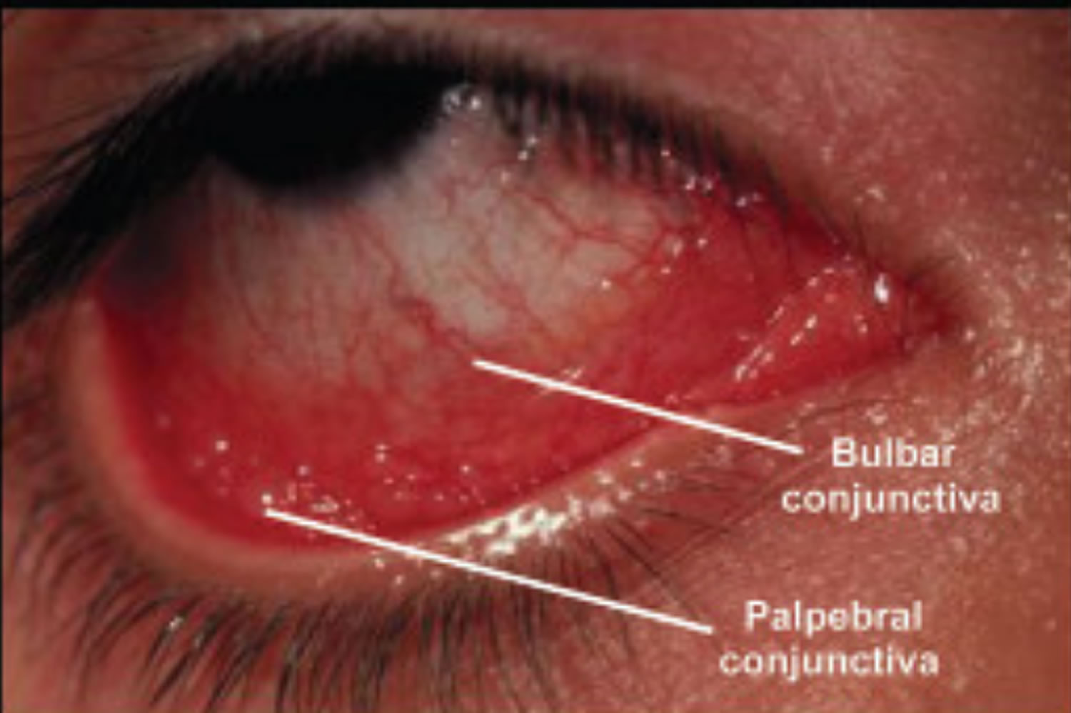
Conjunctivitis also called pink eye, red eye or sticky eye is the inflammation or infection of the conjunctiva, which is the transparent membrane that lines inside your eyelid and covers the white part of your eyeball. The conjunctiva is a thin translucent mucous membrane that can be divided based on the location into palpebral conjunctiva (inside of the eyelids) and bulbar conjunctiva (begins at the edge of the cornea and covers the visible part of the sclera) (Figure 2). The conjunctiva contains nonkeratinizing, squamous epithelium and a thin, richly vascularized substantia propria containing lymphatic vessels and cells, such as lymphocytes, plasma cells, mast cells, and macrophages. The conjunctiva also has accessory lacrimal glands and goblet cells. When the small blood vessels in the conjunctiva become inflamed or infected, they’re more visible. This is what causes the whites of your eyes to appear reddish or pink. Conjunctivitis can be caused by bacterial or viral infection or an allergic reaction. “Pink eye” is most often associated with the bacterial infection or bacterial conjunctivitis. Bacterial conjunctivitis is more common in children, and viral conjunctivitis is more common in adults. Conjunctivitis can cause swelling, itching, burning, discharge, and redness. Though conjunctivitis can be irritating, it rarely affects your vision. Depending on what kind of conjunctivitis you have and how bad it is, treatments can help ease the discomfort of conjunctivitis. Both viral and bacterial conjunctivitis are very contagious and easily spread from one person to another. They are spread through direct or indirect contact with the liquid that drains from the eye of someone who’s infected. One or both eyes may be affected. Therefore, early diagnosis and treatment can help limit its spread. Bacterial and viral conjunctivitis is a common cause of school absences and can spread quickly in schools. Viral conjunctivitis usually gets better in a couple of weeks without treatment. However, you’ll need to see a doctor for bacterial conjunctivitis to get treatment with antibiotic eye drops or ointment. Bacterial conjunctivitis symptoms include yellow discharge, pus that causes the eyelids to stick together, and puffy eyelids. A viral eye infection does not lead to drainage or pus in and around your eye. Viral conjunctivitis main symptom is eye redness. Both viral and bacterial conjunctivitis can occur along with colds or symptoms of a respiratory infection, such as a sore throat. Wearing contact lenses that aren’t cleaned properly or aren’t your own can cause bacterial conjunctivitis.
There are many causes of conjunctivitis. What causes conjunctivitis:
- Bacterial infection. Bacterial conjunctivitis is the second most common cause and is responsible for the majority (50%-75%) of cases in children; it is observed more frequently from December through April 1.
- Viral infection. Viral conjunctivitis is the most common cause of infectious conjunctivitis both overall and in the adult population and is more prevalent in summer 2, 3, 4, 5, 6, 7, 8, 1.
- Allergies. Allergic conjunctivitis is the most frequent cause of conjunctivitis, affecting 15% to 40% of the population and is observed more frequently in spring and summer 1, 9. Allergic conjunctivitis may be seasonal, or triggered by specific allergens, for example, pollen or animal dander (skin cells that are shed by animals with hair, fur or feathers).
- Substances that cause irritation
- Contact lens products, eye drops, or eye ointments
- A chemical splash in the eye
- A foreign object in the eye
- In newborns, a blocked tear duct.
Conjunctivitis can be divided into infectious and noninfectious causes. Viruses and bacteria are the most common infectious causes. Noninfectious conjunctivitis includes allergies, irritation when the eyes are in contact with chemicals and cicatricial (scarring) conjunctivitis, as well as conjunctivitis secondary to immune-mediated diseases and neoplastic processes 10. Conjunctivitis can also be classified into acute, hyperacute, and chronic according to the mode of onset and the severity of the clinical response 11. Furthermore, conjunctivitis can be either primary or secondary to systemic diseases such as gonorrhea, chlamydia, graft-vs-host disease, and Reiter syndrome, in which case systemic treatment is warranted 10. The eye may look similar to you no matter what is causing the conjunctivitis.
Check if you have conjunctivitis
The main symptom of conjunctivitis is red or pink eyes, often with itching, watering or discomfort.
Conjunctivitis (pink eye) usually affects both eyes and makes them:
- Bloodshot
- Puffy eyes or swelling of the eyelids
- Burn or gritty feeling in one or both eyes
- Produce pus that sticks to lashes
- Itchy eyes
- Watery eyes
- Sensitive to light called photophobia.
If you have bacterial conjunctivitis, you may also have yellow or green sticky discharge from the eyes. This can make your eyelids stick together, especially when you wake up from sleep.
If you have viral conjunctivitis, one or both eyes might be affected, and the discharge is likely to be clear.
If you have allergic conjunctivitis, both eyes are usually affected with a clear discharge. You might also have hay fever symptoms, such as an itchy nose, watery eyes and sneezing. Symptoms can be all year round or at certain times of the year (seasonal).
See your doctor or eye doctor (ophthalmologist) right away if:
- You’re in pain or are having trouble seeing
- You become sensitive to light
- Your symptoms have continued for a week or more, or are getting worse
- Your eye is producing a lot of pus or mucus
- You have any other symptoms of an infection, like fever, muscle ache or fatigue.
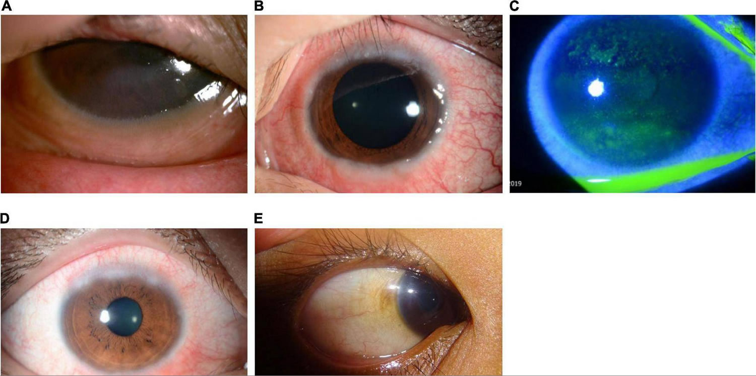
It is important to differentiate conjunctivitis from other sight-threatening eye diseases that have similar clinical presentation and to make appropriate decisions about further testing, treatment, or referral. An algorithmic approach using a focused eye history along with a penlight eye examination may be helpful in diagnosis and treatment (Figure 3). Because conjunctivitis and many other ocular diseases can present as “red eye”, the differential diagnosis of red eye and knowledge about the typical features of each disease in this category are important (see Table 1).
Most cases of conjunctivitis can be categorized as either papillary or follicular. Neither classification is pathognomonic for a particular disease entity 12:
- Papillary conjunctivitis. Papillary conjunctivitis produces a cobblestone arrangement of flattened nodules with central vascular cores 12. It is most commonly associated with an allergic immune response or is a response to a foreign body. Independent of the cause, the histologic appearance of papillary conjunctivitis is the same: closely packed, flat-topped projections, with numerous eosinophils, lymphocytes, plasma cells, and mast cells in the stroma surrounding a central vascular channel.
- Follicular conjunctivitis. Follicular conjunctivitis is seen in a variety of conditions, including inflammation caused by pathogens such as viruses, bacteria, toxins, and topical medications 12. In contrast to papillae, follicles are small, dome-shaped nodules without a prominent central vessel. Histologically, a lymphoid follicle is situated in the subepithelial region and consists of a germinal center with immature, proliferating lymphocytes surrounded by a ring of mature lymphocytes and plasma cells. The follicles in follicular conjunctivitis are typically most prominent in the inferior palpebral and forniceal conjunctiva 13.
Bacterial and viral conjunctivitis is highly contagious. Here’s how to avoid spreading it:
- Basic hygiene is enough to keep from spreading the infection to other people or your other eye.
- Change pillowcases and sheets every day.
- Use a fresh towel every day.
- Wash your hands often, especially after you touch your eyes.
- Don’t wear your contact lenses until your eyes are back to normal.
- Don’t share anything that touches your eyes.
- People who wear contact lenses need to stop wearing their contacts as soon as pink eye symptoms begin. If your symptoms don’t start to get better within 12 to 24 hours, make an appointment with your eye doctor to make sure you don’t have a more serious eye infection related to contact lens use.
Conjunctivitis treatment depends on the cause. If the conjunctivitis is caused by bacteria, antibiotic drops or ointment may be needed. It is important to use any prescription medication for the full
number of days prescribed in order to prevent the infection from coming back and reduce antibiotic resistance in bacteria. In most cases, you won’t need antibiotic eye drops. Since conjunctivitis is usually viral, antibiotics drops or ointments do not help viral, allergic or irritative conjunctivitis.
Viral conjunctivitis, which is often seen with other symptoms like sore throat, runny nose and cough, does not respond to antibiotics and antibiotic medications are not needed. Viral conjunctivitis often begins in one eye and then infects the other eye within a few days. Your symptoms should gradually clear on their own. This typically takes around 2 to 3 weeks. Antiviral medicines may be an option if your viral conjunctivitis is caused by the herpes simplex virus (HSV).
Allergic conjunctivitis is treated with antihistamine eye drops or allergy medicines by mouth and artificial tear drops. Some nasal sprays for hay fever are also helpful. Sometimes your doctor might suggest tests to help you find the allergic trigger.
Bacterial infections may require antibiotic eye drops or ointment. It’s important to keep applying the medicine for several days after your symptoms have improved.
Conjunctivitis treatment is usually focused on symptom relief. Your doctor may recommend:
- Using artificial tears.
- Cleaning your eyelids with a wet cloth.
- Applying cold or warm compresses several times daily.
To help relieve your symptoms and prevent infection:
- Wash your hands thoroughly before touching your eyes.
- Wash your eye gently several times a day with clean cotton wool pad soaked in warm tap water.
- Use a new cotton wool pad for each eye, to prevent passing the infection into your other eye.
- Gently clean any eye discharge from your eye area. Always wipe from the corner of the eye (nearest the nose) outwards.
- If you wear contact lenses and have an infection, throw out your lenses. Wear glasses for at least a week after your symptoms have disappeared.
- Throw out any eye makeup or eyelash extensions used right before or during an eye infection.
If you wear contact lenses, you’ll be advised to stop wearing them until treatment is complete. Your doctor will likely recommend that you throw out soft contacts you’ve already worn. Disinfect hard lenses overnight before you reuse them. Ask your doctor if you should discard and replace your contact lens accessories, such as the lens case used before or during the illness. Also replace any eye makeup used before your illness.
Figure 1. Human eye

Figure 2. Eye anatomy
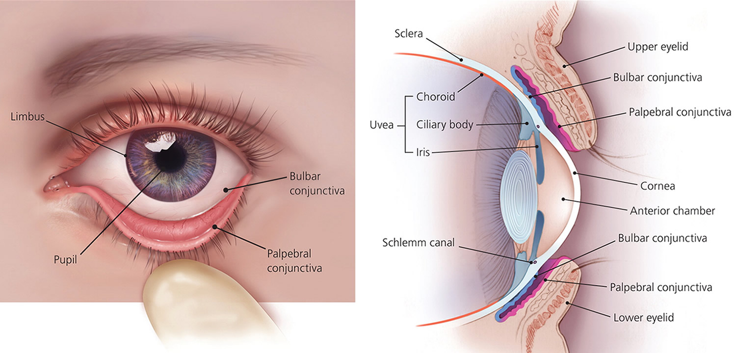
Figure 3. Conjunctivitis
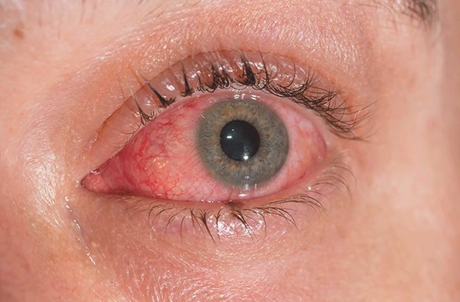
Figure 4. Acute conjunctivitis diagnostic algorithm
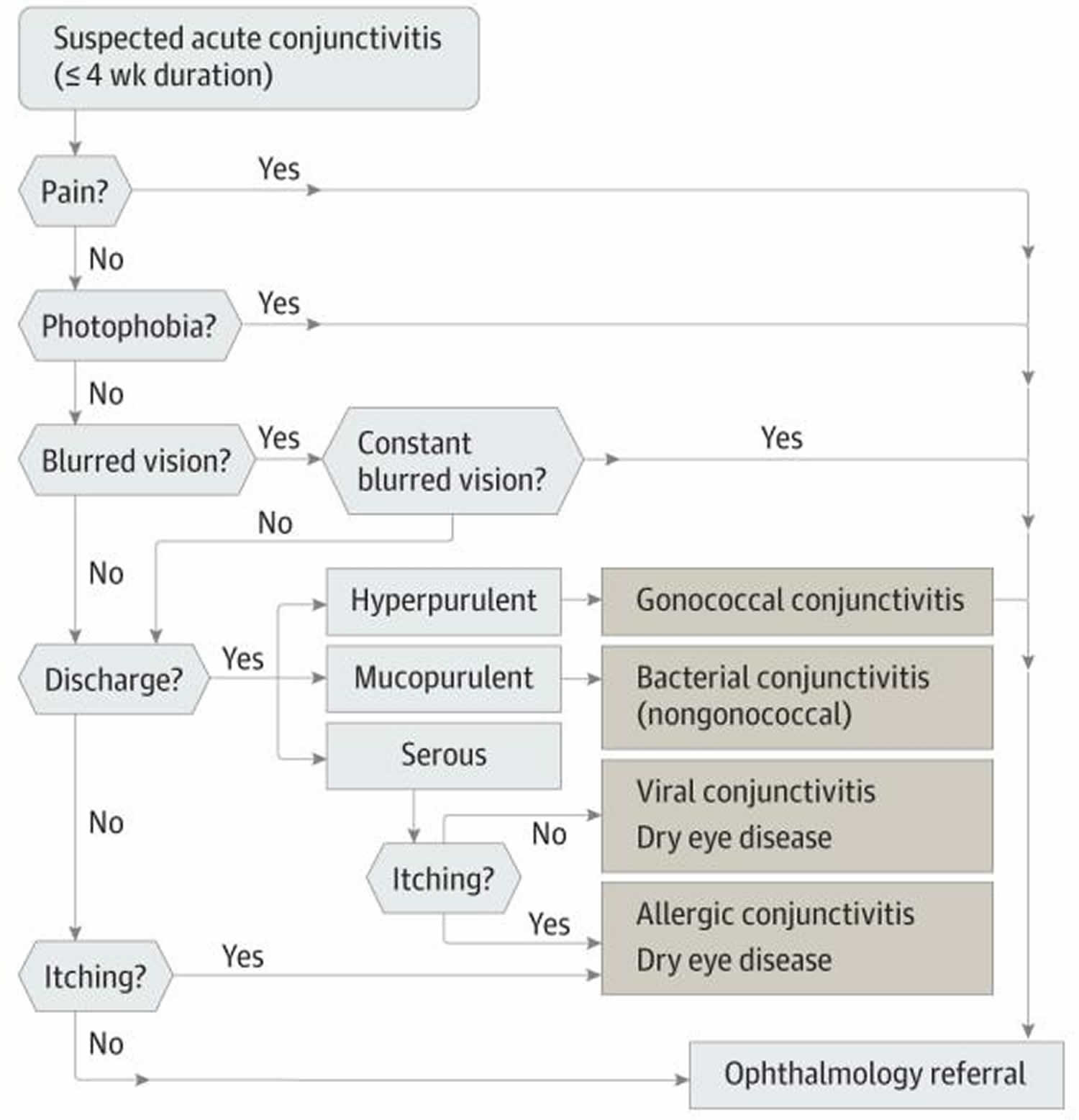
Table 1. Non-conjunctivitis causes of red eye
| Differential Diagnosis | Symptoms | Penlight Examination Findings |
|---|---|---|
| Dry eye disease | Burning and foreign-body sensation. Symptoms are usually transient, worse with prolonged reading or watching television because of decreased blinking. Symptoms are worse in dry, cold, and windy environments because of increased evaporation. | Bilateral redness |
| Blepharitis | Similar to dry eyes | Redness greater at the margins of eyelids |
| Uveitis | Photophobia, pain, blurred vision. Symptoms are usually bilateral. | Decreased vision, poorly reacting pupils, constant eye pain radiating to temple and brow. Redness, severe photophobia, presence of inflammatory cells in the anterior chamber. |
| Angle closure glaucoma | Headaches, nausea, vomiting, ocular pain, decreased vision, light sensitivity, and seeing haloes around lights. Symptoms are usually unilateral. | Firm eye on palpation, ocular redness with limbal injection. Appearance of a hazy/steamy cornea, moderately dilated pupils that are unreactive to light. |
| Carotid cavernous fistula | Chronic red eye; may have a history of head trauma | Dilated tortuous vessels (corkscrew vessels), bruits on auscultation with a stethoscope |
| Endophthalmitis | Severe pain, photophobia, may have a history of eye surgery or ocular trauma | Redness, pus in the anterior chamber, and photophobia |
| Cellulitis | Pain, double vision, and fullness | Redness and swelling of lids, may have restriction of the eye movements, may have a history of preceding sinusitis (usually ethmoiditis) |
| Anterior segment tumors | Variable | Abnormal growth inside or on the surface of the eye |
| Scleritis | Decreased vision, moderate to severe pain | Redness, bluish sclera hue |
| Subconjunctival hemorrhage | May have foreign-body sensation and tearing or be asymptomatic | Blood under the conjunctival membrane |
Footnotes: There are some eye conditions which may look similar to conjunctivitis.
[Sources 15, 16, 14 ]There are serious eye conditions that can cause eye redness. These conditions may cause eye pain, a feeling that something is stuck in your eye (foreign body sensation), blurred vision and light sensitivity. If you experience these symptoms, seek urgent care.
Make an appointment with your doctor if you notice any signs or symptoms you think might be pink eye. Pink eye can be highly contagious for as long as two weeks after signs and symptoms begin. Early diagnosis and treatment can protect people around you from getting pink eye too.
People who wear contact lenses need to stop wearing their contacts as soon as pink eye symptoms begin. If your symptoms don’t start to get better within 12 to 24 hours, make an appointment with your eye doctor to make sure you don’t have a more serious eye infection related to contact lens use.
See your doctor for advice if you have a pink eye that doesn’t start to improve after a few days.
Contact your doctor immediately or go to your nearest emergency room if:
- you have a painful pink eye or red eye
- you have other symptoms, including any changes in your vision like wavy lines or flashing, sensitivity to light (photophobia), a severe headache and feeling sick
- you’ve recently injured your eye, particularly if something has pierced it
- your baby has red eyes – get an urgent medical care if your baby is less than 28 days old
- you wear contact lenses and have conjunctivitis symptoms as well as spots on your eyelids – you might be allergic to the lenses
- your symptoms haven’t cleared up after 2 weeks
- sensitivity to light
- intense redness in one eye or both eyes
Does a red or pink eye always mean infection?
No. A pink or red eye may be a sign of another eye problem such as allergy, foreign body (something stuck in the eye), contact lens reaction, inflammation inside the eye, or glaucoma (high eye pressure).
Can I catch conjunctivitis?
You can catch conjunctivitis from droplets from the eyes, mouth and throat of an infective person. This can happen through touch, coughing or sneezing. You can also catch it from contact with objects that were contaminated with infectious eye secretions, such as towels, face washers and tissues.
Allergic conjunctivitis is caused by exposure to allergens such as:
- dust mites
- pollen
- animal dander (skin cells that are shed by animals with hair, fur or feathers)
- mould spores
- occasionally foods or food additives
Allergic conjunctivitis isn’t contagious so it can’t spread from person to person.
How long is conjunctivitis contagious?
Conjunctivitis (pink eye) generally remains contagious as long as you have tearing and matted eyes. Conjunctivitis (pink eye) is commonly caused by viruses or bacteria. Depending on the cause of your conjunctivitis, signs and symptoms usually improve within a few days to two weeks.
Viral and bacterial conjunctivitis can spread very easily as easily as the common cold. If you have an infection in just one eye, be careful not to spread it to the other eye. And be careful not to spread the infection in public, either.
Good hygiene including washing your hands, avoiding close contact with others, and not sharing towels or pillowcases — is important. It may be okay to return to school or child care if your child does not have a fever, can practice good hygiene, and can avoid close contact with others.
Children who are not able to practice good hygiene or can’t avoid close contact with others should stay home until symptoms clear up.
Things to remember:
- You will be contagious as long as there is a discharge from your eye (usually 10-14 days after symptoms start).
- Do not attend work or school until discharge stops.
Check with your doctor if you have any questions about when your child can return to school or child care.
Can Visine be used for conjunctivitis?
No! Whatever kind of conjunctivitis you have, don’t use red-reducing eye drops, like Visine. These kinds of eye drops may be very uncomfortable if you have an infection. They also could make your symptoms worse.
Can I use breast milk for conjunctivitis?
Breast milk could be more harmful than helpful for conjunctivitis. One of the few studies on whether breast milk can fight infections found that it didn’t cure the most common causes of conjunctivitis and worse, breast milk can introduce new bacteria into the eye and cause serious infection. Eye infections in young children can be very serious even capable of causing blindness. Don’t delay seeing a doctor and don’t rely only on folk remedies.
There is lots of bad advice about conjunctivitis on the internet. Never put anything in your eye that isn’t approved by a doctor. Foods and herbal extracts are not sterile and can make eye conditions much worse. Bloggers who recommend breast milk for conjunctivitis say that substances in breast milk can cure infection and soothe inflammation. But there is no evidence that this helps.
How do I clean my eyes with conjunctivitis?
Gentle cleaning with cotton balls soaked in warm water. Wipe from the inside corner of the eye to the outside corner, dispose of cotton ball, and repeat with a clean cotton ball as necessary. DO NOT try to clean inside the eyelids as this may cause damage to the conjunctiva.
Measles and conjunctivitis
Conjunctivitis can show up before a measles rash or at the same time. Ask these questions about whether conjunctivitis may be a sign of measles:
- Is there a reported outbreak of measles in the area?
- Has the child been vaccinated for measles? If so, then measles conjunctivitis is very unlikely.
- Are there other measles symptoms, like a red, blotchy rash or a high fever (above 104 degrees Farenheit/40 Celsius)? Note that other kinds of conjunctivitis can also cause fever, especially in children. So a mild fever, or fever by itself, isn’t necessarily a sign of measles.
- Is the child sensitive to regular, indoor light? Light-sensitivity is more likely to be a sign of measles-related conjunctivitis. Sensitivity to indoor light is always a sign of a serious eye condition, usually involving sight-threatening damage to the cornea. You should see an eye specialist (ophthalmologist), not just your family doctor or pediatrician.
If you think you or a loved one may have measles-related conjunctivitis, see an ophthalmologist right away and make sure they report it to local health authorities. In some cases, measles can damage the cornea, retina or optic nerve and result in vision loss or blindness.
Allergic conjunctivitis
Allergic conjunctivitis is caused by an allergic reaction, with the majority of cases (90–95%) attributed to seasonal (certain times of the year) allergic conjunctivitis or perennial (all year round) allergic conjunctivitis 17, 18. Allergic conjunctivitis is a common conjunctivitis that is caused by immunoglobulin E (IgE) immune responses affecting more than 40% of the general population and is estimated to occur in up to 30% of children, either alone or in association with allergic rhinitis 19, 20, 21, 22. Allergic conjunctivitis is a reaction of the outer lining of the eyeball (conjunctiva) to things in the environment to which a person is allergic (allergens). Dust, pollen, animal dander, and sometimes even medications can all be allergens. When your eyes are in contact with these allergens, the eyes get red, inflamed, watery, itchy or swollen eyelid 23, 24. Although these symptoms can look like the signs of an infection, allergic conjunctivitis is not an infection and is not contagious. However, these signs and symptoms can be sufficiently bothersome that people with allergic conjunctivitis often experience decreased work productivity, increased work or school absenteeism, limitation of everyday activities, and reduced quality of life 23.
Unlike conjunctivitis that is caused by bacterial or viral infection, allergic conjunctivitis is not contagious, so it cannot be transferred from one person to another.
Allergic conjunctivitis is often accompanied with other signs of hay fever. Allergic conjunctivitis signs can include an itchy, runny nose and sneezing or a history of other allergic conditions. The eyes are itchy and watery.
Allergic conjunctivitis other symptoms can include:
- redness behind the eyelid, spreading up the white of the eye
- swelling of the eye/s making them appear puffy
- excessive tears
- a discharge, yellow or green in color, causing crusting around the eyelids
- a dislike of bright lights (photophobia).
Allergic conjunctivitis symptoms may be:
- Perennial (all year round) due to constant exposure to dust mites, animal dander, indoor and outdoor mould spores and, in some cases, foods or food additives.
- Seasonal (certain times of the year) due to airborne allergens such as mould spores and pollen from grasses, trees, and weeds. The amount of airborne pollen varies from day to day and is dependent on the weather. People with pollen allergies often find their symptoms improve in wet weather and become worse on hot windy days or after thunderstorms.
If allergic conjunctivitis is suspected, allergy testing can help identify the allergen responsible, or “trigger”.
Avoiding or minimizing exposure to known allergens is an important first step in managing allergic conjunctivitis.
Allergic conjunctivitis may be helped by treatments used in conditions such as hay fever e.g. antihistamines. Cool compresses and lubricating eye drops may soothe the eyes.
Figure 5. Allergic conjunctivitis
Allergic conjunctivitis causes
Allergic conjunctivitis is caused by contact with something to which a person may be sensitive or allergic to (allergens), air pollution, atopy, pollen exposure, inflammation, and pet hair 25. Examples of common allergens to the conjunctival surface include tree/grass pollen, house dust mites, animal/pest dander, and mold spores 25, 26. Spring, summer and fall allergies tend to be caused by trees weed, grass, and flower pollen. Some people can have allergies all year round due to other household allergens, including dust, mold and animal dander/hair/fur. Some children may have an underlying medical problem making them more at risk for an allergic eye condition.
Allergic conjunctivitis signs and symptoms
Allergic conjunctivitis symptoms may vary from person to person. Allergic conjunctivitis symptoms can be very mild or very severe. Itching is the most common symptom and eye allergy
is unlikely to be present if itching is not present. Other symptoms may include stinging, tearing, and burning. The conjunctiva is usually pink and/or bloodshot. The white area immediately around the colored part of the eyes can also swell, causing tiny bumps visible on the surface of the eye. Eyelid skin can also be affected, becoming thick, swollen, itchy, or red. Children may frequently rub or roll their eyes if they have allergies. They may even tightly squeeze or blink frequently to help with the itchiness. Symptoms are often worse in the spring and/or summer months, but may stick around throughout the year.
Typical signs and symptoms of allergic conjunctivitis include:
- Redness in both eyes.
- Itching and burning of both the eye and surrounding tissues.
- Watery discharge, often accompanied by acute discomfort in bright light (photophobia).
- Swollen eyelids which may become ‘heavy’ or ‘droopy’. In some severe cases, the eyelids are so swollen that they cannot completely open.
- Swollen conjunctiva which may look light purple and affect vision. Blurred vision or any change in the appearance of the cornea (clear part of the eye that covers the pupil) requires urgent referral to an eye specialist. Speak to your doctor or optometrist for a referral.
Allergic conjunctivitis diagnosis
A diagnosis of allergic conjunctivitis is made by history and examination. Although allergy testing may help pinpoint the specific allergens, it is usually not necessary since the types of allergens that usually cause conjunctivitis are very common, like grass, weed, and tree pollens. Eye drop treatments are the same no matter what allergen is causing the reaction.
If further investigation is needed, even if no identifiable allergens have been found, the second step is skin prick or patch tests 27. Patch tests are preferred in contact blepharoconjunctivitis, while skin prick tests are used in the other diseases 27. These tests are carried out with a standard battery of allergens and sometimes with others that are not normally tested but suspected as the cause of the allergy. If skin testing is indicated but not recommended (e.g., the patient is taking antihistaminic systemic medications), or if results are ambiguous (e.g., presence of dermatographism), or simply to complement the results of previous skin prick test, serum specific IgE measurements for the aeroallergens can be considered 28, 29.
In case of doubt after systemic allergy evaluation tests, a conjunctival allergen provocation test (CAPT) also known as conjunctival allergen challenge or ocular challenge test may be of use to identify the cause 29. In the conjunctival allergen provocation test (CAPT) an allergen is applied to the conjunctival mucosa to evaluate the patients’ immunoreactivity to the allergen. The conjunctival allergen provocation test (CAPT) is used to confirm which allergens the patient is sensitive to and has the same scientific background as other provocation tests used extensively in other mucosae such as nasal or digestive 29, 30. Non-specific or irritant challenges evaluate the hyperreactivity of the ocular mucosa, whilst direct mucosal challenges contain higher concentrations of the allergen encountered in environmental exposure and evaluate patients’ immunoreactivity to the allergen, following the guidelines for standard practice of the European Academy of Allergy and Clinical Immunology 29, 30, 31. A positive test will trigger the same signs and symptoms as those occurring when the allergen is encountered in real life, an IgE-mast cell-dependent immunoreactivity 32, 30. The conjunctival allergen provocation test (CAPT) is also useful to assess the relationship between symptoms and exposure in polysensitized patients and to assess response to therapy 28.
Allergic conjunctivitis treatment
Allergen avoidance is the first line of treatment for allergic conjunctivitis. In the case of pollen allergies, symptoms are often made worse by outdoor activities. Wearing glasses or goggles outdoors can limit contact with allergens. Minimization measures for house dust mites may include removing carpet, using dust mite covers for pillows and mattresses, and washing bedding in hot water are enough to reduce symptoms. Frequent washing of pillowcases and mattress covers as well as vacuuming the carpeting in your room help remove allergens from your surroundings. Regular washing your hair and face can help remove these allergens from the surface of your eyes, hair, and skin. Using artificial tear drops to rinse the eye and remove allergens from the eye can help with symptoms and help calm down the eye inflammation. These drops can provide even more relief when used cold (refrigerated) instead of at room temperature. It is also important to avoid rubbing the eyes, as this can make allergic conjunctivitis worse.
If you have allergic conjunctivitis, both prescription and over-the-counter allergy eye drops can treat allergic conjunctivitis 14. These may include medicines that help control allergic reactions, such as antihistamines and mast cell stabilizers. Or your doctor may recommend medicines to help control inflammation, such as decongestants, steroids and anti-inflammatory drops. Nonprescription versions of these medicines also may be effective. Most of the easily available allergy eye drops work best when used daily for at least a few weeks, and it may take up to a week to get full symptom relief. Some eye drops can be used only on an as needed basis. Allergy eye drops may work better for some than pill or liquid medications as eye drops do not cause any drowsiness or changes in appetite. However, pill or liquid medications may be more helpful if allergies cause a lot of eyelid swelling or affect more than just the eyes. Please speak with your ophthalmologist if you have questions about allergy medications.
Given the different types of over-the-counter eye drops, sometimes what works well for one person may not work as well for another person. You may need to try different types of eye drops before you find one that works for you. If there are still allergic conjunctivitis symptoms even after trying different kinds of allergy eye drops, adding a short-term liquid or pill allergy medication by mouth may help relieve symptoms. Pills and liquid medications by mouth may also a good treatment for people who don’t do well eye drops, or who have other allergy symptoms like a runny nose.
In some cases, steroid eye drops may be needed along with allergy eye drops if the allergic reaction is very severe. However, steroid use needs to be monitored closely by your ophthalmologist and used only as directed to prevent serious eye problems. Use of steroid drops for a long time or at a large amount can cause serious vision problems, including glaucoma, cataracts and eye infections (keratitis). Only ophthalmologists (eye specialists) who can monitor for side effects should prescribe steroids for allergic conjunctivitis. Talk with your ophthalmologist if you have questions about steroid drops.
Topical medications (eye drops) treat the symptoms of allergic conjunctivitis directly. Small drops of medication are delivered straight to the surface of the eye and are available in many different types.
- Antihistamine eye drops – effective but should not be used for longer than 6 weeks without medical advice.
- Antihistamine eye drops containing a vasoconstrictor (substances that cause the walls of blood vessels to narrow, or constrict) – minimize itch and remove redness by narrowing the swollen blood vessels in the eye. They should not be used for longer than 14 days without medical advice.
- Mast cell stabilizer eye drops – best used to prevent symptoms from occurring as they can take three to seven days to work. These can be used as long as necessary.
- Mast cell stabilizer eye drops with antihistamines – fast acting, effective and generally well tolerated.
- Steroid eye drops – effective in relieving symptoms quickly, but are associated with cataract formation, glaucoma and bacterial and viral infections of the cornea and conjunctiva. They should only be used under medical supervision as a short-term treatment and should never be used in the presence of herpes infections.
Antihistamine tablets or syrups help some people when it is difficult to avoid the allergen. Some side effects may include dryness of the eyes, nose, and mouth, and blurred vision. Antihistamines are usually contraindicated for people with glaucoma, advice should be sought from an eye specialist.
Allergen immunotherapy for specific allergens may benefit people with persistent, severe allergic conjunctivitis. However, relief of allergic conjunctivitis symptoms will not happen straight away.
Table 2. Allergic conjunctivitis treatment options
| Drug Class | Mechanism of Action | Target Symptom; Response Phase | Dosing Frequency |
|---|---|---|---|
| Antihistamines | Block histamine H1 receptors | Itching; acute action | 4 times daily |
| Vasoconstrictors (decongestants) | Activation of α-adrenergic receptors | Redness; acute action | 4 times daily |
| Mast cell stabilizers | Prevention of mast cell degranulation | Itching; early and late-phase responses | 2–6 times daily |
| Leukotriene receptor antagonists | Competitive binding to leukotriene receptors | Multiple allergic conjunctivitis signs and symptoms; late-phase responses | 1 time daily |
| Nonsteroidal anti-inflammatory drug (NSAIDs) | Prevention of prostaglandin production | Itching; late-phase response | 4 times daily |
| Corticosteroids | Broad anti-inflammatory action through prevention of proinflammatory mediator synthesis | Multiple allergic conjunctivitis signs and symptoms; early- and late-phase responses | 4 times daily |
| Single-agent antihistamine–mast cell stabilizers | Inverse histamine H1-receptor agonism plus prevention of mast cell degranulation | Itching; acute action and early- and late-phase responses | 1–2 times daily |
Eye drops for allergic conjunctivitis
When you have an allergic reaction your body releases histamine from mast cells, which leads to hay fever. Antihistamines block this reaction. Antihistamines act via histamine receptor antagonism to block the inflammatory effects of histamine and relieve any associated signs and symptoms. Most antihistamines used in the treatment of eye allergy are histamine-1 (H1) receptor antagonists, although some agents have affinity for other subtypes. Histamine-2 (H2) antagonists have been shown to modulate both cell growth and migration. Animal model studies have shown that antihistamines may even reduce infiltration of eosinophils and thus reduce the clinical aspects of the late-phase reaction 33.
First-generation antihistamines are well tolerated and associated with a favorable long-term safety record, but are associated with instillation pain, short duration of action, and limited potency 34. First-generation antihistamines remain available in over-the-counter products, particularly in combination with vasoconstrictors (substances that cause the walls of blood vessels to narrow, or constrict). While newer antihistamines are also H1 antagonists, they have a longer duration of action (4–6 hours) and are better tolerated than their predecessors 33.
Topical antihistamines are widely available without a prescription. Antihistamines competitively block histamine receptors (e.g., H1 or H4) on nerve endings and blood vessels of the mucosal surface, thereby reducing itchiness and conjunctival redness 35. First-generation antihistamines were associated with a range of systemic side effects (e.g., sedation, dizziness, cognitive impairment, blurred vision) caused by anticholinergic actions and nonspecific binding to histamine H2 receptors in addition to drying of the ocular surface. Newer oral, intranasal, and topical ocular antihistamines demonstrate improved H1 receptor selectivity, with fewer adverse effects; however, eye side effects, such as burning and dryness, remain a concern. Topical antihistamines (e.g., levocabastine, emedastine difumarate) are useful for providing rapid relief of allergic conjunctivitis symptoms, but their duration of action is limited; most topical antihistamines require dosing four times.
Antihistamine eye drops
Antihistamines are competitive antagonists of histamine receptors that are present in the conjunctiva and eyelids. Once stimulated, histamine receptors lead to capillary dilation and increased vascular permeability, which leads to common allergic symptoms of itching and edema. Therefore, antihistamines work by preventing the binding of histamine to histamine-1 (H1) receptors and preventing the cascade of inflammatory events. In the eye, only H1 receptors are available 36. Examples include azelastine (Eyezep Eye Drops), levocabastine (Livostin Eye Drops, Zyrtec Levocabastine Eye Drops) and emedastine.
Mast cell stabilizers
Mast cell stabilizers work by preventing the degranulation of sensitized mast cells, thus stopping the release of histamine and other inflammatory mediators 37, 38. Since mast cell stabilizers act before the mast cell is degranulated, they rarely have an impact on the inflammatory mediators once they are already released 39, 38. In other words, mast cell stabilizers are not effective once the patient is symptomatic, and clinical trials have had a difficult time showing their efficacy 39. Since there are other quicker and more effective treatment agents available on the market, mast cell stabilizers are rarely used as monotherapy. The most common mast cell stabilizers used for allergic conjunctivitis are sodium lodoxamide 0.1% (Alomide), cromoglycate 2%, and nedocromil 2% 37, 40. Mast cell stabilizers can be used as a prophylactic measure to prevent mast cell degranulation for repeated exposures to the allergen 41.
Antihistamine and mast cell stabilizer eye drops
When you have an allergic reaction your body releases histamine from mast cells, which leads to hay fever. Mast cell stabilizer medicines help reduce this histamine release after allergen exposure and reduce allergic reactions and hayfever 42. Topical mast cell stabilizers act to prevent mast cell degranulation and subsequent release of proinflammatory molecules triggered by IgE binding to sensitized conjunctival mast cells. Topical mast cell stabilizers (e.g., cromolyn sodium, lodoxamide tromethamine, nedocromil sodium, pemirolast potassium) effectively prevent activation of the early phase response by preventing release of histamine, cytokines, and other inflammatory and chemotactic mediators. Preventing the early phase response blocks downstream inflammation events, including production of prostaglandins and leukotrienes, eosinophil infiltration, chemokine and adhesion molecule expression, and chronic mast cell activation that perpetuate the late-phase response in allergic conjunctivitis. Most mast cell stabilizers require administration four to six times daily; nedocromil sodium can be given twice daily. Because of the required loading time for maximal efficacy of mast cell stabilizers, these medications are most effective when treatment is initiated before symptoms manifest; their effectiveness is limited when allergic conjunctivitis cascades have been activated and mast cell degranulation and histamine release have already occurred.
Dual-Acting Antihistamine–Mast Cell Stabilizing Agents
Agents with dual antihistamine and mast cell stabilizing actions are more suitable for extant allergic conjunctivitis than single-action medications because they block binding of free histamine to receptors and inhibit further release of proinflammatory mediators from mast cells. This dual action rapidly alleviates multiple signs and symptoms of allergic conjunctivitis in the short term and blocks the feed-forward cycle of persistent inflammation caused by continuous mast cell activation in the long term to promote regression of allergic conjunctivitis. Antihistamine–mast cell stabilizing agents (e.g., olopatadine, alcaftadine, epinastine, bepotastine besilate) are currently considered first-line therapeutics for allergic conjunctivitis because they offer acute symptomatic relief and control inflammation, and can be used chronically without long-term safety concerns. Most dual-acting agents require twice-daily dosing. Olopatadine 0.2% and alcaftadine are indicated for once-daily dosing and maintain effectiveness through 16 hours after administration in conjunctival allergen challenge studies 43, 44, 45.
Olopatadine 0.1% (Pataday Twice Daily Relief) was the first topical anti-allergy medication that was approved for twice-daily usage 37. These agents are all preserved with a surfactant called benzalkonium chloride that may cause ocular surface toxicity 46. These are now considered the first line of treatment for allergic eye disease and are the most common ophthalmic agents recommended by eye care practitioners and allergists 37. These agents can be used to prevent mast cell degranulation and acutely following the onset of symptoms 47.
Compared to placebo, olopatadine has been found to improve symptoms of eyelid swelling, eye redness, chemosis (a condition where the conjunctiva, or the outer surface of the eye, swells due to fluid build-up), itch, and overall quality of life. Multiple randomized control trials have compared ketotifen and olopatadine. One meta-analysis found improvement in symptoms of itching after 14 days in favor of olopatadine 0.1% when compared to ketotifen 0.025% 48. Before 2020, olopatadine was only available as a prescription medication, and ketotifen, in the form of Zaditor or Alaway, was clinically commonly prescribed as the first line of relief as an OTC medication. Within the last 4 years, olopatadine became available OTC and has gained popularity to become clinically superior to ketotifen in terms of efficacy.
Leukotriene Receptor Antagonists
Leukotriene receptor antagonists (e.g., montelukast), which are currently available for oral dosing, prevent leukotrienes from binding to their conjunctival receptors to decrease inflammatory signaling and improve multiple ocular symptoms of allergic conjunctivitis. Leukotriene receptor antagonists have a slower onset of action, are less effective than topical antihistamines, and are not used as first-line therapy or monotherapy for allergic conjunctivitis 35.
Topical Vasoconstrictors (Decongestants)
Topical vasoconstrictors (decongestants) were the first ocular medication approved for the treatment of allergic conjunctivitis. Over-the-counter (OTC) topical vasoconstrictors are effective at temporarily decreasing conjunctival hyperemia by stimulating alpha-adrenergic receptors 39. Alpha-adrenergic agonists cause vasoconstriction of conjunctival blood vessels, resulting in decongestion and whitening of the eye 49, 39. However, the use of these agonists can lead to side effects such as rebound hyperemia and tachyphylaxis and, chronically, can lead to conjunctivitis medicamentosa 41, 39. Commonly used topical vasoconstrictors are oxymetazoline, naphazoline, tetrahydrozoline, and phenylephrine. These are best utilized as short-term solutions and should be avoided in narrow-angle glaucoma and cardiovascular issues 49, 39. These should not be recommended as a standalone treatment and used in combination with antihistamines for the treatment of allergic conjunctivitis 49, 50.
Combination eye drops including decongestant (Antihistamine-Vasoconstrictor Combinations)
Some eye drops contain an antihistamine such as pheniramine or antazoline to stop itching, and a vasoconstrictor (substances that cause the walls of blood vessels to narrow, or constrict) such as naphazoline to take away redness through stimulation of vascular alpha-adrenergic receptors e.g. naphazoline + antazoline (Antistine-Privine, Albalon-A), pheniramine + naphazoline (Visine Allergy with Antihistamine, Naphcon-A). Vasoconstrictors are commonly available in nonprescription combination formulations that contain an antihistamine (e.g., naphazoline-antazoline, naphazoline-pheniramine). These formulations exhibit a rapid onset of action and relieve redness and itchiness associated with allergic conjunctivitis. However, they are not recommended for long-term use because of reduced effectiveness over time and a potential rebound effect that can produce persistent red eye on discontinuation 42. As with topical antihistamines, combination antihistamine–vasoconstrictor formulations have a relatively short duration of action and are administered four times daily 24, 14. Limit use of combination eye drops to no more than 5 to 7 days to avoid a ‘rebound’ redness from overuse.
Other eye drops, to prevent allergy symptoms
These eye drops prevent allergic reactions in the eyes and need to be used 4 to 6 times per day, depending on the ingredient, for the entire time you are exposed to triggers, such as during spring e.g. cromoglycate (Cromolux Eye Drops, Opticrom), lodoxamide (Lomide Eye Drops 0.1%)
Oral antihistamines (tablets and syrups)
There are two types of oral antihistamines: newer, less sedating antihistamines, which do not typically cause drowsiness and older sedating antihistamines that cause drowsiness.
Oral antihistamines are good for treating hay fever symptoms, especially if you have a lot of different symptoms. You can also take oral antihistamines in advance if you know you are going to be exposed to allergens or triggers
Newer, less-sedating antihistamines
Newer antihistamines may rarely cause drowsiness; do not drive or operate machinery if you are affected e.g. cetirizine (Zilarex, Zyrtec), desloratadine (Aerius), fexofenadine (Fexotabs, Telfast), loratadine (Claratyne, Lorano).
Cetirizine and loratadine are available as syrups for children; check correct doses for different age groups. Cetirizine is more likely to cause drowsiness than other less sedating antihistamines
Older, sedating antihistamines
Older sedating antihistamines can cause drowsiness, sometimes the next day; it is important you do not drive or operate machinery e.g. chlorpheniramine + pseudoephedrine (Demazin 6 Hour Relief Tablets), dexchlorpheniramine (Polaramine), loratadine + pseudoephedrine (Claratyne-D with Decongestant Repetabs), promethazine (Phenergan, Sandoz Fenezal)
Older antihistamines are not available without a prescription for children under 2 years old. Do not drink alcohol with antihistamines that make you drowsy
If you have other medical conditions, such as glaucoma, epilepsy or prostate problems, or you take antidepressants, check with your doctor before taking these medicines
Topical nonsteroidal anti-inflammatory drugs (NSAIDS)
Topical nonsteroidal anti-inflammatory drugs (NSAIDS) act by blocking the cyclooxygenase enzymes (COX-1 and COX-2) within the cyclooxygenase pathway, resulting in the inhibition of inflammatory mediators such as prostaglandins and leukotrienes 39, 38. These drugs have proven efficacy against conjunctival hyperemia, pruritus, pain, and irritation 39. Topical NSAID agents commonly associated with the relief of ocular allergy symptoms include ketorolac, diclofenac, indomethacin, and flurbiprofen 39, 38. Although ketorolac has been approved for treating allergic conjunctivitis, studies have indicated that it is less effective compared to topical antihistamine agents 39, 38, 51. Moreover, these agents can cause burning and stinging sensations upon instillation, so long-term compliance is an issue. Thus, NSAIDs may be used for temporary relief of itching and hyperemia compared to no treatment; however, they do not aid with symptoms of mucous discharge, chemosis (a condition where the conjunctiva, or the outer surface of the eye, swells due to fluid build-up), and corneal damage, so alternative methods should be considered 39, 38, 51, 52.
Corticosteroids
Corticosteroids prevent production of multiple classes of late-phase response mediators, including prostaglandins, leukotrienes, histamine, and some cytokines. The numerous points of intervention in the inflammatory cascade make glucocorticoids an effective pharmacologic therapy for allergic conjunctivitis, but long-term topical use can lead to serious adverse effects, including increased intraocular pressure and corneal abnormalities 42. Long-term systemic corticosteroid use increases the risk of posterior subcapsular cataract formation 53. For this reason, patients at risk (e.g., those with glaucoma or diabetic retinopathy) or patients who receive higher doses or longer treatment courses of corticosteroids should be monitored by an ophthalmologist.
Corticosteroids (e.g., loteprednol etabonate, given four times daily) are generally not used as primary therapy for allergic conjunctivitis unless there is persistent or moderate-to-severe inflammation that the eye doctor does not feel will respond sufficiently to antihistamine–mast cell stabilizer medications alone 24. When corticosteroids are prescribed, they typically are used for short durations in the early stages of allergic conjunctivitis or during flare-ups until allergic conjunctivitis can be controlled with safer medications such as antihistamines, mast cell stabilizers, or dual-acting, single-molecule antihistamine–mast cell stabilizer agents 54. Topical corticosteroids are important in severe cases of allergic eye disease to break the cycle of inflammation and can be discontinued once the condition is under control. Most cases of seasonal allergic conjunctivitis or perennial allergic conjunctivitis do not often require corticosteroid intervention. For patients who require long-term use of corticosteroids, close observation by an ophthalmologist is warranted.
Supratarsal steroids
Supratarsal steroids are required in patients with the severe palpebral form of the disease who are unresponsive to topical steroids or are non-compliant. The conjunctiva is everted, and injection is given in the supratarsal conjunctiva. An injection of 0.1 ml betamethasone 4 mg/mL, dexamethasone 4 mg/mL, or triamcinolone can be provided 55.
Topical Calcineurin inhibitors
The topical calcineurin inhibitors cyclosporine A and tacrolimus are very effective in the treatment of giant papillary conjunctivitis (GPC), atopic keratocongiuntivitis (AKC), and vernal keratoconjunctivitis (VKC) and may serve as steroid-sparing agents when these forms of chronic allergic conjunctivitis become steroid-dependent 56, 29.
Cyclosporine A (CsA) is also used worldwide for the treatment of dry eye. Suspensions of cyclosporine A may be prepared in pharmacies, although some countries also have commercialized forms 57. The concentrations of cyclosporine A in the different ophthalmic formulations range between 0.01% and 2% and therefore the administration varies between 1 and 6 times per day 31. Tacrolimus may also be prepared as suspension by pharmacies and exists as an ointment for dermatological purposes in most countries at a concentration of 0.03-0.1%. Recent research shows that tacrolimus may have similar if not superior effectivity than cyclosporine A for the treatment of vernal keratoconjunctivitis (VKC). Moreover, dermatologic ointment containing tacrolimus is effective for the treatment of lid eczema in atopic keratocongiuntivitis (AKC) 56, 29. Topical treatment with calcineurin inhibitors has side effects such as stinging/burning sensation and the possibility of molluscum contagiosum virus, papillomavirus, or herpesvirus infection, although there is evidence from studies on dry eye syndrome that treatment with cyclosporine A can be topically administered long term and without systemic absorption 29, 31, 58, 57. At present, tacrolimus is generally administered topically in cases that do not respond to cyclosporine A.1 Finally, in very severe cases, allergic conjunctivitis such as VKC and atopic keratocongiuntivitis (AKC) may require systemic immunosuppression that is usually achieved with cyclosporine A, tacrolimus, or mycophenolate mofetil 56, 29, 59.
Biologicals
In theory, biological treatments could have superior results because they block the underlying inflammation pathways by bonding with specific biological molecules to decrease conjunctival inflammation 60. A few trials have reported the systemic use of the biologicals omalizumab, indicated for severe asthma, and dupilumab, indicated for atopic dermatitis, in vernal keratoconjunctivitis (VKC) and atopic keratocongiuntivitis (AKC). Omalizumab shows generally good results, though it has not yet been approved for allergic conjunctivitis, while dupilumab may increase the risk of blepharoconjunctivitis, which is tacrolimus-responsive in patients with severe atopic disease or previous atopic keratocongiuntivitis (AKC) 56, 61, 62, 63. Benralizumab, mepolizumab, and reslizumab, which are anti IL-5 biologic agents have not been studied in the context of allergic conjunctivitis 28.
Insunakinra (EBI-005) is the first inmunophilin synthesized for topical ophthalmologic use. It is an antagonist of the IL-1 receptor and binds to it, blocking the rest of the pathway. It has been documented to diminish ocular surface symptoms such as itching, inflammation, and discomfort 64.
Another molecule called liftitegrast (Shire Pharmaceuticals) has both activity as an antagonist of the IL-1 receptor and as antagonist of the lymphocyte functional antigen-1 and has proven effective for treatment of ocular surface symptoms 65.
Immunotherapy
The goal of immunotherapy is to diminish the symptoms and signs of rhinitis and conjunctivitis triggered by known allergens and to prevent their recurrence. Allergen-specific immunotherapy may be considered in cases of failure of first line treatments, or as a modifier of the natural course of the disease 56, 66, 67. Changes involve downregulation of Th2 response and upregulation of regulatory T-cells 28. It is carried out by administering gradually increasing amounts of the allergen to induce an immunological tolerance. According to the European Academy of Allergy and Clinical Immunology guidelines, it is indicated in patients with a documented IgE-mediated hypersensitivity to airborne agents, with severe forms of rhinoconjunctivitis that affect their quality of life in spite of allergen avoidance and pharmacotherapy 68, 69, 70. Immunotherapy can also be applied in children, but because it requires a strict regimen of desensitization, it may be difficult to treat children below 6 years of age 71, 67. There are commercial forms of many recognized allergens and the allergist determines the allergen to be prescribed based on previous hypersensitivity tests. Desensitization consists of two phases, an induction phase that lasts 5-8 months and a maintenance phase that last 3-5 years 67, 70. The standard method of administering the antigen has been subcutaneous injection, but recently other less invasive methods have been developed, such as sublingual or epicutaneous administration, with good results 58, 67. Adherence to sublingual is deemed better because it does not involve injections but has not been studied as exhaustively as subcutaneous injection; more randomized controlled trials are needed. Other forms of immunotherapy such as intralymphatic administration or edible vaccines are still being studied 58, 70.
In isolated allergic conjunctivitis (IgE- and non-IgE-mediated), allergen immunotherapy may be considered on the same premise as in rhinoconjunctivitis. However, there is less evidence of its beneficial effects and a few studies have documented an improvement of the clinical symptoms in vernal keratoconjunctivitis (VKC) but not in atopic keratocongiuntivitis (AKC) 58, 72, 59, 70.
Surgery
In very recalcitrant cases of atopic keratocongiuntivitis (AKC) and vernal keratoconjunctivitis (VKC), eye surgery may be needed. Papillae resection, in some cases with grafting of autologous conjunctiva, amniotic membrane or mucous membrane are effective in the treatment of severe forms of vernal keratoconjunctivitis (VKC) with corneal ulcers 69, 73, 74. Plaque resection may be necessary for subepithelial deposits in vernal keratoconjunctivitis (VKC) 75, 76. In atopic keratocongiuntivitis (AKC), surgery may be needed for eyelid and conjunctival scarring.
Atopic disease and atopic keratocongiuntivitis (AKC) can be complicated by subcapsular cataracts and/or severe ocular surface disease that may require complex surgery such as superficial keratectomy, limbal transplantation, or keratoprosthesis implantation 77.
Allergic conjunctivitis home remedies
If your conjunctivitis is caused by allergies, stopping the source of the allergy is important. Allergic conjunctivitis will continue as long as you’re in contact with whatever is causing it. Allergic conjunctivitis is not contagious. You can still go to work or school with allergic conjunctivitis and no one else will catch it.
To reduce your symptoms of allergic conjunctivitis you can:
- Take allergy medicine or use allergy eye drops.
- Put a cool, damp washcloth over your eyes for a few minutes.
- Use over-the-counter lubricating eye drops (artificial tears).
Allergic conjunctivitis prognosis
Most people with allergic conjunctivitis the prognosis for allergic conjunctivitis is good 78, 25. However, visual impairment can occur related to certain types of severe eye allergies, too much eye rubbing, eye infections, corneal damage or steroid use. Additionally, the medications used to manage allergic conjunctivitis can be associated with the development of cataracts 25.
Vernal conjunctivitis
Vernal conjunctivitis also called vernal keratoconjunctivitis is a severe type of seasonal allergic conjunctivitis that results in small, raised lumps on the inside of the upper eyelid called Horner Tranta dots and a stringy (ropey), mucus discharge (Figures 6, 7 and 8) 79, 80, 81. Large bumps or papillae on the conjunctiva are a classic sign of vernal conjunctivitis. Histopathology shows eosinophils in the conjunctival secretions. Vernal conjunctivitis is frequently associated with allergic rhinitis, atopic dermatitis, or asthma. Vernal conjunctivitis may be associated with a single allergen but more usually with multiple sensitivities. Vernal conjunctivitis is often seen in areas of the world where the weather is hot and dry than in cold climates, most commonly in Asia, Central and West Africa and South America 82, 83, 84, 85, 86, 81. Vernal conjunctivitis is also seen commonly in the Middle East, Japan, India, the Mediterranean area, North America, and Australia 87, 88. At least one study showed prevalence ranges from 4.0 to 11.1% prevalence of vernal keratoconjunctivitis in African countries schoolchildren 84, 89. Vernal conjunctivitis is thought to be relatively unusual in North America and Western Europe 90, 87. One European study demonstrated the prevalence of vernal conjunctivitis was between 1.2 to 10.6 per 10,000 and 0.8/10,000 with corneal complications has been reported 85. The increased incidence in hot regions is speculated to be secondary to a higher level of pollution by pollens and various other allergens 81.
Vernal conjunctivitis is a T-helper-2 (Th2) lymphocyte-driven disease characterized by infiltration of the conjunctiva by a number of inflammatory cell types, including eosinophils, mast cells, and T lymphocytes 91, 92. This differs from atopic keratoconjunctivitis, which has been shown to involve both Th1 and Th2 inflammatory cascades 93. Increased levels of tumor necrosis factor (TNF) alpha, histamine, tryptase, IgE, and IgG antibodies are observed on pathologic examination of tears 94. It is believed that the exaggerated immunoglobulin E (IgE) response observed with vernal conjunctivitis in response to common allergens may be a secondary event 92. Mast cells and basophils cause the immediate reaction through the release of histamine and the recruitment of inflammatory cells lymphocytes and eosinophils 79. This results in the release of a number of pro-inflammatory cytokines, including but not limited to interleukin (IL)-4, IL-5, and IL-13, as well as other toxic cell mediators such as eosinophil cationic protein, eosinophil-derived neurotoxin/eosinophil protein X (EDN/EPX) that result in corneal damage 92, 95. Release of these factors mediates the remodeling, eye inflammation, and itch that are commonly associated with vernal conjunctivitis.
Diagnosis and treatment of vernal conjunctivitis is a challenge for many ophthalmologists, since no precise diagnostic criteria have been established, the pathogenesis of the disease is unclear, and anti-allergic treatments are often ineffective in patients with moderate or severe disease 79.
Vernal conjunctivitis usually affects both eyes and is severe, occurring seasonally and mainly in children and more common in young boys than girls, but this difference becomes smaller as age increases 96. Often, patients with vernal conjunctivitis also have asthma or eczema. The majority of vernal conjunctivitis occurs in patients between the ages of 5 to 25 years of age with an age of onset between 10 to 12 years old; however there are reports of patients as young as 5-months-old 88, 97. Symptoms typically occur throughout the year, but are worse in spring (vernal means springtime in Latin) and summer time. 23% of patients may have a perennial (all year round) form of them disease and many may have recurrences outside of the springtime 88, 96. Symptoms can be so bad that children need to be treated with topical (eye drops) or systemic (oral tablets) steroids in addition to allergy eye drops such as topical cromolyn or antihistamine preparations. However, vernal conjunctivitis is more difficult to treat than other types of allergic conjunctivitis, and may need special immune based medications such as cyclosporine drops to control the eye inflammation and prevent other eye problems. Sleeping in an air-conditioned room, ice packs and cold compresses can help with symptom relief. Furthermore, without adequate treatment, the seasonal vernal conjunctivitis can evolve into a chronic perennial vernal conjunctivitis after a mean of 3 years from disease onset 92.
Most vernal conjunctivitis patients eventually do grow out of vernal conjunctivitis usually lasts between five to ten years and usually resolves after puberty or adolescence and is rarely seen after the age of thirty. In contrast, children diagnosed with atopic keratoconjunctivitis will suffer from signs and symptoms throughout their life, and may develop more severe complications, as the atopic keratoconjunctivitis is progressive 98.
Figure 6. Vernal conjunctivitis
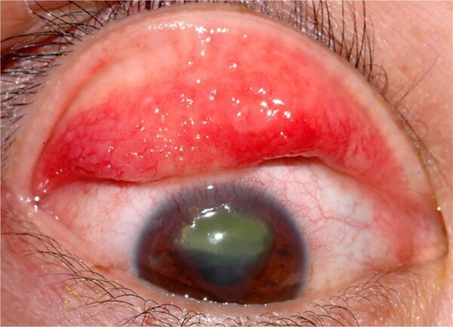
Figure 7. Vernal keratoconjunctivitis
Footnote: Large cobblestone papillae. Upper tarsal giant papillae are typical of vernal conjunctivitis. These have characteristically flattened tops which sometimes demonstrate stain with fluorescein. Giant papillae can sometimes be seen near the limbus and, while relatively uncommon, symblepharon formation (an eye condition that causes the conjunctiva of the eye to stick to itself or the cornea) and conjunctival fibrosis (scar) can occur.
[Source 81 ]Figure 8. Horner–Trantas dots

Footnote: Peri-limbal Horner–Trantas dots are focal white dots consisting of degenerated epithelial cells and eosinophils and are indicative of vernal keratoconjunctivitis.
[Source 79 ]Figure 9. Vernal conjunctivitis treatment options
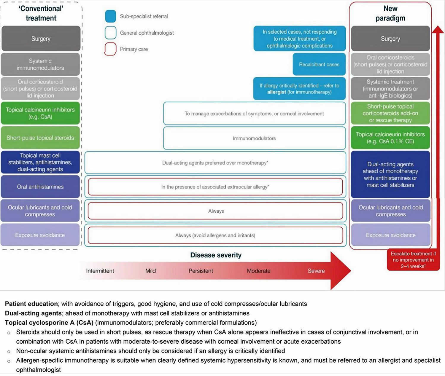
Footnotes: Treating vernal keratoconjunctivitis should entail a stepwise approach, identifying triggers, educating patients/caregivers on good ocular health, and addressing symptoms. In a new paradigm of management, the use of immunomodulators (e.g., topical cyclosporine A) should be considered early to tackle the inflammatory and chronic nature of vernal keratoconjunctivitis, with topical corticosteroids reserved as an add-on, short-pulse therapy for persistent disease, during acute exacerbations, or in patients with corneal involvement. Any use of corticosteroids requires tapering once symptoms have been controlled to avoid adverse events. For patients with an identified allergy, referral to an allergist is recommended for additional systemic treatment. In the rare patients who do not respond to medical treatment, surgery may be required.
*In the case of associated rhinitis, consider treatment according to Allergic Rhinitis and its Impact on Asthma (ARIA) protocol;
†No improvement is defined as no improvement in symptoms or changes in conjunctival, papillary or ocular surface clinical signs.
Abbreviations: CE = cationic emulsion; CsA = cyclosporine A; IgE = immunoglobulin E.
[Source 79. Adapted from Leonardi et al. 99]Vernal conjunctivitis causes
Vernal conjunctivitis is a severe type of seasonal allergic conjunctivitis that results in small, raised lumps on the inside of the upper eyelid called Horner Tranta dots and a stringy, mucus discharge 100, 101. A personal or family history of atopy is seen in a large proportion of vernal conjunctivitis patients 102. Atopy is a genetic tendency to develop allergic diseases, such as asthma, allergic rhinitis, and atopic dermatitis (eczema). Vernal conjunctivitis was originally thought to be due to a solely immunoglobulin E (IgE) mediate reaction via mast cell release and cell-mediated immune mechanisms 103. It has now been shown that IgE is not enough to cause the varied inflammatory response that is seen with vernal conjunctivitis 90. Activated eosinophils have also been implicated to play a significant role in the pathophysiology of vernal conjunctivitis and these can be shown consistently in conjunctival scrapings of vernal conjunctivitis patients; however mononuclear cells and neutrophils are also seen 103, 90. Additionally, the role of type 4 hypersensitivity mediated by CD4 T-helper-2 (Th2) lymphocytes with immunomodulators such as IL-4, IL-5, and bFGF has also been highlighted through a few studies 91, 92, 88, 90. This differs from atopic keratoconjunctivitis, which has been shown to involve both Th1 and Th2 inflammatory cascades 93. Immunomodulators like interleukins 4 (IL-4), IL-5, IL-13, and fibroblast growth factors have also been implicated. Increased levels of tumor necrosis factor (TNF) alpha, histamine, tryptase, IgE, and IgG antibodies are observed on pathologic examination of tears 94. There is also reported over-expression of cytokines and chemokines in the conjunctiva of these patients 104. Thought has been given to a possible endocrine predisposing factor as well as there is a decrease in symptoms and prevalence after puberty 103, 88.
Apart from personal allergy history, other predisposing factors include male gender, close contact with animals, and increased exposure to dust and sunlight 105.
A hereditary association or genetic link has been suggested, but no direct genetic associations have been made. Vernal conjunctivitis is seen more often in patients who have family history of atopy and might be positive in close to 49% of these cases. But no clear correlation with specific genetic loci has been identified 88.
Vernal conjunctivitis types
Vernal conjunctivitis or vernal keratoconjunctivitis is a subtype of allergic conjunctivitis 100. The episodes are often periodic and have seasonal recurrences. Seasonal exacerbations characterize vernal keratoconjunctivitis in the initial stages with a peak incidence during spring and summer. Over time, vernal conjunctivitis tends to become perennial (all year round).
Additional types of vernal conjunctivitis include perennial and seasonal rhinoconjunctivitis, atopic keratoconjunctivitis, and giant papillary conjunctivitis 80.
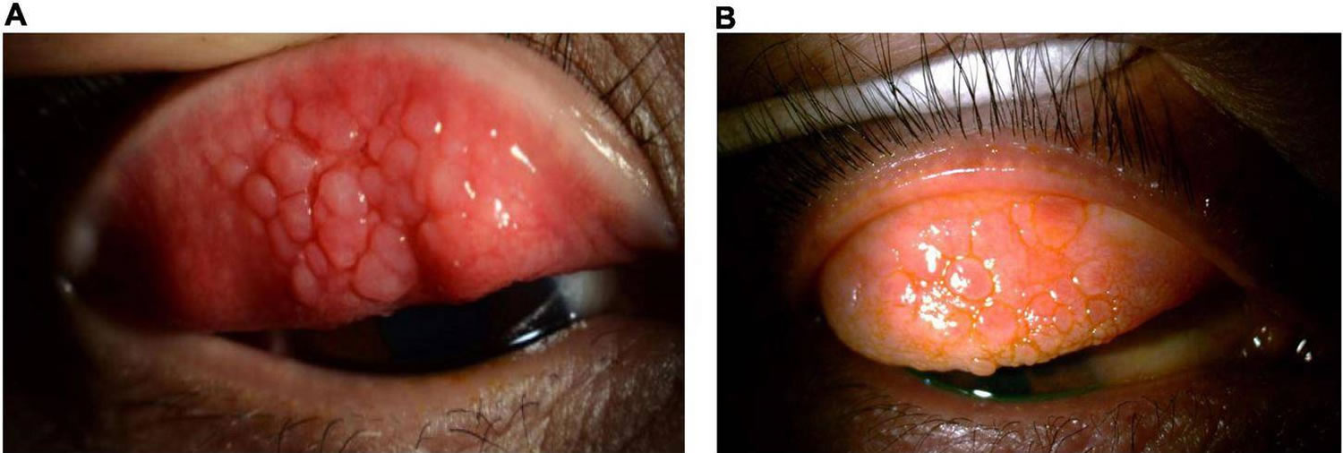
Vernal conjunctivitis is classified into three clinical subtypes based on the location of the papillae into palpebral (tarsal), limbal (bulbar), and mixed forms 91, 106, 79:
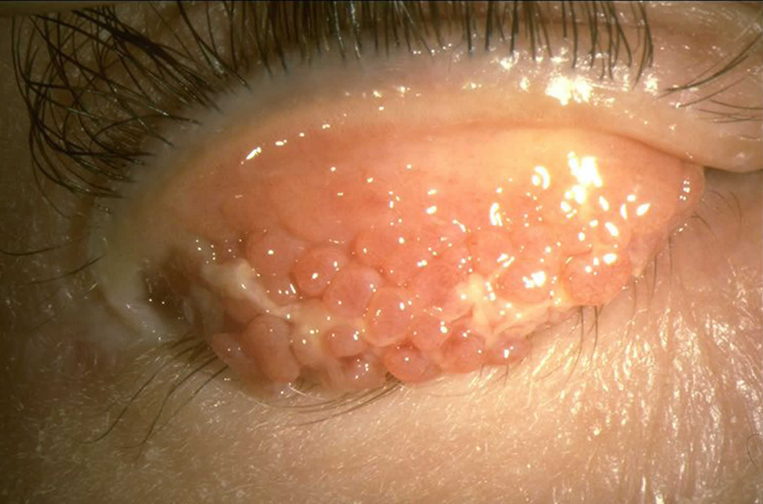
- Palpebral vernal conjunctivitis (tarsal vernal conjunctivitis). Palpebral vernal conjunctivitis is characterized by large, cobblestone-like papillae on the upper tarsal conjunctiva. These can differ in shape and size, but are usually defined as >1.0 mm in diameter 33, 91. There is a close association between the inflamed conjunctiva and the corneal epithelium, often leading to significant corneal disease.
- Limbal vernal conjunctivitis (bulbar vernal conjunctivitis). Limbal vernal conjunctivitis typically involves Horner–Trantas dots indicating lymphocytic and eosinophilic infiltration of the limbal conjunctiva 33, 91. Limbal vernal conjunctivitis typically affects the Black and Asian populations.
- Mixed vernal conjunctivitis. Mixed vernal conjunctivitis has features of both the palpebral (tarsal) and limbal (bulbar) subtypes in only one eye (as signs are often heterogeneous between eyes) 33.
According to the Management of Vernal Keratoconjunctivitis in Asia (MOVIA) Expert Working Group, the most common form of vernal conjunctivitis seen in clinics across Asia is the tarsal form; however, up to one-third of patients are assumed to have the mixed form 79. The limbal form of vernal conjunctivitis is considered less common in Asia (based on clinical experience) 79.
Figure 10. Palpebral vernal conjunctivitis
Footnote: Palpebral vernal conjunctivitis also called tarsal vernal conjunctivitis is characterized by large, cobblestone-like papillae on the upper tarsal conjunctiva.
[Source 79 ]Figure 11. Limbal vernal conjunctivitis
Footnote: Limbal vernal conjunctivitis typically involves Horner–Trantas dots, indicating lymphocytic and eosinophilic infiltration of the limbal (bulbar) conjunctiva. The mixed form is characterized by the presence of both tarsal and limbal subtypes in only one eye.
[Source 79 ]Figure 12. Shield ulcer
Footnote: Shield ulcer formation. Shield ulcers usually form on the upper third of the cornea. Plaques can also form when inflammatory debris accumulates at the base of a shield ulcer.
[Source 79 ]Vernal conjunctivitis symptoms
The most common eye symptoms of vernal conjunctivitis are initially severe itching, redness, and tearing. Other common symptoms include blurred vision, photophobia, foreign body sensation, burning, blinking or eye twitching (blepharospasm) and a characteristic ropey, stringy mucus, and/or serous discharge 33, 107, 103, 88. Other typical signs and symptoms include moderate-to-intense conjunctival hyperemia, mild-to-moderate chemosis, foreign-body sensation, and pain, all of which can be very intense upon awakening, causing what is called “the morning misery” 33. Vernal conjunctivitis typically affect both eyes but may also affect one eye only.
Clinical signs of vernal conjunctivitis include a papillary reaction of the upper tarsal conjunctiva and throughout the limbus. The signs of vernal conjunctivitis can be divided into conjunctival, limbal and corneal signs 81. Moreover, the clinical examination findings also vary dependent on the geographical location 108.
- Conjunctival signs include diffuse conjunctival injection and upper tarsal giant papillae. These are discrete >1mm in diameter that characteristically have flattened tops which sometimes demonstrate stain with fluorescein 103, 109. Additionally, these giant papillae can sometimes be seen near the limbus and, while relatively uncommon, symblepharon formation (an eye condition that causes the conjunctiva of the eye to stick to itself or the cornea) and conjunctival fibrosis can occur 110.
- Palpebral disease: In the early stages, there is conjunctival hyperemia and velvety papillary hypertrophy of the superior tarsal plate. In the intense disease, flat polygonal macropapillae, which are <1mm in size, are seen along with the whitish inflammatory disease. With further disease progression, these can form giant papillae that are>1mm in size and result from rupture of the dividing septa. Mucoid deposits can occur between giant papillae. The milder form of the pathology is characterized by minimal conjunctival congestion and decreased mucus production 111.
- Bulbar disease: The bulbar disease is also called limbal disease and is particularly more common and severe in tropical regions. This is characterized by congestion of the bulbar conjunctiva in the interpalpebral area. Gelatinous thickened papillae can form around the limbus and are associated with apically located whitish cellular collections known as Horner Tranta Spots 112.
- Limbal signs include thickening and opacification of the limbal conjunctiva as well as gelatinous appearing and sometime confluent limbal papillae. Peri-limbal Horner-Trantas dots are focal white limbal dots consisting of degenerated epithelial cells and eosinophils 103. Limbal disease can result in a limbal stem cell deficiency which can lead to pannus formation with corneal neovascularization 110.
- Corneal signs vary according to the severity of the disease process 103. Punctate epithelial erosions or keratitis can coalesce into macro-erosions of the epithelium 90.
- Plaques containing fibrin and mucous can accumulate into macro-erosions forming Shield ulcers (see Figure 12). Corneal neovascularization can ensue and resolution can leave a characteristic ring-like scar 109, 87.
- A waxing and waning gray-white lipid depositing in the peripheral, superficial stroma can occur and is known as pseudogerontoxon 103. Pseudogerontoxon is characterized by a paralimbal band of superficial scarring adjacent to the inflamed limbus resulting from recurrent limbal disease. It usually mimics arcus senalis.
- Vernal keratoconjunctivitis is also associated with corneal ectasias, particularly keratoconus, which results from persistent eye rubbing and occasionally superficial vascularization. Keratoconus has been shown to be more prominent in vernal conjunctivitis patients as well; possibly due to increased eye rubbing of chronically irritated patients 113.
- Associated bilateral herpes simplex keratitis has also been reported 114
- Lid signs. Blepharitis is often associated with patients with vernal keratoconjunctivitis. Some cases might be seen with eczema or maceration of the lid. Subconjunctival fibrosis and symblepharon can rarely develop due to the inflammatory complications 115.
Severe vernal conjunctivitis can result in sight-threatening complications 79. Eye surface damage as a result of repetitive eyelid trauma and vernal conjunctivitis-associated inflammatory activity can lead to corneal complications such as superficial punctate keratopathy, shield ulcers, corneal scarring, keratoconus, dry eyes, limbal stem-cell deficiency, and secondary infections 33, 82, 116, 117. Shield ulcers, which can be self-limiting or associated with bacterial keratitis, usually form on the upper third of the cornea, and can lead to loss of vision (see Figure 12) 91. Plaques can also form when inflammatory debris accumulates at the base of a shield ulcer 116 and can be particularly resistant to topical therapy or require surgical intervention 91. Limbal stem-cell deficiency can occur with longstanding inflammation 91. Keratoconus and irregular astigmatism can result from frequent eye rubbing in the pediatric population 118, 119, 120. In these patients, there is a fine balance between the benefits of corticosteroids and the risk for vision loss as a result of overtreatment. Patients with severe vernal conjunctivitis may also develop lid complications and acquire ptosis often with atopic dermatitis 79. The variable severity of these complications in each patient presents a challenge to ophthalmologists to not only manage acute episodes, but also to prevent reappearances.
Vernal conjunctivitis complications
The ocular complications reported among vernal keratoconjunctivitis patients include steroid-induced cataract, steroid-induced glaucoma, irregular astigmatism, keratoconus, acute hydrops, shield ulcer, central corneal scars, and limbal tissue hyperplasia 80.
Vernal conjunctivitis differential diagnosis
The close differentials of vernal keratoconjunctivitis include all types of allergic conjunctivitis, including seasonal allergic conjunctivitis, perennial allergic conjunctivitis, atopic keratoconjunctivitis (AKC) and giant papillary conjunctivitis 80. Most of these are IgE mediated responses except giant papillary conjunctivitis. None have gender predilection, except atopic keratoconjunctivitis (AKC) and vernal keratoconjunctivitis, which are more prevalent among males. History and physical examination often help in distinguishing between these clinical entities.
Atopic keratoconjunctivitis (AKC) typically has an older age of onset in 20-50 years of age, as opposed to onset prior to age 10 years with vernal conjunctivitis 80. Conjunctival involvement is classically on the upper tarsus in vernal conjunctivitis and on the lower tarsus in atopic keratoconjunctivitis. Additionally, atopic keratoconjunctivitis is typically more chronic in nature and more commonly results in scarring of the cornea and conjunctival cicatrization, whereas vernal conjunctivitis is typically more self-limiting and do not result in severe vision-threatening complications 78, 109. Atopic keratoconjunctivitis (AKC) is more commonly associated with asthma, rhinitis, dermatitis. Vernal keratoconjunctivitis is associated with increased goblet cells compared to decrease goblet cells in atopic keratoconjunctivitis.
Vernal conjunctivitis treatment
Management should follow a stepwise approach with active patient education 79:
- Address triggers and aggravating factors (e.g., environment and allergens).
- Maintain eye health, including frequent hand, face, and hair washing.
- Use of eye lubricants and cold compresses should always be considered.
- For the use of any eye drops on the eye surface, it is recommended to use preservative-free compounds, where possible, to minimize eye surface toxicity.
- Conventional topical anti-allergic drugs
- Dual-acting agents, antihistamines, and mast cell stabilizers are all effective for reducing signs and symptoms of mild or moderate vernal conjunctivitis.
- Dual-acting agents should be considered ahead of monotherapy with antihistamines or mast cell stabilizers.
- Cyclosporine A 0.1% cationic emulsion
- Topical cyclosporine A 0.1% cationic emulsion should be considered for patients with in moderate-to-severe or persistent vernal conjunctivitis.
- Patients should be instructed on how to apply cyclosporine A eye drops to minimize stinging or burning on instillation, such as using artificial tears prior to instillation.
- Topical corticosteroid eye drops
- In patients with only conjunctival involvement, topical corticosteroids should be reserved for use after loss of control or persistence of symptoms with immunomodulators such as cyclosporine A.
- Topical corticosteroids are effective for the management of acute exacerbations, or when the cornea is involved, and preferably only introduced in patients with more severe disease. In these individuals, corticosteroids should be used in combination with cyclosporine A to account for the fact that cyclosporine A may require ≥1 week to act.
- Because of an increased risk of adverse events/or vision loss with chronic use, topical corticosteroid eye drops should be used in short pulses alone or in combination with cyclosporine A under the supervision of an ophthalmologist and tapered rapidly.
- Tacrolimus
- In regions where available, tacrolimus should be reserved for patients with severe vernal conjunctivitis that is refractory to cyclosporine A.
- Tacrolimus can be considered as a treatment for moderate-to-severe vernal conjunctivitis in patients with allergy of the eyelid, but note this may be off-label.
- Vasoconstrictors
- Vasoconstrictors are not recommended for the treatment of vernal conjunctivitis. Topical vasoconstrictors can be effective at alleviating hyperemia but offer little to no relief from itchiness and have a short duration of action. If used to address hyperemia, vasoconstrictors should be used with caution and only for a short period (no longer than 5–7 days) due to adverse events and tachyphylaxis 121.
- Vasoconstrictors may cause several side effects including rebound redness, chronic follicular conjunctivitis, and tachyphylaxis. Vasoconstrictors are rarely used in the pediatric population. It is recommended that these agents should be avoided.
- Topical decongestants do not reduce the allergic response because they do not antagonize any of the mediators of allergic inflammation. Burning or stinging on instillation is a common adverse event, and prolonged use and/or discontinuation following longer-term use can lead to rebound hyperemia and conjunctivitis medicamentosa 122. These events are usually associated with topical combinations of vasoconstrictors and first-generation antihistamines, such as pheniramine and antazoline, which are available as over-the-counter products.
- Non-steroidal anti-inflammatory drugs (NSAIDs)
- Non-steroidal anti-inflammatory drugs (NSAIDs) are not recommended as they do not target the specific inflammatory mechanism associated with vernal conjunctivitis.
- Non-steroidal anti-inflammatory drugs (NSAIDs) used in the treatment of eye allergy typically inhibit cyclooxygenase (COX)-1 and COX-2 enzymes 33. Some have demonstrated a slight effect in the treatment of ocular allergy, by targeting itching, intercellular adhesion molecule-1 expression, and tear tryptase levels 33, 121. However, use of NSAIDs is not encouraged in vernal keratoconjunctivitis because of their local side effects, such as burning or stinging after application, increased risk of inducing keratitis on the ocular surface, and because they do not target the specific inflammatory mechanism associated with vernal keratoconjunctivitis.
- Oral antihistamines
- Second-generation systemic non-sedative antihistamines are preferred over older first-generation antihistamines.
- Allergen-specific immunotherapy
- Allergen-specific immunotherapy is only recommended when clearly defined systemic hypersensitivity to an identified allergen exists.
- Patients requiring allergen-specific immunotherapy should be referred to an allergist or specialist ophthalmologist.
- In selected patients with ocular complications or persisting symptoms following prior treatments
- Surgery, oral corticosteroids (short pulses) or corticosteroid lid injection, or systemic treatment with immunomodulators or biologics may be appropriate options for use by corneal specialists.
In patients with a history of vernal conjunctivitis, drug treatment should be planned early and started at the beginning of spring or continued all year, depending on allergen exposure and duration of symptoms 33.
Table 3. Overview of medications currently available across Asia for the management of vernal conjunctivitis
| Class | Drug | Standard dosing | Indication | Considerations |
|---|---|---|---|---|
| Topical antihistamines (second generation) | Antazoline Emedastine Levocabastine | QID | Relief of itching Relief of signs and symptoms | • Short duration of action • Frequently not enough alone to treat the entire disease |
| Mast cell stabilizers | DSCG Lodoxamide NAAGA Pemirolast potassium | BID to QID | Relief of signs and symptoms | • Long-term use • Slow onset of action • Prophylactic dosing • Frequently not enough alone to treat the entire disease |
| Dual-acting agents (antihistamine + mast cell stabilizers) | Alcaftadine Azelastine Bepotastine Epinastine Ketotifen Olopatadine | QD QD to BID BID BID BID to QID BID | Relief of itching Relief of signs and symptoms | • Side effects • Bitter taste (azelastine) • Frequently not enough alone to treat the entire disease |
| Immunomodulators (calcineurin inhibitors) | Cyclosporine A Tacrolimus | QD to QID QD | Treatment of severe vernal keratoconjunctivitis and AKC not responding to anti-allergic drugs | • Pharmacy-compounded preparations vary from center to center • Quality control and availability of pharmacy-compounded preparations are poor • Tacrolimus is largely used off-label in ocular allergy (tacrolimus is approved for vernal keratoconjunctivitis only in Japan) |
| Corticosteroids | Betamethasone Desonide Dexamethasone Fluorometholone Hydrocortisone Loteprednol Prednisolone Rimexolone | As required (up to 7 days) | Treatment of allergic inflammation Use in moderate-to-severe forms | • Risk for long-term adverse events • No mast cell stabilization • Potential for inappropriate patient use • Requires close monitoring |
| Vasoconstrictors (vasoconstrictor–antihistamine combinations) | Naphazoline/ pheniramine | BID to QID | Episodic itching and redness | • Rapid onset but short duration of action • Only addresses hyperemia • Associated with a range of side effects • Potential for inappropriate patient use |
Footnotes: * Availability and access to treatments may vary across clinics, hospitals, regions, and countries. Each treatment option should be considered in accordance with the level of evidence available at the time.
Abbreviations: AKC = atopic keratoconjunctivitis; BID = twice daily; DSCG = disodium cromoglycate; NAAGA = N-acetyl-aspartyl glutamic acid; QD = once daily; QID = four times daily; VKC = vernal keratoconjunctivitis.
[Source 79 ]Vernal conjunctivitis treatment options
Removal of any and all possible allergens as well as conservative management such as cool compresses and lid scrubs make up the first line of therapy 123. A topical antihistamine only may work in mild cases 123. Topical mast cell stabilizers (cromolyn sodium, nedocromil sodium, and lodoxamide) are typically used with topical antihistamines and have been shown to be effective in moderate presentations of vernal conjunctivitis 123. Mast-cell stabilizers have a loading period to reach their full therapeutic effect 90. Overall, topical antihistamines (instilled four times daily) appear to be safe and well tolerated in patients with vernal conjunctivitis 79. Alongside mast cell stabilizers and dual-acting agents, they are often the first-choice treatment and are effective in reducing the signs and symptoms of vernal conjunctivitis 121. Antihistamine drugs with eye activity are a good therapeutic option for patients with allergic conjunctivitis, including vernal conjunctivitis, since they can inhibit proinflammatory cytokine secretion from conjunctival epithelial cells 124.
Alongside antihistamines and dual-acting agents, mast cell stabilizers (instilled two to four times daily) form the first line of treatment and are effective in reducing the signs and symptoms of vernal keratoconjunctivitis. Mast cell stabilizers have been shown to act on multiple cells involved in allergic inflammation, including eosinophils, neutrophils, macrophages, mast cells, and monocytes. For example, lodoxamide is known to have an effect on eosinophil activation (shown in studies by measuring tear eosinophil cationic protein before and after therapy), while N-acetyl-aspartyl glutamic acid (NAAGA) is known to inhibit leukotriene synthesis and release of histamine by mast cells 33. Overall, mast cell stabilizers are generally well tolerated especially preservative-free options; however, there are tolerability concerns with some agents, including burning and stinging sensations upon instillation, blurred vision, and an undesirable aftertaste. Data on long-term efficacy and safety of mast cell stabilizersare lacking 121.
Dual-acting agents are the combination of mast cell stabilizing properties and histamine receptor antagonism (instilled twice daily) has shown clear benefits in treating forms of eye allergy 79. Dual-acting agents are well tolerated and are not associated with significant ocular drying effects 33, and should be preferred over monotherapy with antihistamines or mast cell stabilizers alone. A key benefit of dual-acting agents is the provision of rapid symptomatic relief by alleviating itching and redness 33. Olopatadine and ketotifen, for example, have shown to be effective in relieving itching, tearing, conjunctival hyperemia, mucus discharge, and photophobia 125.
If seasonal recurrence is known, it is suggested that mast-cell stabilization therapy be initiated prior to the season in which symptoms are encountered and continued throughout the season 90. Dual-action agents with both histamine-1 (H1) blocking mechanism and mast-cell stabilization have the benefits of working immediately and having long-term disease modifying effects. Topical corticosteroids are typically the most effective. High pulse dose with quick tapering and use of low-absorptions corticosteroids (fluoromethelone, loteprednol, remexolone, etc.) is preferred when using topical corticosteroid therapy 123. Oral corticosteroids can be considered in sight threatening conditions 103, 87. Supratarsal injection of local corticosteroid (triamcinolone or dexamethasone) into the upper tarsal papillae can sometimes offer short term relief as well 126.
Long term immunomodulation (a process in which the immune system is modified or modulated in a beneficial way) with steroid sparing agents such as cyclosporine and tacrolimus is often needed. Topical calcineurin inhibitors (topical immunomodulators) are frequently prescribed for patients with vernal conjunctivitis 121. Topical calcineurin inhibitors are recommended in patients with acute-phase or persistent, moderate or severe vernal conjunctivitis that is not responding to anti-allergic drugs 121. Topical Cyclosporine A is effective in controlling ocular surface inflammation in vernal conjunctivitis and is thought to work by inhibiting Th2 proliferation and IL-2 production, and by reducing levels of immune cells and mediators acting on the ocular surface and conjunctiva 127. Different concentrations of cyclosporine A, ranging from 0.05 to 2.0%, are currently available in different countries with different clinical indications 128, 129. The 0.1% cationic emulsion (CE) formulation increases the bioavailability of cyclosporine A in the tear film compared with available oil-based (often used when cyclosporine A is pharmacy-compounded) or anionic emulsion cyclosporine A formulations 130. Clinical studies on the efficacy of topical cyclosporine A 0.1% cationic emulsion (CE) for treating vernal conjunctivitis have consistently shown a beneficial effect of cyclosporine A, with safety and efficacy demonstrated for up to 7 years and comparable with those seen in patients with dry eye disease 127, 131, 132, 133.
There is no consensus on the minimum effective concentration of cyclosporine A to treat vernal conjunctivitis, as this can depend on the emulsion vehicle 79. Topical cyclosporine A in concentrations of 0.05% to 2% has been shown to decrease inflammatory cytokines and the signs and symptoms of treated vernal conjunctivitis patients 134, 135. Currently, cyclosporine A 0.1% cationic emulsion (CE) is approved based on significant improvements in signs, symptoms, and quality of life in patients with severe vernal conjunctivitis, and is commercially available across Asia. It is recommended that cyclosporine A 0.1% CE should be initiated twice daily for patients with mild-to-moderate disease and four times daily for those with moderate-to-severe disease for up to 6 months. Other commercially available cyclosporine A formulations exist but are not approved for use in vernal conjunctivitis (as of December 2020) 79. In addition, cyclosporine A may be compounded in hospital-based pharmacies to provide variable concentrations and formulations; however, its quality may be compromised, and availability is limited to specialist centers 79.
The most commonly associated side effects with cyclosporine A is a stinging or burning sensation on instillation, and no new safety issues were identified in long-term follow-ups 79. Instillation pain can be minimized by following a standard technique for eye drop instillation, which includes using artificial tears prior to instillation (avoiding any potential washout effect of the subsequently administered cyclosporine A, by waiting at least 10 minutes between administrations), followed by one drop into the lower conjunctival sac of each eye in the morning, at noon, in the afternoon, and in the evening, approximately 4 hours apart 133. Patients should also be counseled to expect stinging and burning when the drops are applied, but that the sensation should taper off with regular use as prescribed.
After long cycles of treatment, cyclosporine A can allow control of symptoms without corticosteroids 79. There is a trend in the Asia-Pacific region, in using cyclosporine A as a first-line treatment for patients who present with moderate-to-severe or persistent vernal conjunctivitis 79. In patients presenting with conjunctival involvement only, cyclosporine A alone is recommended for treatment, while corticosteroids should only be prescribed (as short pulses) if there is an inadequate response to cyclosporine A 79. Otherwise, cyclosporine A can be used in combination with corticosteroids in patients with corneal complications (or limbitis) or acute exacerbations, in the understanding that cyclosporine A may take over a week to act due to its immunosuppressive mechanism of action 79.
Tacrolimus is a strong, non-steroidal immunosuppressant that binds to FK506-binding proteins in T lymphocytes and inhibits calcineurin activity 136. Tacrolimus has been investigated in a small randomized controlled trial in vernal keratoconjunctivitis, where it demonstrated improvements in symptoms of itching, foreign-body sensation, and photophobia, as well as in signs of limbal inflammatory activity and keratitis, with maintenance of disease control 137, 138. Tacrolimus may be administered as eye drops or an ointment. The use of tacrolimus (0.03%) ointment in ophthalmic and dermatologic form is currently off-label in Asia for patients with vernal keratoconjunctivitis. However, there are findings in published studies of small population sizes demonstrating effectiveness and tolerability 137, 138, 139, 140, 141, 142.
Tacrolimus 0.1% topically has also shown to improved signs and symptoms of vernal conjunctivitis, with one study showing improvement even in patients unresponsive to 0.1% topical cyclosporine A 143, 144. A study comparing the use of 0.1% tacrolimus ophthalmic ointment dosed twice a day versus 2% cyclosporine A eye drops four times a day for the treatment of vernal conjunctivitis, did not find a statistically significant difference in signs or symptoms or side effects between the two groups 145. Additionally, adult patients with vernal conjunctivitis may respond more favorably to topical cyclosporine A therapy 146. Oral anti-histamines are sometimes used, but there is no real evidence in their support 81.
Similar to cyclosporine A, tacrolimus may be associated with instillation pain 136. As a strong immunosuppressant, topical tacrolimus may be associated with an increased risk for corneal infections with prolonged use, and close monitoring of patients on long-term therapy is necessary 142. Topical tacrolimus (0.02–0.1%) should be reserved for patients with severe vernal keratoconjunctivitis involving allergy of the eyelid or whose disease is refractory to cyclosporine A 121.
Topical corticosteroid eye drops should be used as short, pulsed therapy to provide symptomatic relief in patients with more persistent vernal keratoconjunctivitis or acute exacerbations, or when the cornea is involved (e.g., patients with shield ulcers or hyperplasia in limbal vernal keratoconjunctivitis), and under an ophthalmologist’s supervision 121. Topical corticosteroids can be administered at a low or high dose, depending on the severity of vernal keratoconjunctivitis, and can be given up to four times daily with or without cyclosporine A. Corticosteroids act by suppressing the late-phase reaction in both experimental and clinical settings. Corticosteroids, in part, limit the inflammatory cascade by inhibiting phospholipase A2, and consequently act to prevent migration of leukocytes, release of hydrolytic enzymes, growth of fibroblasts, and lead to changes in vascular permeability 147.
Corticosteroids with low intraocular absorption (“soft steroids”), such as hydrocortisone, fluorometholone and loteprednol, may be preferred 33. Dosages are chosen based on the inflammatory state of the eye, with therapy prescribed in pulses of 3–5 days (1). Loteprednol is usually indicated for 7–8 days in the treatment of the acute phase 33. Prednisolone, dexamethasone, and betamethasone are sometimes considered only as second-line options, or as first-line treatment in more severe cases, due to their potential effect on intraocular pressure 33. Steroid–antibiotic combination eye drops are not recommended, since vernal keratoconjunctivitis is an allergic inflammation and not an infection 33.
Moderate-to-severe vernal keratoconjunctivitis may require repeated topical corticosteroid treatment to downregulate conjunctival inflammation 33. Persistent and severe symptoms, thick mucus discharge with moderate-to-severe corneal involvement, numerous and inflamed limbal infiltrates, and/or giant papillae may indicate a need for corticosteroids in addition to cyclosporine A use 33. However, use of corticosteroids as first-line therapy is not recommended in those with only conjunctival involvement 33.
Corticosteroids are not recommended for long-term use because of the increased risk for ocular adverse events, including increased intraocular pressure (IOP), glaucoma, cataracts, and susceptibility to infection 33. These adverse events depend, in part, on the structure of the steroid, the dose, and duration of treatment 148. Treatment with corticosteroids requires careful monitoring for the development of intraocular pressure (IOP), which should be promptly brought under control to prevent deterioration or loss of vision 149.
Supratarsal corticosteroid injections have demonstrated some value in treating adults with vernal keratoconjunctivitis but may not be an appropriate approach for children or for application by general ophthalmologists 149, 150. For more specialist ophthalmologists, this may be an option in children with refractory, severe, or challenging vernal keratoconjunctivitis.
Systemic treatment with oral antihistamines or antileukotrienes can reduce the severity of flare-ups and generalized hyperreactivity 33. Newer second-generation antihistamines are preferred over older first-generation antihistamines, in order to avoid the sedative and anticholinergic effects that are associated with first-generation agents 151.
Systemic immunotherapy is also an option, especially for those patients with underlying systemic atopic disease such as atopic dermatitis and asthma. There are case reports of the successful use of omalizumab, an anti-IgE monoclonal antibody, in patients with vernal conjunctivitis recalcitrant to other treatment modalities, but this remains an off-label use 152, 153. Dupilumab is a human monoclonal antibody against interleukin-4 receptor alpha used in the treatment of atopic dermatitis. There is currently a multi-center, double-masked, randomized, and placebo-controlled, parallel-group study that is evaluating the safety and efficacy of dupilumab in the treatment of signs and symptoms of individuals with atopic keratoconjunctivitis (NCT04296864), and may also play a role in treating vernal disease in those patients older than 12 years 154. However, dupilumab has been found to itself induce atopic keratoconjunctivitis compared to placebo in treated patients, so its role as a primary treatment is debatable 81.
Allergen-specific immunotherapy is indicated only when a clearly defined systemic hypersensitivity to an identified allergen exists 121 and should be managed by an allergist or specialist ophthalmologist. The combination of clinical history with the results of a skin-prick test and specific serum IgE should be taken into consideration when the choice of immunotherapy is made 121. There are currently no robust studies of allergen-specific immunotherapy in vernal keratoconjunctivitis 121. A meta-analysis of several double-blind, randomized, placebo-controlled trials investigating sublingual immunotherapy for allergic conjunctivitis, reported a significant reduction in total ocular symptom scores and ocular signs (redness, itchiness, and tearing) vs. placebo in pollen-induced allergic conjunctivitis, but not in allergic conjunctivitis associated with house dust mites, in the pediatric population 155.
Case reports and ongoing clinical practice have indicated that, in rare instances, specialist surgical management may be beneficial in patients with vernal keratoconjunctivitis 79. Surgical approaches that focus on conjunctival cobblestone papillae, shield ulcers, corneal plaques, limbal insufficiency, and other ocular surface presentations that do not respond to medical treatment can be beneficial 156, 157, 158.
Surgical approaches can include excision of giant papillae, debridement of the corneal plaque to remove cytotoxic cells, and amniotic membrane transplantation 159. Surgical treatment, especially corneal plaque removal, may be appropriate for patients with ulcers of moderate-to-severe severity, because short-term re-epithelialization rates are higher and the number of complications is lower than that associated with medical therapy 159. Other reports suggest surgical treatment, such as giant papillae excision, may be appropriate in cases of corneal involvement and in the presence of coarse giant tarsal papillae resulting in ptosis (or mechanical pseudoptosis) 33, 160. Amniotic membrane transplantation following keratectomy has been described as a successful treatment for deep ulcers, in severe allergic patients with slight stromal thinning 161.
Patients with vernal keratoconjunctivitis who may benefit from surgical intervention should be referred to a corneal specialist first. Leonardi et al. 33 have suggested that cryotherapy and/or giant papillae excision papillae should otherwise be avoided because they only treat the complications of vernal keratoconjunctivitis and not the underlying disease, and may induce unnecessary scarring.
Vernal conjunctivitis prognosis
Generally vernal conjunctivitis is a rather benign and self-limiting disease that may resolve with age or spontaneously at puberty 103, 162, 163. Sometimes the debilitating nature of vernal conjunctivitis when it is active necessitates therapy to control symptoms. Complications typically arise from occasional corneal scarring and the unsupervised used of topical corticosteroids can result in cataracts, or glaucoma can lead to permanent visual impairment 103, 162. Rarely, complications in the form of keratoconus, shield ulcers, corneal scarring, and vascularisation may significantly affect visual acuity. Corneal ulcers are reported in close to 9.7% of patients 164, 165. In some patients (as many as 12% of cases) symptoms may persist beyond childhood, which in some cases may represent a conversion to an adult form of atopic keratoconjunctivitis 103. This persistence into adulthood has been shown to be as high as 12% 166.
Bacterial conjunctivitis
Bacterial conjunctivitis is conjunctivitis caused by bacteria and is very contagious. The most common bacteria are Staphylococcus species, Streptococcus pneumoniae, Haemophilus influenzae and Moraxella catarrhalis 167, 168. Bacterial conjunctivitis presents with conjunctival injection, mucopurulent discharge, and crusty eyelids. The diagnosis is usually clinical. The condition is often self-limiting, but there is good evidence that antibiotics improve remission rates. Most of the current evidence suggests that the choice of topical antibiotics and the treatment regimen do not significantly affect the rate of recovery from infection. Failure to recognize and treat bacterial conjunctivitis may lead to complications, such as keratitis or anterior uveitis. Bacterial conjunctivitis is commonly associated with hyperacute (<24 hours) onset of severe and very rapid progressing conjunctivitis. Bacteria rank second among the most common causes of infectious conjunctivitis. Bacterial conjunctivitis almost always affects both eyes, although it may start in just one eye. Symptoms include gritty feeling like sand in your eye and a thick yellow discharge (pus) that crust over your eyelashes, especially after sleep. With bacterial conjunctivitis, you have sore, red eyes with a lot of sticky pus in the eye. Some bacterial infections, however, may cause little or no discharge. Sometimes the bacteria that cause pink eye are the same that cause strep throat.
Bacterial conjunctivitis has an incubation period and communicability of 1-7 days and is commonly caused by 169:
- Neisseria gonorrhoeae
- Neisseria meningitidis
Bacterial conjunctivitis with acute or subacute onset (hours-days) of moderate to severe severity are commonly caused by 169:
- Haemophilus influenzae biotype III (previously known as Haemophilus aegyptius)
- Haemophilus influenzae
- Streptococcus pneumoniae
- Streptococcus viridans
- Staphylococcus aureus
Chronic bacterial conjunctivitis is used to describe any conjunctivitis lasting more than 4 weeks. It has an incubation period of 2-7 days 170.
Clinically distinguishing bacterial conjunctivitis from other conjunctivitis is essential as it can help direct therapies and potentially curb unnecessary empiric antibiotic administration. Traditionally, a purulent or mucopurulent discharge has been associated with the diagnosis of bacterial conjunctivitis while watery discharge has been more consistent with viral or allergic conjunctivitis 14. A 2003 study contested this assertion based on a lack of evidence to support discharge characteristics correlating with the cause of conjunctivitis 14, 171. A later study by the same authors concluded that three findings were significantly predictive of bacterial conjunctivitis, including glued eyes, lack of itching, and no previous history of conjunctivitis 14, 171. A 2006 prospective study of children with conjunctivitis described five clinical history and physical examination variables associated with bacterial culture-positive infections. These included a history of gluey or sticky eyelids in the morning, examination findings of mucoid or purulent discharge, eyelids or eyelashes crusting or gluing on examination, absence of burning sensation, and lack of watery discharge 172. The inconsistencies of these findings concerning discharge as a predictor of bacterial infection might be attributable to the 2003 survey focusing on history, while the 2006 study included in-office physical examination findings. These differences reinforce the idea of variability in the presentation of bacterial conjunctivitis.
Gonococcal infection of the eye, when it does occur, typically presents as conjunctivitis (Figure 13). Gonococcal conjunctivitis in adults most often results from the accidental transfer of microorganisms from one part of the body to another (autoinoculation) in persons with genital gonococcal infection. Persons with gonococcal conjunctivitis may initially develop mild, nonpurulent conjunctivitis, that, if untreated, typically progresses to marked conjunctival redness, copious purulent discharge, and conjunctival edema 173. Less often, ulcerative keratitis develops. Untreated gonococcal conjunctivitis can cause complications that may include corneal perforation, endophthalmitis (an infection of the tissues or fluids inside the eyeball) and blindness.
Bacterial conjunctivitis may need antibiotic ointment or drops prescribed by a doctor. Treatment should be applied to both eyes, even if only one eye appears to be infected. Always treat the unaffected eye first. The findings of 2023 Cochrane review suggest that the use of topical antibiotics is associated with a modestly improved chance of resolution in comparison to the use of placebo 174. Since no evidence of serious side effects was reported, use of antibiotics may therefore be considered to achieve better clinical and microbiologic efficacy than placebo.
Figure 13. Gonococcal conjunctivitis
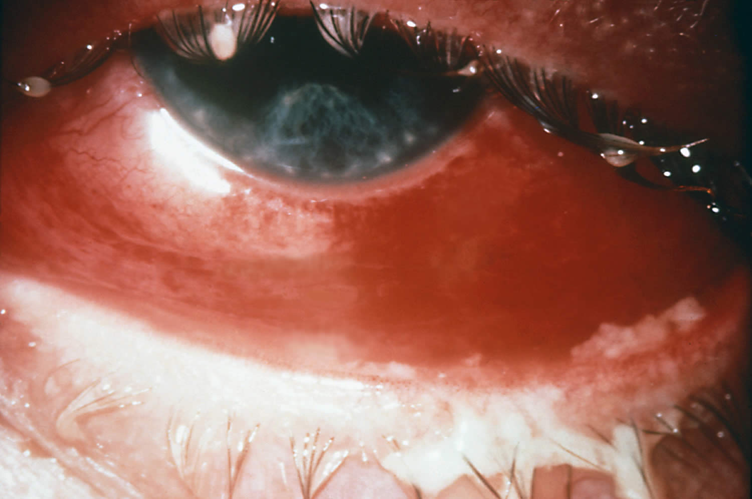
Footnote: Severe case of gonococcal conjunctivitis. Note the purulent material on the upper and lower lids.
[Source 175 ]Figure 14. Chlamydial conjunctivitis
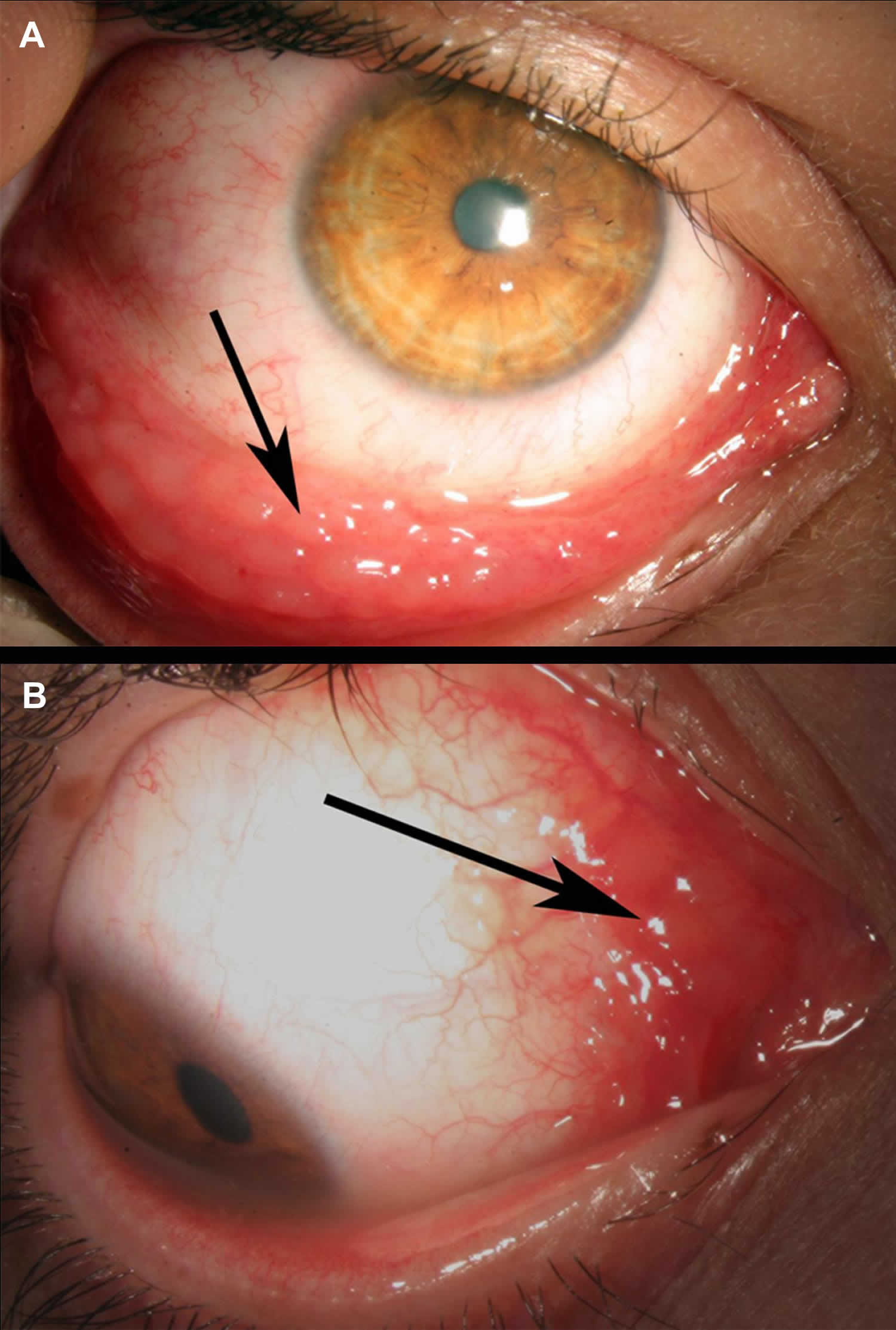
Footnotes: 22-year-old man presenting with a long-standing (>4 months) history of redness in his right eye accompanied by scant mucopurulent discharge was treated by several courses of topically administered antibiotics. Owing to the poor response to therapy, he was referred to our ocular inflammatory clinic. External examination of the right eye revealed prominent conjunctival follicles almost exclusively located at the inferior fornix of the conjunctiva (A) and prominent conjunctival bulbar follicles at the semilunar fold (B). The rest of the examination was unremarkable except for a symptom of mild burning on urination for the past 3 months. Nucleic acid amplification testing (NAAT) on a conjunctival swab confirmed the presence of Chlamydia trachomatis in both samples and excluded the presence of Neisseria gonorrhoeae. The patient was prescribed a 1-week course of oral doxycycline 100 mg twice a day (because topical antibiotics are ineffective). Symptoms and signs resolved completely within 1-month thereafter.
[Source 176 ]Bacterial conjunctivitis causes
Bacterial conjunctivitis is inflammation of the conjunctiva caused by direct contact with infected secretions.
The most common bacteria that cause bacterial conjunctivitis are 168:
- Staphylococcus, the same family of bacteria that cause staph infections.
- Streptococcus, the bacteria family that causes strep throat and pneumococcal disease.
- Haemophilus influenzae, a bacteria family best known for causing meningitis in young children.
- Sexually transmitted infections (STIs), like Chlamydia trachomatis (chlamydia), Neisseria gonorrhoeae (gonorrhea) and Treponema pallidum (syphilis). When these infections pass from birthing parent to child during birth, it can lead to neonatal conjunctivitis, a condition that can cause permanent eye damage and blindness.
Bacterial pathogens in adults are more often staphylococcal species with Haemophilus influenzae and Streptococcus pneumoniae responsible for a smaller percentage of cases 177. Staphylococcus aureus is more commonly found in adults and the elderly but is also present in pediatric cases of bacterial conjunctivitis 172. There has also been an increase in the frequency of conjunctivitis secondary to methicillin-resistant Staphylococcus aureus (MRSA) 177. Contact lens wearers are more susceptible to gram-negative infections 172. Pseudomonas aeruginosa is more likely to be the isolate from critically ill, hospitalized patients 172. Neonates can be affected by the vertical, oculogenital transmission of Neisseria gonorrhoeae and Chlamydia trachomatis resulting in acute bacterial conjunctivitis 178. Neisseria gonorrhoeae and Chlamydia trachomatis organisms can also cause a hyperacute infection in sexually active adolescents and adults 178.
The most common causative organism of bacterial conjunctivitis in children is Haemophilus influenzae, followed by Streptococcus pneumoniae and Moraxella catarrhalis 179, 180, 181.
Bacterial conjunctivitis can be quite contagious. The most common ways to get the contagious bacterial conjunctivitis include:
- Direct contact with an infected person’s bodily fluids, usually through hand-to-eye contact.
- Contact with contaminated fingers, fomites or oculo-genital contact with someone infected. Young, sexually active adults below the age of 25 years, have a high risk, especially if they do not use condoms on sexual encounters.
- Spread of the infection from bacteria living in the person’s own nose and sinuses.
- Not cleaning contact lenses properly. Using poorly fitting contact lenses or decorative contacts are risks as well.
- Compromised tear production or drainage
- Disruption of the natural epithelial barrier
- Abnormality of adnexal structures
- Trauma
- Immunosuppressed status.
Children are the people most likely to get pink eye from bacteria or viruses. This is because they are in close contact with so many others in school or day care centers. Also, they don’t practice good hygiene.
In children, the disease is often caused by Haemophilus influenzae, Streptococcus pneumoniae and Moraxella catarrhalis.
The most common pathogens for bacterial conjunctivitis in adults are Staphylococcal species, followed by Streptococcus pneumoniae and Haemophilus influenzae.
The course of bacterial conjunctivitis usually lasts 7-10 days.
- Hyperacute bacterial conjunctivitis is often caused by Neisseria gonorrhoeae. When the infection does not respond to standard antibiotic therapy in sexually active patients, Chlamydia trachomatis should be suspected.
- Chronic bacterial conjunctivitis is commonly caused Staphylococcus aureus, Moraxella lacunata, and enteric bacteria.
Bacterial conjunctivitis signs and symptoms
Bacterial conjunctivitis symptoms may include:
- the feeling that something is in your eye, or a gritty sensation in your eye
- red eyes
- burning eyes
- itchy eyes
- painful eyes (this is usually with the bacterial form)
- watery eyes
- puffy eyelids
- blurry or hazy vision
- sensitivity to light (photophobia)
- lots of mucus, pus, or thick yellow discharge from your eye. There can be so much that your eyelashes stick together.
Bacterial conjunctivitis complications
Complications from bacterial conjunctivitis are uncommon; however, severe infections can result in keratitis, corneal ulceration and perforation, and blindness 14, 172, 180.
Bacterial conjunctivitis diagnosis
In most cases, your doctor can diagnose bacterial conjunctivitis by asking about your recent health history and symptoms and examining your eyes.
Classic physical examination findings of bacterial conjunctivitis are conjunctival erythema and purulent discharge 178. A complete eye examination should include assessment of visual acuity and corneal involvement 14, 182. Although slit lamps are beneficial to a comprehensive eye assessment, they are not routinely available in primary care offices 14. An otoscopic examination is warranted if ear symptoms are reported in children to diagnose concurrent acute otitis media 180.
Rarely, your doctor may take a swab to test for sample of the liquid that drains from your eye for laboratory analysis, called a culture 168. A culture may be needed if your symptoms are severe or if your doctor suspects a high-risk cause, such as:
- A foreign body in your eye.
- A serious bacterial infection.
- A sexually transmitted infection (STI).
Conjunctival cultures are also the recommended in cases where newborn conjunctivitis (ophthalmia neonatorum) is suspected, or where copious purulent discharge makes the diagnosis of gonococcal or chlamydial infection more likely 178, 14.
Your doctor can use the results to guide your treatment.
In the following days or weeks, your doctor may recommend that you have a follow-up visit with an eye care specialist to check on how your eye is healing and adjust your treatment if necessary.
Bacterial conjunctivitis differential diagnosis
The differential diagnosis for bacterial conjunctivitis includes viral and allergic conjunctivitis 172. Clear discharge and itching are more characteristic of allergies and viral infections 14, 172. Trauma can also present with similar symptoms to conjunctivitis of bacterial origin. Keratitis and iridocyclitis should be ruled out as corneal infections, and iris inflammation can lead to significant morbidity 172.
Bacterial conjunctivitis treatment
Bacterial conjunctivitis can resolve without treatment; however, your doctor may prescribe antibiotic eye drops, depending on how severe your symptoms are and with the consideration of benefits of treatment, the natural course of the disease if left untreated, antibiotic resistance, and the philosophy of antibiotic stewardship 168. And distinguishing bacterial conjunctivitis from other causes of conjunctivitis can be difficult, and doctors often err on the side of empiric antibiotic therapy 182. And antibiotic eye drops or ointments may speed up your recovery.
Studies have indicated that around 50 percent of pediatric infectious conjunctivitis presentations are attributed to bacteria, while physicians prescribe antibiotics in up to 80 to 95 percent of these cases 182, 183. Ophthalmologists use antibiotic therapy in a smaller percentage of cases than general practitioners 182, 183. Treatment with topical antibiotics has demonstrated a decrease in symptoms, improved resolution times, decreased transmission, and hastened return to school or work 14, 180, 181, 182.
Conjunctivitis usually goes away on its own within 1 to 2 weeks. If your symptoms last longer than that, you should see your ophthalmologist. He or she can make sure you don’t have a more serious eye problem.
The natural course of untreated bacterial conjunctivitis is the resolution of the infection within one week 172. Another consideration is the continued resistance patterns of the eye bacterial pathogens 180. With an understanding of the described management variables, uncomplicated bacterial conjunctivitis can be treated empirically with topical antibiotics, or managed expectantly without antibiotics 14. In complicated cases involving patients with immunocompromise, contact lens use, and suspected gonococcal or chlamydial infections, antibiotic therapy should be provided 14. If the decision is made to initiate empiric treatment, the antibiotic chosen should be broad-spectrum and include coverage of gram-positive and gram-negative ocular bacteria 180. Topical aminoglycosides, polymyxin B combination drugs, macrolides, and fluoroquinolones are the most commonly prescribed ophthalmic agents 184, 180, 14. The duration of treatment is generally five to seven days 180.
Recently, data has shown recorded emerging resistance to most classes of these drugs 180. Topical erythromycin has been a therapeutic choice for years; however, microbial resistance and unsatisfactory coverage for Haemophilus influenzae have limited its usefulness. Topical polymyxin B/trimethoprim and several fluoroquinolones effectively manage most cases of acute bacterial conjunctivitis 178, 180. Newer fluoroquinolones have the least documented resistance; however, they are costly 180. They should be considerations in areas of increased local antibiotic resistance 178, 184. Bacterial conjunctivitis secondary to gonococcal or chlamydial infections requires systemic treatment 14. Oral antibiotics are also indicated in cases of bacterial conjunctivitis with concurrent acute otitis media 180. Ophthalmia neonatorum secondary to Chlamydia trachomatis requires oral or intravenous erythromycin in addition to topical erythromycin for 14 days 178, 185, 186.When gonorrhea is the cause of the newborn infection, hospital admission, a single dose of intravenous or intramuscular ceftriaxone, and eye irrigation are the indicated therapy until resolution of the infection 14.
Follow up for acute bacterial conjunctivitis should be encouraged if there is no improvement in symptoms after one to two days 172.
Bacterial conjunctivitis prognosis
The prognosis for uncomplicated bacterial conjunctivitis is good with complete resolution and rare adverse events with both antibiotic treatment and expectant management strategies 14, 180.
Viral conjunctivitis
Viral conjunctivitis is conjunctivitis caused by viruses and is very contagious. Virus is the most common cause of infectious conjunctivitis in the adult population (80%) and is more prevalent in the summer 187, 188. Viral conjunctivitis may involve one or both eyes, causing red itchy eyes with a ‘watery’ discharge, redness, blood vessel engorgement, pain, photophobia, and pseudomembranes 12. Viral conjunctivitis is highly contagious and means you are likely to have come into contact with someone who already has conjunctivitis. Sometimes viral conjunctivitis is accompanied by cold or flu symptoms. Children are most susceptible to viral infections, and adults tend to get more bacterial infections.
Viral conjunctivitis symptoms include:
- red, sore, watery or gritty eyes
- itchy and swollen eyes
- crusty eyelids.
Between 65% and 90% of cases of viral conjunctivitis are caused by adenoviruses. The adenovirus is part of the Adenoviridae family that consists of a nonenveloped, double-stranded DNA virus. Frequently associated infections caused by the adenovirus include upper respiratory tract infections (URTI), eye infections, and diarrhea in children. Viral conjunctivitis caused by adenoviruses produce two of the common clinical entities: pharyngoconjunctival fever, and epidemic keratoconjunctivitis 170.
- Pharyngoconjunctival fever is characterized by abrupt onset of high fever, pharyngitis, and bilateral conjunctivitis, and by periauricular lymph node enlargement.
- Epidemic keratoconjunctivitis is more severe and presents with watery discharge, hyperemia, chemosis, and ipsilateral periauricular lymphadenopathy. Lymphadenopathy (enlarged lymph glands) is observed in up to 50% of viral conjunctivitis cases and is more prevalent in viral conjunctivitis compared with bacterial conjunctivitis.
- Acute hemorrhagic conjunctivitis
- Acute follicular conjunctivitis
Viral conjunctivitis secondary to adenoviruses is highly contagious, and the risk of transmission has been estimated to be 10-50% 170. Incubation and communicability are estimated to be 5-12 days and 10-14 days, respectively 170.
Viral conjunctivitis secondary to herpes simplex virus (HSV) also known as herpes comprises 1.3-4.8% of all cases of acute conjunctivitis and is usually unilateral 189, 170. Primary herpes simplex virus type 1 (HSV-l) infection in humans occurs as a non-specific upper respiratory tract infection. Herpes simplex virus (HSV) spreads from infected skin and mucosal epithelium via sensory nerve axons to establish latent infection in associated sensory cranial nerve 5 (trigeminal nerve) and its ganglia. Latent infection of the trigeminal ganglion occurs in the absence of recognized primary infection, and reactivation of the virus may follow any of the three branches.
There is no specific treatment for most viral conjunctivitis and it usually will get better on its own.
Viral conjunctivitis causes
The most common cause of viral conjunctivitis is adenoviruses. The adenovirus is part of the Adenoviridae family that consists of a nonenveloped, double-stranded DNA virus. Frequently associated infections caused by the adenovirus include upper respiratory tract infections, eye infections, and diarrhea in children.
Viral conjunctivitis can be contracted by direct contact with the virus, airborne transmission, and reservoir such as swimming pools 190, 191.
Most cases of viral conjunctivitis are highly contagious for 10-14 days. Washing hands and avoidance of eye contact are key to preventing transmission to others.
Up to 90% of viral conjunctivitis cases are caused by adenoviruses 14. In children pharyngoconjunctival fever due to human adenovirus (HAdV) types 3, 4, and 7 results in acute follicular conjunctivitis with fever, sore throat (pharyngitis) and periauricular lymphadenopathy 12. Epidemic keratoconjunctivitis is the most severe eye infection caused by adenovirus and is classically associated with human adenovirus (HAdV) serotypes 8, 19, and 37 12. The cornea can be affected by the viral replication in the epithelium and anterior stroma leading to superficial punctate keratopathy and subepithelial infiltrates 192.. Monotherapy with povidone-iodine 2% have demonstrated the resolution of symptoms 193. Further combinations of povidone-iodine with corticosteroids are undergoing phase 3 randomized controlled studies 12. Visual symptoms caused by subepithelial infiltrates can be debilitating and the use of tacrolimus, 1% and 2% cyclosporine A eye drops have been shown to effective 194, 195, 196.
Herpetic conjunctivitis
Herpes simplex virus (HSV) is estimated to be responsible for 1.3 – 4.8% of acute conjunctivitis cases 189. Herpes conjunctivitis is common in adults and children and associated with follicular conjunctivitis. Herpetic conjunctivitis treatment with topical antiviral agents is aimed at reducing virus shedding and the development of keratitis.
Varicella-zoster virus
Varicella-zoster virus, a virus that causes chickenpox or varicella, can cause conjunctivitis either by direct contact of the eye or skin lesions or inhalation of infected aerosolized particles, especially with the involvement of the first and second branches of the trigeminal nerve.
Acute Hemorrhagic Conjunctivitis (AHC)
Acute hemorrhagic conjunctivitis (AHC) is a very contagious form of viral conjunctivitis and symptoms include foreign body sensation, epiphora, eyelid edema, conjunctival chemosis, and subconjunctival hemorrhage 12. A small proportion of patients experience systemic symptoms of fever, fatigue, limb pain. Picornaviruses EV70 and coxsackievirus A24 variant (CA24v) are thought to be the responsible viruses 197. Transmission is primarily via hand to eye contact and fomites 198.
Figure 15. Acute hemorrhagic conjunctivitis
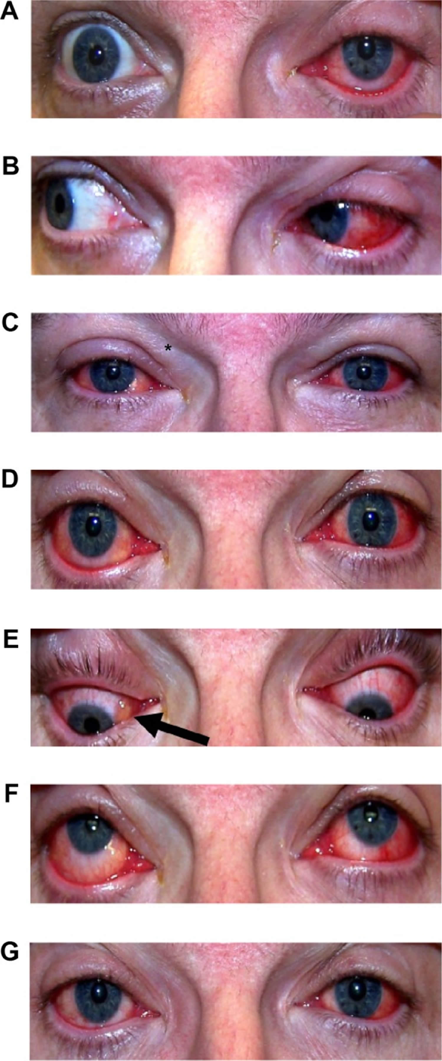
Footnotes: Acute hemorrhagic conjunctivitis caused by coxsackievirus A24 variant (CA24v) infection. (A) First day morning; (B) first day afternoon; (C) second day am; (D) second day pm; (E) third day am; (F) fourth day pm; (G) sixth day pm. The bilateral lid edema (*), vascular dilation, and conjunctival erythema were noted on the second day, and the inferior-nasal chemosis (arrow) was present on the third day post-acute hemorrhagic conjunctivitis. The clinical signs were less and resolved more quickly in the right (second infected) than the left (first affected) eye.
[Source 198 ]COVID-19 conjunctivitis
COVID-19 has been reported to cause conjunctivitis along with fever, cough, respiratory distress, and death 199. Retrospective and prospective studies show that 1% to 6% of patients display COVID-19 related conjunctivitis, with conjunctival swabs being positive in 2.5% of cases 200. Transmission through ocular tissues is incompletely understood. Ophthalmologists are at higher risk for COVID-19 infection due to the close proximity to patients, equipment intense clinics, direct contact with patients’ conjunctival mucosal surfaces, and high-volume clinics. Systemic COVID-19 is contracted through direct or airborne inhalation of respiratory droplets. Additional protective measures such as social distancing, wearing masks, reduced clinic volumes, and sterilization of surfaces should be practiced to reduce transmission risk.
Viral conjunctivitis signs and symptoms
Viral conjunctivitis can lead to one or more of these symptoms:
- eye irritation and redness
- excessive tearing from eyes
- discharge from the eyes
- swelling of the eyelids
- photophobia (unable to tolerate looking into light)
- often associated with an upper respiratory tract infection (cold).
Patients with viral conjunctivitis present with sudden onset foreign body sensation, red eyes, itching, light sensitivity, burning, and watery discharge. Whereas with bacterial conjunctivitis, patients present with all the above symptoms, but with pus (mucopurulent discharge) and sticky eyelids upon waking.
Visual acuity is usually at or near their baseline vision. The cornea can have subepithelial infiltrates that can decrease the vision and cause light sensitivity. The conjunctiva is injected (red) and can also be edematous. In some cases, a membrane or pseudomembrane can be appreciated in the tarsal conjunctiva. These are sheets of fibrin-rich exudates that are devoid of blood or lymphatic vessels. True membranes can lead to the development of subepithelial fibrosis and symblepharon, and also bleed heavily on removal 201. Follicles, small, dome-shaped nodules without a prominent central vessel, can be seen on the palpebral conjunctiva. The majority of viral conjunctivitis patients will have follicles present, but the presence of papillae does not rule out a viral etiology 202.
Fever, malaise, fatigue, presence of enlarged lymph nodes (lymphadenopathy) and other constitutional signs help to differentiate viral conjunctivitis from other causes. Unequal pupil size (anisocoria) and photophobia are associated with serious eye conditions including anterior uveitis, keratitis, and scleritis 203.
When primary eye herpes simplex virus (HSV) infection occurs, the patient typically manifests unilateral, thin, and watery discharge and sometimes vesicles may appear on the face or eyelids and vision may be affected. Corneal involvement may occur. With herpes zoster, there is a linear dermatomal pattern of vesicles. The conjunctiva is often red with a mucopurulent discharge. In a small percentage of patients, there is a history of external eye HSV infection that may lead to the diagnosis.
Conjunctivitis caused by adenoviruses:
- Pharyngoconjunctival fever presents by abrupt onset of high fever, pharyngitis, subconjunctival hemorrhage, bilateral conjunctivitis, and by preauricular lymph node enlargement.
- Epidemic keratoconjunctivitis is more severe and presents with watery discharge, conjunctival membranes or pseudo membranes, hyperemia, chemosis, and ipsilateral preauricular lymphadenopathy. Can involve both epithelial and subepithelial corneal infiltrates.
- Can affect the cornea, look for subepithelial infiltrates.
- Lymphadenopathy is observed in up to 50% of viral conjunctivitis cases and is more prevalent in viral conjunctivitis compared with bacterial conjunctivitis.
Herpes virus conjunctivitis:
- The eyelids often are swollen with small bruise. Watery discharge and lymphadenopathy in front of the ear may be present. Usually unilateral.
- Skin or eyelid margin vesicles, or ulcers on the bulbar conjunctiva
- The cornea often demonstrates a punctate epitheliopathy. In severe cases, there can be a corneal epithelial defect (Dendritic epithelial keratitis). It typically begins in one eye and progresses to the fellow eye over a few days.
- It is important to note that herpes virus conjunctivitis does not form conjunctival membranes or pseudo membranes.
- Herpes zoster virus can involve ocular tissue, especially if the first and second branches of the trigeminal nerve are involved. Eyelids (45.8%) are the most common site of ocular involvement, followed by the conjunctiva. Corneal complication and uveitis may be present in 38.2% and 19.1% of cases, respectively. Severe forms include those presenting with the Hutchinson sign (vesicles at the tip of the nose, which has a high correlation with corneal involvement).
Viral conjunctivitis complications
Viral conjunctivitis complications may include 12:
- Punctate keratitis
- Bacterial superinfection
- Conjunctival scarring
- Corneal ulceration
- Chronic infection
Viral conjunctivitis diagnosis
In most cases, your doctor can diagnose viral conjunctivitis by asking about your recent health history and symptoms and examining your eyes. Palpation of the preauricular lymph nodes may reveal a reactive lymph node that is tender to the touch and will help differentiate viral conjunctivitis versus bacterial conjunctivitis.
Viral conjunctivitis secondary to adenoviruses is highly contagious, and the risk of transmission has been estimated to be 10-50%. Patients commonly report recent contact with an individual with a red eye, or they may have a history of recent symptoms of an upper respiratory tract infection (URTI). Incubation and communicability are estimated to be 5-14 days for viral conjunctivitis secondary to adenoviruses.
Laboratory testing is typically not indicated unless the symptoms are not resolving and infection last longer than 4 weeks 12. Laboratory testing can be indicated in certain situations such as a suspected chlamydial infection in a newborn, an immune-compromised patient, excessive amounts of discharge, or suspected gonorrhea co-infection 12. In the office, doctors can run tests to positively identify adenovirus with a specificity and sensitivity of 89% and 94%, respectively. However, ophthalmologists usually can make the diagnosis clinically with confirmational additional testing 204.
Regarding adenovirus testing, the gold standard has traditionally been cell culture because once the virus is isolated, the diagnosis is definitive and the characterization of the virus can be undertaken 205. Disadvantages include cost and increased time associated with cell culture-based testing. The mainstay of viral conjunctivitis testing in the developed world is the detection of viral DNA by polymerase chain reaction (PCR). PCR for adenovirus has been shown to be 93% sensitive and 97.3% specific from conjunctival swabs, with similar values, found for HSV diagnosis 205. A recent development has been the creation of a rapid detection testing platform for adenovirus, the AdenoPlus assay (Rapid Pathogen Screening Inc., Sarasota, Florida, USA). This is a swift office-based test designed to detect 53 serotypes of adenovirus and provide a result within 10 minutes 206. Studies have demonstrated high specificity values of 92% to 98% in detecting adenoviral conjunctivitis 207, 208, 209. However, the test has lower sensitivity reported compared with PCR analysis 207, 208, 209. Rapid detection testing can be more useful in the future once viral conjunctivitis treatments reach the clinical setting 210.
Viral conjunctivitis treatment
Viral conjunctivitis treatment is aimed at symptomatic relief and not to eradicate the self-limiting viral infection 12. The resolution of viral conjunctivitis can take up to 3 weeks. Treatment includes using artificial tears for lubrication four times a day or up to ten times a day with preservative-free tears. Cool compresses with a wet washcloth to the periocular area may provide symptomatic relief. Preventing the spread of infection to the other eye or other people requires the patient to practice good hand hygiene with frequent washing, avoidance of sharing towels or linens, and avoiding touching their eyes. A person is thought to shed the virus while their eyes are red and tearing.
If a membrane or pseudomembrane is present, it can be peeled at the slit lamp to improve patient comfort and prevent any scar formation from occurring 12. These membranes can either be peeled with a jeweler forceps or a cotton swab soaked with topical anesthetic 12. Topical steroids can help with the resolution of symptoms. However, they can also cause the shedding of the virus to last longer. Patients should be informed that they are highly contagious and should refrain from work or school until their symptoms resolve. While using steroids, they may still shed the virus without the visual symptoms that would indicate that they have an infection. Steroids should be reserved for patients with decreased vision due to their subepithelial infiltrates or severe conjunctival injection causing more the expected discomfort 211, 212.
The use of povidone-iodine, a non-specific disinfectant, is a promising new treatment for adenoviral conjunctivitis 206. This is an inexpensive and widely available antiseptic solution which is used as part of the aseptic preparation for ocular surgery. It is able to kill extracellular organisms but has no effect intracellularly. It does not induce drug resistance because its mechanism of action is not immunologically dependent. A single dose of 2.5% povidone-iodine in infants with adenoviral conjunctivitis resulted in a reduction in symptom severity and reduced recovery time without significant side effects 213.
Topical corticosteroids alone are contraindicated in epithelial herpes simplex keratitis and are associated with prolonged viral shedding and infection 210. When used in combination with an anti-infective, corticosteroids have demonstrated good tolerability and efficacy in treating the inflammatory and infectious components of conjunctivitis 214, 215.
Viral conjunctivitis prognosis
The majority of cases of virus conjunctivitis resolve on their own 12. In rare cases, chronic infection may occur. Most cases resolve within 14-30 days. In some patients, photophobia, diminished vision and glare may be a problem 12.
Newborn conjunctivitis
Newborn babies can develop conjunctivitis or an infection of the eye surface that affects newborn babies within the first 30 days of life also called neonatal conjunctivitis or ophthalmia neonatorum, caused by bacterial or viral infection, irritation or a blocked tear duct 216, 217, 218. If your baby has a discharge from their eye, seek advice from your doctor or pediatrician. Newborn conjunctivitis if left untreated can cause blindness and serious health problems for newborns if it’s not treated. Newborns should be seen by a doctor right away if they have conjunctivitis symptoms. Newborns with conjunctivitis can develop watery eyes within a few days to several weeks after birth. Their eyelids become puffy, red, and tender.
Conjunctivitis in a newborn may be caused by:
- A blocked tear duct. Babies are often born with blocked tear ducts. This can cause a watery or sticky discharge in their eyes. It usually goes away without treatment. Occasionally it can lead to an infection that needs antibiotics. If your baby has a discharge from their eye, seek advice from your doctor, maternal health nurse or paediatrician.
- Irritation produced by the topical antibiotics given at birth
- An infection passed from the mother to baby during childbirth
Even mothers without symptoms at the time of delivery can carry and pass bacteria or viruses to babies during birth.
To prevent newborn conjunctivitis, most states have laws requiring doctors to put drops or ointment in a newborn’s eyes. This typically happens within 2 to 3 hours of birth. In the past, hospitals used silver nitrate 1% solution (Crede’s method) 219. Now, hospitals mostly use antibiotic eye drops or ointment, such as tetracycline hydrochloride 1% eye ointment, erythromycin 0.5% eye ointment, chloramphenicol 1% eye ointment or povidone‐iodine 2.5% solution (water‐based) 220. Erythromycin is the only ophthalmic ointment recommended for use among neonates in the United States 221. The recommended regimen to prevent newborn conjunctivitis caused by Neisseria gonorrhoeae bacteria is erythromycin 0.5% ophthalmic ointment in each eye in a single application at birth 221. Silver nitrate and tetracycline ophthalmic ointments are no longer manufactured in the United States, bacitracin is ineffective, and povidone iodine has not been studied adequately 222, 223. Gentamicin ophthalmic ointment has been associated with severe ocular reactions 224, 225. If erythromycin ointment is unavailable, infants at risk for exposure to gonococcal infection, especially those born to a mother at risk for gonococcal infection or with no prenatal care, can be administered ceftriaxone 25–50 mg/kg body weight IV or IM, not to exceed 250 mg in a single dose 221.
Prevalence of newborn conjunctivitis has decreased significantly in developed countries since the abandonment of silver nitrate as topical prophylaxis. Current estimates of prevalence of newborn conjunctivitis in developed countries are typically < 0.5%. However, a higher incidence of newborn conjunctivitis is still found certain regions of the world, particularly in developing countries. A recent study found an estimated prevalence of 17% among nearly 1000 newborn infants in Pakistan 226. Incidence of neonatal conjunctivitis remains high in Africa 227. Furthermore, studies from sub-Saharan Africa suggest that Chlamydia trachomatis accounted for up to 33% of neonatal conjunctivitis 228.
Aseptic neonatal conjunctivitis most often is a chemical conjunctivitis that is induced by silver nitrate solution, which is used at birth for prophylaxis of infectious conjunctivitis. Chemical conjunctivitis is becoming less common owing to the use of erythromycin ointment or povidone iodide in place of silver nitrate solution for the prophylaxis of infectious conjunctivitis.
Bacterial and viral infections are major causes of septic newborn conjunctivitis, with Chlamydia being the most common infectious agent. Infants may acquire these infective agents as they pass through the birth canal during the birth process 229.
The incidence of infectious ophthalmia neonatorum or newborn conjunctivitis ranges from 1-2%, depending on the socioeconomic character of the area. Chlamydia is the most common infectious agent that causes ophthalmia neonatorum in the United States, where 2%-40% of neonatal conjunctivitis cases are caused by Chlamydia 230.
In contrast, the incidence of gonococcal newborn conjunctivitis has been reduced dramatically and causes less than 1% of cases of neonatal conjunctivitis 231.
The pathology of newborn conjunctivitis is influenced by the anatomy of the conjunctival tissues in the newborn. The inflammation of the conjunctiva may cause blood vessel dilation, potentially dramatic swelling of the conjunctiva (chemosis) and excessive secretion. This infection tends to be more serious in neonates owing to their lack of immunity, lack of lymphoid tissue in the conjunctiva, and absence of tears at birth.
Prompt diagnosis is key in establishing proper treatment and minimizing potential serious complications of neonatal conjunctivitis.
Figure 16. Newborn conjunctivitis
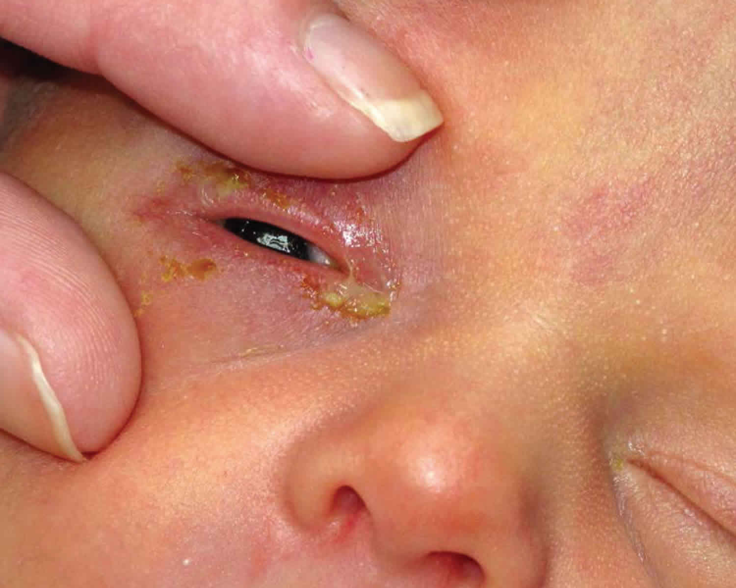
Newborn conjunctivitis causes
The cause of newborn conjunctivitis is often difficult to determine because symptoms are often very similar for each cause.
The most common causes of newborn conjunctivitis include 218:
- Chemical: Classically, the most common cause of neonatal conjunctivitis due to use of post-delivery use of ophthalmic silver nitrate used in the prevention of gonococcal eye infections. However, the incidence of chemical conjunctivitis in the United States has significantly decreased since replacement of silver nitrate with erythromycin ointment
- Bacterial (Chlamydia trachomatis trachomatis most common). Bacterial cause of neonatal conjunctivitis include:
- Chlamydia trachomatis (most common). Newborn chlamydial conjunctivitis is much more prevalent than newborn gonococcal conjunctivitis and has historically been underdiagnosed due to lack of accurate diagnostic techniques 232, 233.
- Neisseria gonorrhoeae. Neisseria gonorrhoeae is one of the most severe and feared causes of neonatal conjunctivitis, requiring prompt diagnosis and treatment.
- Staphylococcus aureus 226, 234, 235, 236, 237.
- Pseudomonas aeruginosa. Pseudomonas, although rare, may lead to potentially blinding complications such as rapid corneal ulceration and perforation.
- Streptococcus spp. (including Streptococcu haemolyticus, Streptococcu pneumonia)
- Other bacteria include Klebsiella, Proteus, Enterobacter, Serratia, and Eikenella corrodens 238
- Viral. Herpes simplex virus (HSV).
Often, no causative pathogen can be found in newborns with conjunctivitis due to methods for obtaining and culturing for bacteria, or due to causes other than bacteria such as chemical conjunctivitis or nasolacrimal duct obstruction 239.
Chlamydial conjunctivitis
- Cause: A mother with untreated chlamydia can pass the Chlamydia trachomatis bacteria to the baby during childbirth (chlamydia-infected cervix during the birth process). Chlamydial infections occur in 4–10% of pregnant women in the United States. Infants whose mothers have untreated chlamydial infections have a 30–40% chance of developing conjunctivitis (incidence of 6.2 per 1000 live births). The risk of chlamydial transmission from an infected mother to newborns is 15% on average (range = 8% to 44%) 240. Furthermore, an increased prevalence of chlamydial infection in some high‐income countries is associated with a commensurate rise in risk of chlamydial conjunctivitis 241. Pooled mean prevalence of Chlamydia trachomatis bacteria was higher at 6.9% in pregnant women in Eastern and Southern Africa, and 6.1% in pregnant women in West and Central Africa 242.
- Symptoms: Redness of the eyes, swelling of the eyelids, and discharge of pus. Some newborns with chlamydial conjunctivitis can have the infection in other parts of their bodies. The bacteria can infect the lungs and nasopharynx (the area at the back of the nose to the mouth).
Newborn chlamydial conjunctivitis or chlamydial ophthalmia neonatorum symptoms are likely to appear 5 to 12 days after birth and is associated with a high risk of corneal and conjunctival scarring, hemorrhagic conjunctivitis, and rarely, loss of vision if left untreated 243, 232, 244. Symptoms can develop earlier if the amniotic sac is ruptured during delivery.
The presentation of chlamydial conjunctivitis may range from mild hyperemia with scant mucoid discharge to eyelid swelling, swelling of the conjunctiva (chemosis) and pseudomembrane formation. Patients typically present with unilateral or bilateral watery discharge, which may become more copious and purulent later.
Although most cases are mild and self-limited, chlamydial conjunctivitis occasionally may be severe. Pseudomembranes, thickened palpebral conjunctiva, significant peripheral pannus, and corneal opacification may be present.
Blindness, although rare and much slower to develop than in gonococcal conjunctivitis, is generally not due to corneal involvement as in gonococcal conjunctivitis. Instead, eyelid scarring and corneal pannus can gradually progress to central corneal opacification by mechanisms reminiscent of trachoma.
A follicular reaction does not occur, because newborns have no requisite lymphoid tissue present in the conjunctiva.
Like gonococcal conjunctivitis, chlamydial conjunctivitis also may be associated with extraocular involvement, including pneumonitis, otitis, and pharyngeal and rectal colonization.
Gonococcal conjunctivitis
- Cause: A mother with untreated gonorrhea can pass the Neisseria gonorrhoeae bacteria to the baby during childbirth. Newborn gonococcal conjunctivitis is mainly contracted from the mother’s infected birth canal during delivery, but can also be contracted in utero by ascending infections. Neonates born to gonorrhoea‐infected mothers have a 30% to 50% risk of developing gonococcal conjunctivitis 245. Furthermore, there is increasing incidence of drug‐resistant strains of Neisseria gonorrhoeae bacteria globally 246, 247, 248. The pooled mean prevalence of Neisseria gonorrhoeae bacteria was estimated at 3.7% in pregnant women in Eastern and Southern Africa, and 2.7% in pregnant women in West and Central Africa 242.
- Symptoms: Red eyes, thick pus in the eyes, and swelling of the eyelids. It can also progress to serious infections of the bloodstream (bacteremia) and lining of the brain and spinal cord (meningitis) in newborns.
Newborn gonococcal conjunctivitis or gonococcal ophthalmia neonatorum tends to be the most serious form of neonatal conjunctivitis. Untreated or inappropriately treated gonococcal conjunctivitis can result in corneal perforation and vision loss in 24 hours 249, 250. In one case series, the mean duration of corneal perforation from untreated gonococcal conjunctivitis was 11 days 251. In areas with low incidence of gonococcal neonatal conjunctivitis or limited access to appropriate health care, appropriate clinical diagnosis and appropriate therapy may be delayed, which can lead to loss of vision 252, 253, 254, 173.
Newborn gonococcal conjunctivitis symptoms are likely to appear 2 to 5 days after birth. Gonococcal conjunctivitis presents with the most rapid onset, usually occurring 24-48 hours following birth. Typically, patients develop a hyperacute conjunctivitis, associated with marked lid edema, swelling of the conjunctiva (chemosis) and purulent discharge (pus). The classic presentation is severe bilateral purulent conjunctivitis.
Corneal ulceration may occur, particularly in the periphery, where massive limbal conjunctival chemosis traps inflammatory mediators and organisms, with rapid progression to perforation the cornea if treatment is delayed and endophthalmitis.
Patients also may have systemic manifestations, including rhinitis, stomatitis, arthritis, meningitis, anorectal infection, and septicemia.
Chemical conjunctivitis
- Cause: When eye drops are given to newborns to help prevent a bacterial infection, the newborn’s eye may become irritated.
- Symptoms: Mildly red eye and some swelling of the eyelids. Symptoms are likely to last for only 24 to 36 hours.
Chemical conjunctivitis secondary to silver nitrate solution application usually occurs in the first day of life, disappearing spontaneously within 2-4 days.
Neonatal conjunctivitis due to other microbial agents is usually milder.
Other neonatal conjunctivitis
- Causes: Viruses that cause genital or oral herpes (herpes simplex virus 1 and 2), and bacteria other than chlamydia and gonorrhea, can cause conjunctivitis and severe eye damage in newborns. This includes bacteria that normally live in a mother’s vagina and are not sexually transmitted. They may pass such viruses to the baby during childbirth. However, herpes conjunctivitis is less common than conjunctivitis caused by gonorrhea and chlamydia.
- Symptoms: Red eye and swollen eyelids with some pus.
Herpes simplex virus (HSV) is a rare cause of neonatal keratoconjunctivitis, found in less than 1% of cases 255 and can be associated with a generalized herpes simplex infection.
Herpes simplex keratoconjunctivitis often presents in infants with generalized herpes simplex infections, characterized by corneal epithelial involvement or vesicles on the periocular skin. Serious systemic complications, such as encephalitis, may also occur in these neonates owing to their poor immunologic response.
Most infants with such an infection acquire the infection during the birth process. Caesarean delivery is strongly considered when active maternal genital disease is recognized at term since the risk of transmitting HSV (herpes simplex virus) to the neonate during vaginal delivery is 25 to 60% 256.
During pregnancy and prior to giving birth, mothers with genital herpes should consult with their doctor about ways to minimize the chances of spread to their newborn baby.
Risk factors for developing newborn conjunctivitis
Risk factors of neonatal conjunctivitis may include:
- Maternal infections harbored in the mother’s birth canal
- HIV-infected mothers 257
- Exposure of the infant to infectious organisms
- Increased birth weight 257
- Inadequacy of ocular prophylaxis immediately after birth
- Premature rupture of membranes (PROM) 258
- Ocular trauma during delivery
- Mechanical ventilation
- Prematurity
- Poor prenatal care
- Poor hygienic delivery conditions
- Post-delivery infection due to direct contact with health care workers or by aerosolization
- Silver nitrate exposure.
Newborn conjunctivitis prevention
Newborn conjunctivitis prevention through good prenatal care and treatment of chlamydial, gonococcal, or herpetic infections during pregnancy remains the best preventative method 218.
Topical prophylaxis
Use of topical silver nitrate to prevent neonatal gonococcal conjunctivitis was first introduced by Credé in 1880 and has been classically been cited as the most common cause of neonatal chemical conjunctivitis 219. However, the incidence of chemical conjunctivitis has declined as the use of silver nitrate as prophylaxis has been abandoned in many modern countries in favor of topical medications with a more favorable side effect profile such as erythromycin. However, countries often vary in their accessibility to these more favorable topical antibiotics. One study evaluating the current methods of treatment in maternity hospitals in Croatia showed that 75% of all maternity hospitals in Croatia routinely administered preventive topical treatment, including topical tobramycin (83.3%), povidone-iodine (8.3%), erythromycin (4.2%), and silver nitrate (4.2%), partially due to the lack of availability of erythromycin and tetracycline in Croatia 259. Data supporting the superiority of one topical antibiotic over another seem to remain clear cut. A recent systematic review including 30 trials found little evidence that any prophylaxis involving tetracycline 1%, erythromycin 0.5%, povidone-iodine 2.5%, or silver nitrate 1% provided better prevention from serious outcomes such as blindness or any adverse visual outcomes 220.
Systemic prophylaxis
Infants with possible infectious exposure in utero or during birth process should receive appropriate prophylaxis following birth in attempt to prevent ocular and systemic complications 218. Gonococcal prophylaxis includes single injection of ceftriaxone 50 mg/kg IM or IV in those neonates born to mothers with untreated or suspected gonococcal infection 218. Other preventative measures include proper hand-washing techniques by peripartum and nursery staff.
Maternal screening
The United States Preventive Services Task (USPSTF) currently recommends routine screening for chlamydial and gonorrheal infection in all sexually active or pregnant women if they are 24 years or younger, or at least 25 years and at increased risk for infection 218. These preventative screening measures have significantly decreased perinatal chlamydial and gonorrheal infections in the United States. One study done at a medical center in the United States reports a decrease from 15.6% positivity rate of Chlamydia trachomatis eye cultures before the implementation of universal prenatal screening (1986-1993), to 1.8% during the screening period (1994-2002) 260.
Newborn conjunctivitis signs and symptoms
Non-specific signs of neonatal conjunctivitis include conjunctival injection, tearing, mucopurulent or non-purulent discharge, chemosis, and eyelid swelling.
Chemical conjunctivitis
Typically results in mild conjunctival injection accompanied by tearing, spontaneously resolving within 2-4 days
Chlamydia trachomatis conjunctivitis
Presentation may range from mild hyperemia with scant mucoid discharge to eyelid swelling, chemosis, and pseudomembrane formation
Neisseria gonorrhoeae conjunctivitis
- Typically, patients present with acute conjunctivitis, associated with chemosis, severe lid edema, and mucopurulent discharge
- Corneal involvement is the most serious complication, involving diffuse epithelial edema and ulceration that may progress to perforation of the cornea and endophthalmitis
- Initially, superficial keratitis gives the corneal surface a lackluster appearance followed by marginal and central infiltrates appear, which then ulcerate
HSV conjunctivitis
- Typically, patients present with unilateral or bilateral lid edema, moderate amount of conjunctival injection, and non-purulent, serosanguinous discharge
- Other signs include vesicles on the skin surrounding the eye and corneal epithelial involvement with microdendrites or geographic ulcers (in contrast to typical dendrites as seen in adults)
Newborn conjunctivitis complications
Eye complications of neonatal conjunctivitis include pseudomembrane formation, corneal edema, thickened palpebral conjunctivia, peripheral pannus formation, corneal opacification, staphyloma, corneal perforation, endophthalmitis, loss of eye, and blindness. Risk of complications can minimized with prompt diagnosis and appropriate antibiotic therapy.
If untreated, peripheral corneal ulceration may occur in Neisseria gonorrhoeae infection and rapidly progress to corneal perforation. Complications of gonococcal conjunctivitis and subsquent systemic involvement include arthritis, meningitis, anorectal infection, septicemia, and death.
When unrecognized and not immediately treated, Pseudomonas infection may lead to endophthalmitis and subsequent death.
Systemic complications of chlamydia conjunctivitis include pneumonitis, otitis, and pharyngeal and rectal colonization. Pneumonia has been reported in 10-20% of infants with chlamydial conjunctivitis 261.
HSV keratoconjunctivitis can cause corneal scarring and ulceration. Additionally, disseminated HSV infection often includes central nervous system involvement 256.
Newborn conjunctivitis diagnosis
History
Time frame of signs/symptoms following birth play an important role in determining the most likely cause and subsequent proper diagnosis and treatment 218:
- Chemical conjunctivitis (typically presents within first 24 hours following birth)
- Neisseria gonorrhea (3-5 days after birth)
- Chlamydia trachomatis (5-14 days)
- HSV or herpes simplex virus (1-2 weeks)
Physical examination
Your baby’s doctor will perform an eye exam on the baby. A thorough examination of the globe and periocular structures of a neonate suspected to have neonatal conjunctivitis is crucial. Corneal involvement should be investigated closely with and without fluorescein and blue cobalt light. A complete systemic examination should be performed by trained physician familiar with the physical exam of a neonate.
If the eye does not appear normal, the following tests may be done:
- Culture of the drainage from the eye to look for bacteria or viruses
- Slit-lamp exam to look for damage to the surface of the eyeball
Laboratory studies for neonatal conjunctivitis should include the following:
- Conjunctival scraping for Gram stain or Giemsa stain
- Conjunctival scraping for polymerase chain reaction (PCR) assay to detect chlamydia and gonorrhea
- Culture on chocolate agar and/or Thayer-Martin for Neisseria gonorrhoeae bacteria
- Culture on blood agar for other bacteria
- Culture of corneal epithelial cells for HSV if cornea is involved; PCR should also be considered in cases of possible HSV conjunctivitis
Culture and histology
Bacterial cultures on blood and chocolate agar are indicated in every case of neonatal conjunctivitis and remain the criterion standard despite newer diagnostic methods.
Since Chlamydia bacteria are obligate intracellular organisms, the culture specimens need to contain epithelial cells and not just exudative material. PCR is generally accepted as the most useful test for chlamydial conjunctivitis owing to its high sensitivity 261.
In cases in which gonorrhea is suspected, the agar should be inoculated immediately since Ngonorrhoeae is very sensitive to moisture and temperature changes.
Laboratory evaluation for the presence of HSV infection is indicated if a corneal epithelial defect is present, if vesicles are present on the eyelids or other parts of the body, and if the diagnosis cannot be made on ocular examination. The presence of HSV in tissue culture remains the criterion standard in the diagnosis of HSV, despite a high false-negative rate. HSV infections may be more rapidly diagnosed with PCR, and PCR testing for HSV is more sensitive than viral culture 261. Laboratory evaluation for suspected HSV becomes more important in neonatal disease because the clinical presentation may be highly atypical in an immunologically immature newborn.
Cytologic findings for various forms of conjunctivitis are as follows:
- Chemical conjunctivitis – Neutrophils, occasional lymphocytes on Gram stain
- Bacterial conjunctivitis – Bacteria, neutrophils on Gram stain
- Gonococcal conjunctivitis – Neutrophils, Gram-negative intracellular diplococci on Gram stain
- Chlamydial conjunctivitis – Neutrophils, lymphocytes, plasma cells on Gram stain; basophilic intracytoplasmic inclusions in epithelial cells on Giemsa stain
- Herpetic conjunctivitis – Lymphocytes, plasma cells, multinucleate giant cells on Gram stain; eosinophilic intranuclear inclusions in epithelial cells on Papanicolaou smear, but with low sensitivity
Newer diagnostic techniques
Nucleic acid amplification tests such as polymerase chain reaction (PCR) and transcription-mediated amplification are more sensitive than culture in detecting chlamydial and gonorrheal organisms 231.
PCR assays may have a higher sensitivity and similar specificity in diagnosing neonatal chlamydial conjunctivitis, compared with conventional methods 262.
PCR for HSV from conjunctival scrapings has high sensitivity and specificity, but it is expensive, not always readily available, and is usually reserved for the diagnosis of encephalitis. Direct florescent antibody (DFA) studies are useful for rapid detection, have high sensitivity and specificity, and can be used to type the virus 256.
Newborn conjunctivitis treatment
Doctors may treat conjunctivitis in infants caused by a bacterial infection with antibiotics. It will depend on the severity of the infection and the bacteria that caused it. Rinsing the newborn’s infected eye with a saline solution will remove any debris that may develop in response to the infection.
How antibiotics are applied:
- As an eye drop or ointment in the eye (topical)
- By mouth (orally)
- Through a vein (intravenous or IV)
- As a shot (intramuscular)
- Doctors may combine eye antibiotic eye drop or ointment with one of the other methods
If your baby has a blocked tear duct that causes conjunctivitis, a gentle, warm massage between the eye and nasal area may help. If the blocked tear duct does not clear by 1 year of age, the newborn may require surgery.
The treatment prior to laboratory results should include topical erythromycin ointment and an IV or IM third-generation cephalosporin. Prompt treatment of gonococcal conjunctivitis is important, since this organism can penetrate an intact corneal epithelium and rapidly cause corneal ulceration. Because of the rapid progression of gonococcal conjunctivitis, patients with acute neonatal conjunctivitis should be treated for gonococcal conjunctivitis until culture results are available; the treatment is altered according to the laboratory results.
In cases of chlamydial conjunctivitis, systemic treatment is necessary because of the significant risk for life-threatening pneumonia.
Infants with a potentially sexually transmitted disease, such as gonorrhea or chlamydia, should undergo evaluation for other sexually transmitted diseases, such as syphilis and HIV 263, as should the mother and her sexual partner(s).
Newborns with conjunctivitis are at risk for secondary infections, such as pneumonia, meningitis, and septicemia, which can lead to sepsis and death and thus should be admitted for full workup and treatment.
Bacterial conjunctivitis rarely fails to respond to treatment.
A consultation can be made with a pediatrician or pediatric infectious specialist in neonatal conjunctivitis, and the patient should be seen daily until response to treatment is confirmed.
Discharged patients should continue the treatment, according to clinical presentations and available culture results. Treatment may be modified later per culture results.
Avoid eye patching
Treatment of neonatal chemical conjunctivitis is not necessary. Lubrication with artificial tear preparations may ease mild discomfort.
Chemical conjunctivitis
No treatment required; supportive care only (may use artificial tears four times daily). Lubrication with artificial tear preparations may ease mild discomfort.
Typically disappears spontaneously within 2-4 days.
Neonatal Chlamydial conjunctivitis
Treatment should be initiated upon a positive diagnostic test. Neonatal Chlamydial conjunctivitis is treated with erythromycin drops four times daily plus oral erythromycin (50 mg/kg/day divided four times daily) for 14 days 218. Alternatively, oral azithromycin may be used for both chlamydial conjunctivitis and pneumonia, 20 mg/kg once daily for three days 218.
While outpatient treatment is an option, hospitalization may be required. Evaluation of systemic involvement needed.
Topical therapy for chlamydial conjunctivitis has proven to be relatively ineffective compared to oral therapy, particularly in treating nasopharyngeal infection 264. Topical erythromycin ointment may be beneficial as an adjunctive therapy. Doctors usually use oral antibiotics to treat inclusion conjunctivitis.
Since the efficacy of systemic erythromycin therapy is approximately 80%, a second course sometimes is required.
Systemic treatment is important in cases of chlamydial conjunctivitis since topical therapy is ineffective in eradicating the bacteria in the nasopharynx of the infant, which could cause a life-threatening pneumonia if left untreated.
The use of azithromycin and erythromycin are associated with increased risk of infantile hypertrophic pyloric stenosis (IHPS) in infants within 2 weeks of life. Infants should be closely monitored for potential signs of infantile hypertrophic pyloric stenosis when started on azithromycin or erythromycin 218.
Neonatal Gonococcal conjunctivitis
Newborns who have gonococcal conjunctivitis should be managed in consultation with an infectious disease specialist. Topical irrigation with normal saline should be used to remove mucopurulent discharge, as Gonococcal infection may produce more mucopurulent discharge than other pathogens. Ceftriaxone in a single dose (Ceftriaxone 25–50 mg/kg body weight IV or IM in a single dose, not to exceed 250 mg) 221. One dose of ceftriaxone is adequate therapy for gonococcal conjunctivitis 221. Ceftriaxone should be administered cautiously to neonates with hyperbilirubinemia, especially those born prematurely 221. Cefotaxime 100 mg/kg body weight IV or IM as a single dose can be administered for those neonates unable to receive ceftriaxone because of simultaneous administration of IV calcium 221. Topical antibiotic therapy alone is inadequate and unnecessary if systemic treatment is administered 221.
If there is systemic disease, treatment is required for 7 to 14 days, depending on the nature of the invasive infection. Bacitracin or erythromycin ointment every 2 to 4 hours in addition to systemic treatment of ceftriaxone 218. Hospitalization and evaluation for disseminated Neisseria gonorrhoeae infection should be initiated. Topical saline drops to remove discharge. Topical atropine should be considered for pain relief if there is corneal involvement.
- Note: Chlamydial testing should be performed simultaneously from the inverted eyelid specimen. Mothers of newborns with gonococcal conjunctivitis caused by Neisseria gonorrhoeae should be evaluated, tested, and presumptively treated for gonorrhea, along with their sex partners. All neonates with gonococcal conjunctivitis should also be treated for chalmydia. Mother and sexual partner should be treated as well.
Other bacterial neonatal conjunctivitis
- Gram positive: Bacitracin ointment four times daily for 2 weeks
- Gram negative: Gentamicin, tobramycin or ciprofloxacin four times daily for 2 weeks
Neonatal herpetic conjunctivitis
Neonates with a suspected herpes simplex infection should be treated with systemic acyclovir to reduce the risk of a systemic infection. An effective dose is Acyclovir 45mg/kg/day IV plus vidarabine 3% ointment 5x/day 218. The recommended minimal duration is 14 days, but a course as long as 21 days may be required depending on presence of central nervous system involvement.
Infants with neonatal HSV keratitis should also receive a topical ophthalmic drug, most commonly 1% trifluridine drops or 3% vidarabine ointment 5x/day for 14-21 days depending on presence or absence of central nervous system involvement. Topical ganciclovir 0.15% gel is now also available, although none of these topical agents is specifically approved for neonatal use 265.
Topical antibiotics can also be considered to prevent secondary bacterial infections in cases with significant epithelial defects.
Medical follow up
- Patients with neonatal conjunctivitis should be followed daily for signs of improvement or worsening, especially acutely due to concerns of rapidly progressing infectious complications.
- Patient should be followed closely by pediatrician for evaluation and treatment of potential systemic infection.
Newborn conjunctivitis prognosis
The prognosis of neonatal conjunctivitis is generally considered to be good as long as early diagnosis is made and prompt medical therapy is initiated 218. Most cases of infectious conjunctivitis respond to appropriate treatment. However, morbidity and mortality increases in cases of systemic involvement requiring hospitalization and intensive monitoring.
Conjunctivitis causes
Causes of conjunctivitis include:
- Bacterial infection. Bacterial conjunctivitis is the second most common cause and is responsible for the majority (50%-75%) of cases in children; it is observed more frequently from December through April.
- Viral infection. Viral conjunctivitis is the most common cause of infectious conjunctivitis both overall and in the adult population and is more prevalent in summer.
- Allergies. Allergic conjunctivitis is the most frequent cause, affecting 15% to 40% of the population and is observed more frequently in spring and summer.
- Substances that cause irritation
- Contact lens products, eye drops, or eye ointments
- A chemical splash in the eye
- A foreign object in the eye
- In newborns, a blocked tear duct.
Viral or bacterial conjunctivitis is highly contagious and spread from person. You could develop viral or bacterial conjunctivitis if you come into contact with:
- Discharge from the eyes, nose or throat of an infected person through touch, coughing or sneezing.
- Contaminated fingers or contact with contaminated surfaces or objects.
- Contaminated water while swimming.
Viral or bacterial conjunctivitis can also be transmitted from an infected mother to her baby during vaginal delivery.
Allergic conjunctivitis occurs more commonly among people who already have seasonal allergies. They develop it when they come into contact with a substance that triggers an allergic reaction in their eyes or because of a foreign body.
Viral conjunctivitis
Most cases of viral conjunctivitis are caused by adenovirus but also can be caused by other viruses, including herpes simplex virus (HSV) and varicella-zoster virus (VZV).
Bacterial conjunctivitis
Bacteria rank second among the most common causes of infectious conjunctivitis. The most common bacteria are:
- Staphylococcus, the same family of bacteria that cause staph infections.
- Streptococcus, the bacteria family that causes strep throat and pneumococcal disease.
- Haemophilus influenzae, a bacteria family best known for causing meningitis in young children.
- Sexually transmitted infections (STIs), like chlamydia, gonorrhea and syphilis. When these conditions pass from birthing parent to child during birth, it can lead to neonatal conjunctivitis, a condition that can cause permanent eye damage and blindness.
Bacterial conjunctivitis can be contracted directly from infected individuals, by an abnormal proliferation of the native conjunctival flora, or from the spread of infection from the organisms colonizing the patient’s nasal and sinus mucosa. Bacteria infiltrate the conjunctival epithelial layer and sometimes the substantia propia as well.
Direct infection and inflammation of the conjunctival surface, bystander effects on adjacent tissues such as the cornea, and the host’s acute inflammatory response and long-term reparative response all contribute to the pathology.
In children, the disease is often caused by Haemophilus influenzae, Streptococcus pneumoniae and Moraxella catarrhalis.
The most common pathogens for bacterial conjunctivitis in adults are Staphylococcal species, followed by Streptococcus pneumoniae and Haemophilus influenzae.
The course of bacterial conjunctivitis usually lasts 7-10 days.
- Hyperacute bacterial conjunctivitis is often caused by Neisseria gonorrhoeae. When the infection does not respond to standard antibiotic therapy in sexually active patients, Chlamydia trachomatis should be suspected.
- Chronic bacterial conjunctivitis is commonly caused Staphylococcus aureus, Moraxella lacunata, and enteric bacteria.
Allergic conjunctivitis
Allergic conjunctivitis affects both eyes and is a response to an allergy-causing substance such as pollen. In response to allergens, your body produces an antibody called immunoglobulin E (IgE). Immunoglobulin E (IgE) triggers special cells in the mucous lining of your eyes and airways to release inflammatory substances, including histamines. Your body’s release of histamine can produce a number of allergy symptoms including runny nose, sneezing, itchy skin, raised rash (hives) and conjunctivitis.
If you have allergic conjunctivitis, you may experience intense itching, tearing and inflammation of your eyes as well as sneezing and watery runny nose. Most allergic conjunctivitis can be controlled with allergy eye drops. Allergic conjunctivitis is not contagious.
Conjunctivitis resulting from irritation
Irritation from a chemical splash or foreign object in your eye also is associated with conjunctivitis. Sometimes flushing and cleaning the eye to wash out the chemical or object causes redness and irritation. Symptoms, which may include watery eyes and a mucous discharge, usually clear up on their own within about a day.
If flushing doesn’t resolve the symptoms, or if the chemical is a caustic one such as lye, see your health care provider or eye specialist as soon as possible. A chemical splash into the eye can cause permanent eye damage. Ongoing symptoms could indicate that you still have the foreign body in your eye. Or you also could have a scratch on the cornea or the membrane covering the eyeball, called the conjunctiva.
Risk factors for getting conjunctivitis
Risk factors for conjunctivitis include:
- Exposure to someone infected with the viral or bacterial form of conjunctivitis.
- Exposure to something you’re allergic to, for allergic conjunctivitis.
- Using contact lenses, especially extended-wear lenses.
Conjunctivitis prevention
To prevent the spread of infective conjunctivitis, practice good hygiene. For example:
- Don’t touch your eyes with your hands, unless you have just washed your hands.
- Wash your hands often with soap and warm water or alcohol-based hand sanitiser especially after touching their eyes or putting in eye drops. Hand washing is the best way to avoid getting conjunctivitis and avoid spreading it to others.
- Use a clean towel and washcloth daily.
- Don’t share towels or washcloths.
- Change your pillowcases every day.
- Throw away old eye cosmetics, such as mascara.
- Throw away tissues or cotton balls that have touched your eyes.
- Don’t share eye cosmetics or personal eye care items or eye drops with others.
Keep in mind that conjunctivitis is no more contagious than the common cold. It’s okay to return to work, school or child care if you’re able to practice good hygiene and avoid close contact. However, if work, school or child care involves close contact with others it may be best to stay home until you or your child’s symptoms clear up.
Preventing conjunctivitis in newborns
Newborns’ eyes are susceptible to bacteria present in the mother’s birth canal. These bacteria often cause no symptoms in the mother. In some cases, these bacteria can cause infants to develop a serious form of conjunctivitis known as ophthalmia neonatorum, which needs immediate treatment to preserve sight. That’s why shortly after birth, an antibiotic ointment is applied to every newborn’s eyes. The ointment helps prevent eye infection.
Recommendations for contact lenses
If you wear contact lenses, wash your hands before putting in and taking out your lenses. Regularly cleaning and throwing out your lenses can also help keep infections away. Always store your
contact lenses in solutions recommended by contact lens guidelines. Never swim in a lake or hot tub with your contact lenses on. If you have been diagnosed with conjunctivitis, do NOT wear your contact lenses until you have healed, and then, start with a fresh pair of lenses.
Conjunctivitis signs and symptoms
The most common symptoms of conjunctivitis include:
- Redness in one or both eyes.
- Itchiness in one or both eyes.
- A gritty feeling in one or both eyes.
- A discharge in one or both eyes that forms a crust during the night that may prevent your eye or eyes from opening in the morning.
- Tearing.
- Sensitivity to light called photophobia.
Unequal pupil size (anisocoria) and photophobia are associated with serious eye conditions including anterior uveitis, keratitis, and scleritis 203.
Systemic diseases associated with conjunctivitis include skin and mucous membrane diseases (acne rosacea, ichthyosis, xeroderma pigmentosum), collagen vascular diseases (systemic lupus erythematosus, rheumatoid arthritis, Sjogren’s disease, granulomatosis with polyangiitis), and autoimmune disease (graft versus host disease, Steven-Johnson syndrome, and ocular cicatricial pemphigoid).
Conjunctivitis complications
In both children and adults, conjunctivitis can cause inflammation in the cornea called keratitis or corneal ulcer that can cause permanent eye damage and even blindness if they go too long without treatment. Prompt evaluation and treatment by your doctor or eye doctor can reduce your risk of complications. Most people who have conjunctivitis recover within 2-5 days. Rarely, complications can occur.
Conjunctivitis complications can include:
- Trachoma
- Uveitis
- Corneal inflammation (keratitis) and cornea-conjunctiva inflammation (keratoconjunctivitis).
- More severe corneal diseases, especially corneal ulcers and recurrent corneal erosions.
Because of the risk of permanent damage, you shouldn’t ignore pink eye symptoms if they’re still getting worse after more than a few days.
See your doctor if you have:
- Eye pain.
- A feeling that something is stuck in your eye.
- Blurred vision.
- Light sensitivity.
Conjunctivitis diagnosis
In most cases, your doctor can diagnose conjunctivitis by asking about your recent health history and symptoms and examining your eyes. Rarely, your doctor may take a sample of the liquid that drains from your eye for laboratory analysis, called a culture.
A culture may be needed if your symptoms are severe or if your doctor suspects a high-risk cause, such as:
- A foreign body in your eye.
- A serious bacterial infection.
- A sexually transmitted infection.
Conjunctivitis treatment
Conjunctivitis treatment depends on what type of conjunctivitis you have. Conjunctivitis treatment is usually focused on symptom relief. Your doctor may recommend using artificial tears, cleaning your eyelids with a wet cloth, and applying cold or warm compresses several times daily.
If you have bacterial conjunctivitis, you may need antibiotic ointment or drops. Put the drops in both eyes. Keep using the drops until 2 days after the discharge has gone.
If you have viral conjunctivitis, you doesn’t need antibiotics. There is no specific treatment for viral conjunctivitis, it generally improves on its own without treatment.
If you have allergic conjunctivitis, the following things might help:
- avoiding anything that triggers the allergic reaction
- antihistamine eye drops or syrup — ask your doctor what antihistamines to take
- lubricating eye drops
- allergen immunotherapy for severe cases.
To reduce the symptoms of bacterial or viral conjunctivitis you can:
- Take ibuprofen or another over-the-counter pain killer.
- Use over-the-counter lubricating eye drops (artificial tears).
- Put a warm, damp washcloth over your eyes for a few minutes. To make this warm compress:
- Soak a clean washcloth in warm water then wring it out so it’s not dripping.
- Lay the damp cloth over your eyes and leave it in place until it cools.
- Repeat this several times a day, or as often as is comfortable.
- Use a clean washcloth each time so you don’t spread the infection.
- Use a different washcloth for each eye if you have infectious conjunctivitis in both eyes.
If your eyelids are sticking together, a warm washcloth can loosen the dried mucus so you can open your eyes.
Use clean cotton wool (one piece for each eye). Boil water and then let it cool down before you:
- gently rub your eye lashes to clean off crusts
- hold a cold flannel on your eyes for a few minutes to cool them down
Stop wearing contact lenses. If you wear contact lenses, you’ll be advised to stop wearing them until treatment is complete. Your doctor will likely recommend that you throw out contact lens you’ve worn if your lenses are disposable. Use a new pair when you go back to wearing your contacts. Your old contacts are likely infected and could get you sick again if you wear them again.
Disinfect hard lenses overnight before you reuse them. Ask your doctor if you should discard and replace your contact lens accessories, such as the lens case used before or during the illness.
Stop wearing eye makeup. Throw out your old eye makeup and get new makeup once your eyes are healthy.
Viral conjunctivitis is like a common cold in the eye. There is no treatment for the virus and usually you just have to let it heal on its own. Viral conjunctivitis should go away within a week or two without treatment. Since viral conjunctivitis is caused by virus, antibiotics won’t help, and antibiotics may even cause harm by reducing their effectiveness in the future or causing a medication reaction.
Bacterial conjunctivitis usually produces more mucus or pus than viral or allergic conjunctivitis. Bacterial conjunctivitis can be treated with antibiotics prescribed by a doctor.
Gonococcal conjunctivitis treatment
In the only published study of the treatment of gonococcal conjunctivitis in adults, all 12 study participants responded to a single 1 gram intramuscular injection of ceftriaxone 266. Based on this study, the recommended treatment for gonococcal conjunctivitis is a single ceftriaxone 1 gram intramuscular dose 267, 268. In addition, a one-time lavage of the infected eye with saline should be considered 267, 268.
Chlamydial conjunctivitis treatment
The recommended treatment regimen for the baby with chlamydial conjunctivitis is a 14-day course of oral erythromycin base or erythromycin ethylsuccinate 50 mg/kg/day orally divided into 4 doses daily for 14 days 269. Treatment with oral erythromycin or oral azithromycin in infants during the first 6 weeks of life has been associated with an increased risk of infantile hypertrophic pyloric stenosis (IHPS) 270, 271, 272, 273. Therefore, infants treated with either of these antibiotics should be followed for infantile hypertrophic pyloric stenosis signs and symptoms 272, 269. The use of topical erythromycin alone is not effective for baby with chlamydial conjunctivitis and it is not recommended for use in combination with oral antibiotics 269.
Data on oral azithromycin for the treatment of neonatal chlamydial infection are limited, but a small study suggested a short 3-day course of azithromycin may be effective 274. The use of a recommended or alternative therapy has an efficacy of only approximately 80% and some infants require treatment with a second course of antibiotics.
Chemical conjunctivitis treatment
Since this type of pink eye is caused by chemical irritation (like from eye drops), treatment is usually not required. Chemical conjunctivitis will usually get better in 24 to 36 hours.
Doctors usually give antibiotic drops or ointments to treat conjunctivitis caused by other bacteria. For both bacterial and viral conjunctivitis, a warm compress to the eye may relieve swelling and irritation. Be sure to wash hands before and after touching the infected eyes.
Allergic conjunctivitis treatment
Allergen avoidance is the first line of treatment for allergic conjunctivitis. In the case of pollen allergies, symptoms are often made worse by outdoor activities. Wearing glasses or goggles outdoors can limit contact with allergens. Frequent washing of pillowcases and mattress covers as well as vacuuming the carpeting in your room help remove allergens from your surroundings. Regular washing your hair and face can help remove these allergens from the surface of your eyes, hair, and skin. Using artificial tear drops to rinse the eye and remove allergens from the eye can help with symptoms and help calm down the eye inflammation. These drops can provide even more relief when used cold (refrigerated) instead of at room temperature. It is also important to avoid rubbing the eyes, as this can make allergic conjunctivitis worse.
If you have allergic conjunctivitis, both prescription and over-the-counter allergy eye drops can treat allergic conjunctivitis 14. These may include medicines that help control allergic reactions, such as antihistamines and mast cell stabilizers. Or your doctor may recommend medicines to help control inflammation, such as decongestants, steroids and anti-inflammatory drops. Nonprescription versions of these medicines also may be effective. Most of the easily available allergy eye drops work best when used daily for at least a few weeks, and it may take up to a week to get full symptom relief. Some eye drops can be used only on an as needed basis. Allergy eye drops may work better for some than pill or liquid medications as eye drops do not cause any drowsiness or changes in appetite. However, pill or liquid medications may be more helpful if allergies cause a lot of eyelid swelling or affect more than just the eyes. Please speak with your ophthalmologist if you have questions about allergy medications.
Given the different types of over-the-counter eye drops, sometimes what works well for one person may not work as well for another person. You may need to try different types of eye drops before you find one that works for you. If there are still allergic conjunctivitis symptoms even after trying different kinds of allergy eye drops, adding a short-term liquid or pill allergy medication by mouth may help relieve symptoms. Pills and liquid medications by mouth may also a good treatment for people who don’t do well eye drops, or who have other allergy symptoms like a runny nose.
In some cases, steroid eye drops may be needed along with allergy eye drops if the allergic reaction is very severe. However, steroid use needs to be monitored closely by your ophthalmologist and used only as directed to prevent serious eye problems. Use of steroid drops for a long time or at a large amount can cause serious vision problems, including glaucoma, cataracts and eye infections (keratitis). Only ophthalmologists (eye specialists) who can monitor for side effects should prescribe steroids for allergic conjunctivitis. Talk with your ophthalmologist if you have questions about steroid drops.
Home remedies for conjunctivitis
To help you cope with the symptoms of conjunctivitis until it goes away, try to:
- Apply a compress to your eyes. To make a compress, soak a clean, lint-free cloth in water and wring it out before applying it gently to your closed eyelids. Generally, a cool water compress will feel the most soothing, but you also can use a warm compress if that feels better to you. If conjunctivitis affects only one eye, don’t touch both eyes with the same cloth. This reduces the risk of spreading conjunctivitis from one eye to the other.
- Try eye drops. Nonprescription eye drops called artificial tears may relieve symptoms. Some eyedrops contain antihistamines or other medicines that can be helpful for people with allergic conjunctivitis.
- Stop wearing contact lenses. If you wear contact lenses, you may need to stop wearing them until your eyes feel better. How long you’ll need to go without contact lenses depends on what’s causing your conjunctivitis. Ask your doctor if you should discard and replace your contact lens accessories, such as the lens case used before or during the illness. If your lenses aren’t disposable, clean them thoroughly before reusing them.
Stop infectious conjunctivitis from spreading
DO
- wash hands regularly with warm soapy water
- wash pillows and face cloths in hot water and detergent
DON’T
- wear contact lenses until your eyes are better
- share towels and pillows
- rub your eyes
Staying away from work or school: Conjunctivitis is no more contagious than the common cold. It’s okay to return to work, school or child care if you’re able to practice good hygiene and avoid close contact. However, if work, school or child care involves close contact with others it may be best to stay home until you or your child’s symptoms clear up.
- When a child has viral conjunctivitis, he/she is at risk for getting bacterial conjunctivitis, too. Many kids end up having both types of eye infection at the same time. To help prevent that, during the time the eyes are red, gently wipe the eyelids with a cotton ball soaked in warm water about every 1 to 2 hours while the child is awake.
- Bacterial conjunctivitis can be spread to others if they come in contact with pus from the eyes. For that reason, people who have bacterial conjunctivitis should not share towels, washcloths or other personal hygiene items that may be used on the eyes or face. Washing hands often can also decrease the risk. Unless the pus is excessive, the risk of spreading bacterial conjunctivitis is low after the first 24 hours of using antibiotic eye drops. Children can return to child care or school at that time.
Conjunctivitis prognosis
Most forms of conjunctivitis caused by infections clear without causing any eye problems. However, some serious infections and certain types of viral eye infections, such as herpes, can cause vision problems if not treated appropriately.
- Høvding G. Acute bacterial conjunctivitis. Acta Ophthalmol. 2008;86(1):5–17. doi: 10.1111/j.1600-0420.2007.01006.x[↩][↩][↩]
- Hørven I. Acute conjunctivitis: a comparison of fusidic acid viscous eye drops and chloramphenicol. Acta Ophthalmol (Copenh) 1993;71(2):165–168. doi: 10.1111/j.1755-3768.1993.tb04983.x[↩]
- Stenson S, Newman R, Fedukowicz H. Laboratory studies in acute conjunctivitis. Arch Ophthalmol. 1982;100(8):1275–1277. doi: 10.1001/archopht.1982.01030040253009[↩]
- Rönnerstam R, Persson K, Hansson H, Renmarker K. Prevalence of chlamydial eye infection in patients attending an eye clinic, a VD clinic, and in healthy persons. Br J Ophthalmol. 1985;69(5):385–388. doi: 10.1136/bjo.69.5.385[↩]
- Harding SP, Mallinson H, Smith JL, Clearkin LG. Adult follicular conjunctivitis and neonatal ophthalmia in a Liverpool eye hospital, 1980-1984. Eye (Lond) 1987;1(pt 4):512–521. doi: 10.1038/eye.1987.77[↩]
- Uchio E, Takeuchi S, Itoh N, et al. Clinical and epidemiological features of acute follicular conjunctivitis with special reference to that caused by herpes simplex virus type 1. Br J Ophthalmol. 2000;84(9):968–972. doi: 10.1136/bjo.84.9.968[↩]
- Woodland RM, Darougar S, Thaker U, et al. Causes of conjunctivitis and keratoconjunctivitis in Karachi, Pakistan. Trans R Soc Trop Med Hyg. 1992;86(3):317–320. doi: 10.1016/0035-9203(92)90328-a[↩]
- Fitch CP, Rapoza PA, Owens S, et al. Epidemiology and diagnosis of acute conjunctivitis at an inner-city hospital. Ophthalmology. 1989;96(8):1215–1220. doi: 10.1016/s0161-6420(89)32749-7[↩]
- Bielory BP, O’Brien TP, Bielory L. Management of seasonal allergic conjunctivitis: guide to therapy. Acta Ophthalmol. 2012;90(5):399–407. doi: 10.1111/j.1755-3768.2011.02272.x[↩]
- American Academy of Ophthalmology. Cornea/External Disease Panel . Preferred Practice Pattern Guidelines: Conjunctivitis-Limited Revision. American Academy of Ophthalmology; San Francisco, CA: 2011.[↩][↩]
- Mannis MJ, Plotnik RD. Bacterial conjunctivitis. In: Tasman W, Jaeger EA, editors. Duanes Ophthalmology on CD-ROM. Lippincott Williams & Wilkins; 2006.[↩]
- Solano D, Fu L, Czyz CN. Viral Conjunctivitis. [Updated 2023 Aug 28]. In: StatPearls [Internet]. Treasure Island (FL): StatPearls Publishing; 2024 Jan-. Available from: https://www.ncbi.nlm.nih.gov/books/NBK470271[↩][↩][↩][↩][↩][↩][↩][↩][↩][↩][↩][↩][↩][↩][↩][↩]
- Alfonso SA, Fawley JD, Alexa Lu X. Conjunctivitis. Prim Care. 2015 Sep;42(3):325-45. doi: 10.1016/j.pop.2015.05.001[↩]
- Azari AA, Barney NP. Conjunctivitis: a systematic review of diagnosis and treatment. JAMA. 2013 Oct 23;310(16):1721-9. doi: 10.1001/jama.2013.280318. Erratum in: JAMA. 2014 Jan 1;311(1):95. Dosage error in article text.[↩][↩][↩][↩][↩][↩][↩][↩][↩][↩][↩][↩][↩][↩][↩][↩][↩][↩][↩][↩][↩]
- Leibowitz HM. The red eye. N Engl J Med. 2000;343(5):345–351. doi: 10.1056/NEJM200008033430507[↩]
- Cronau H, Kankanala RR, Mauger T. Diagnosis and management of red eye in primary care. Am Fam Physician. 2010 Jan 15;81(2):137-44. https://www.aafp.org/pubs/afp/issues/2010/0115/p137.html[↩]
- Butrus S, Portela R. Ocular allergy: diagnosis and treatment. Ophthalmol Clin North Am. 2005 Dec;18(4):485-92, v. doi: 10.1016/j.ohc.2005.07.007[↩]
- Gomes PJ. Trends in prevalence and treatment of ocular allergy. Curr Opin Allergy Clin Immunol. 2014 Oct;14(5):451-6. doi: 10.1097/ACI.0000000000000100[↩]
- Leonardi A, Castegnaro A, Valerio AL, Lazzarini D. Epidemiology of allergic conjunctivitis: clinical appearance and treatment patterns in a population-based study. Curr Opin Allergy Clin Immunol. 2015 Oct;15(5):482-8. doi: 10.1097/ACI.0000000000000204[↩]
- What is allergic conjunctivitis? https://aapos.org/glossary/allergic-conjunctivitis[↩]
- Carr W, Schaeffer J, Donnenfeld E. Treating Allergic Conjunctivitis: A Once-daily Medication that Provides 24-hour Symptom Relief. Allergy & Rhinology. 2016;7(2). doi:10.2500/ar.2016.7.0158[↩][↩][↩]
- Singh K, Axelrod S, Bielory L. The epidemiology of ocular and nasal allergy in the United States, 1988-1994. J Allergy Clin Immunol. 2010 Oct;126(4):778-783.e6. doi: 10.1016/j.jaci.2010.06.050[↩]
- Chigbu D.I. The pathophysiology of ocular allergy: A review. Cont Lens Anterior Eye 32: 3–15; quiz 43–44, 2009.[↩][↩]
- Bielory L., Meltzer E.O., Nichols K.K.et al. An algorithm for the management of allergic conjunctivitis. Allergy Asthma Proc 34: 408–420, 2013.[↩][↩][↩]
- Baab S, Le PH, Gurnani B, et al. Allergic Conjunctivitis. [Updated 2024 Jan 26]. In: StatPearls [Internet]. Treasure Island (FL): StatPearls Publishing; 2024 Jan-. Available from: https://www.ncbi.nlm.nih.gov/books/NBK448118[↩][↩][↩][↩]
- Wise S.K., Lin S.Y., Toskala E., Orlandi R.R., Akdis C.A., Alt J.A., Azar A., Baroody F.M., Bachert C., Canonica G.W., et al. International consensus statement on allergy and rhinology: Allergic rhinitis—2023. Int. Forum. Allergy Rhinol. 2023;13:293–859. doi: 10.1002/alr.23090[↩]
- Villegas BV, Benitez-Del-Castillo JM. Current Knowledge in Allergic Conjunctivitis. Turk J Ophthalmol. 2021 Feb 25;51(1):45-54. doi: 10.4274/tjo.galenos.2020.11456[↩][↩]
- Dupuis P, Prokopich CL, Hynes A, Kim H. A contemporary look at allergic conjunctivitis. Allergy Asthma Clin Immunol. 2020;16:5. doi: 10.1186/s13223-020-0403-9[↩][↩][↩][↩]
- Fauquert JL, Jedrzejczak-Czechowicz M, Rondon C, Calder V, Silva D, Kvenshagen BK, Callebaut I, Allegri P, Santos N, Doan S, Perez Formigo D, Chiambaretta F, Delgado L, Leonardi A Interest Group on Ocular Allergy (IGOA) from the European Academy of Allergy and Clinical Immunology. Conjunctival allergen provocation test : guidelines for daily practice. Allergy. 2016;72:43–54. doi: 10.1111/all.12986[↩][↩][↩][↩][↩][↩][↩][↩]
- Pepper AN, Ledford DK. Nasal and ocular challenges. J Allergy Clin Immunol. 2018;141:1570–1577. doi: 10.1016/j.jaci.2017.11.066[↩][↩][↩]
- Leonardi A, Bogacka E, Fauquert JL, Kowalski ML, Groblewska A, Jedrzejczak-Czechowicz M, Doan S, Marmouz F, Demoly P, Delgado L. Ocular allergy: recognizing and diagnosing hypersensitivity disorders of the ocular surface. Allergy. 2012;67:1327–1337. doi: 10.1111/all.12009[↩][↩][↩]
- Bilkhu PS, Wolffsohn JS, Naroo SA, Robertson L, Kennedy R. Effectiveness of nonpharmacologic treatments for acute seasonal allergic conjunctivitis. Ophthalmology. 2014;121:72–78. doi: 10.1016/j.ophtha.2013.08.007[↩]
- Leonardi A. Management of vernal keratoconjunctivitis. Ophthalmol Ther. (2013) 2:73–88. 10.1007/s40123-013-0019-y[↩][↩][↩][↩][↩][↩][↩][↩][↩][↩][↩][↩][↩][↩][↩][↩][↩][↩][↩][↩][↩][↩][↩][↩][↩]
- Yanni JM, Sharif NA, Gamache DA, Miller ST, Weimer LK, Spellman JM. A current appreciation of sites for pharmacological intervention in allergic conjunctivitis: effects of new topical ocular drugs. Acta Ophthalmol Scand Suppl. (1999) 228:33–7. 10.1111/j.1600-0420.1999.tb01171.x[↩]
- Gane J, Buckley R. Leukotriene receptor antagonists in allergic eye disease: a systematic review and meta-analysis. J Allergy Clin Immunol Pract. 2013 Jan;1(1):65-74. doi: 10.1016/j.jaip.2012.07.001[↩][↩]
- Simons F.E., Simons K.J. Histamine and H1-antihistamines: Celebrating a century of progress. J. Allergy Clin. Immunol. 2011;128:1139–1150.e4. doi: 10.1016/j.jaci.2011.09.005[↩]
- Bielory L. Ocular allergy overview. Immunol. Allergy Clin. N. Am. 2008;28:1–23. doi: 10.1016/j.iac.2007.12.011[↩][↩][↩][↩]
- Sanchez-Hernandez M.C., Campo P., Benitez del Castillo J.M., Merayo-Lloves J., Montero J., Valero A., Antón E., Rondon C., Matheu V., Fernández-Parra B., et al. Consensus document on allergic conjunctivitis (DECA) J. Investig. Allergol. Clin. Immunol. 2015;25:94–106.[↩][↩][↩][↩][↩][↩]
- Abelson M.B., Shetty S., Korchak M., Butrus S.I., Smith L.M. Advances in pharmacotherapy for allergic conjunctivitis. Expert. Opin. Pharmacother. 2015;16:1219–1231. doi: 10.1517/14656566.2015.1040760[↩][↩][↩][↩][↩][↩][↩][↩][↩][↩][↩]
- Palmares J., Delgado L., Cidade M., Quadrado M.J., Filipe H.P. Allergic conjunctivitis: A national cross-sectional study of clinical characteristics and quality of life. Eur. J. Ophthalmol. 2010;20:257–264. doi: 10.1177/112067211002000201[↩]
- La Rosa M., Lionetti E., Reibaldi M., Russo A., Longo A., Leonardi S., Tomarchio S., Avitabile T., Reibaldi A. Allergic conjunctivitis: A comprehensive review of the literature. Ital. J. Pediatr. 2013;39:18. doi: 10.1186/1824-7288-39-18[↩][↩]
- Bielory B.P., O’Brien T.P., and Bielory L. Management of seasonal allergic conjunctivitis: Guide to therapy. Acta Ophthalmol 90: 399–407, 2012.[↩][↩][↩]
- Abelson M.B., and Gomes P.J. Olopatadine 0.2% ophthalmic solution: The first ophthalmic antiallergy agent with once-daily dosing. Expert Opin Drug Metab Toxicol 4: 453–461, 2008.[↩]
- Greiner JV, Edwards-Swanson K, Ingerman A. Evaluation of alcaftadine 0.25% ophthalmic solution in acute allergic conjunctivitis at 15 minutes and 16 hours after instillation versus placebo and olopatadine 0.1%. Clin Ophthalmol. 2011 Jan 13;5:87-93. doi: 10.2147/OPTH.S15379[↩]
- Ackerman S, D’Ambrosio F Jr, Greiner JV, Villanueva L, Ciolino JB, Hollander DA. A multicenter evaluation of the efficacy and duration of action of alcaftadine 0.25% and olopatadine 0.2% in the conjunctival allergen challenge model. J Asthma Allergy. 2013 Apr 8;6:43-52. doi: 10.2147/JAA.S38671[↩]
- Kari O., Saari K.M. Updates in the treatment of ocular allergies. J. Asthma Allergy. 2010;3:149–158. doi: 10.2147/JAA.S13705[↩]
- Berger W., Gomes P.J., Beck M., Kimura S., Westbrook T., Storms W., Galant S. Effects of adjuvant therapy with 0.1% olopatadine hydrochloride ophthalmic solution on quality of life in patients with allergic rhinitis using systemic or nasal therapy. Ann. Allergy Asthma Immunol. 2005;95:361–371. doi: 10.1016/S1081-1206(10)61155-6[↩]
- Kam K.W., Chen L.J., Wat N., Young A.L. Topical Olopatadine in the Treatment of Allergic Conjunctivitis: A Systematic Review and Meta-analysis. Ocul. Immunol. Inflamm. 2017;25:663–677. doi: 10.3109/09273948.2016.1158282[↩]
- Dordal M.T., Lluch-Bernal M., Sánchez M.C., Rondón C., Navarro A., Montoro J., Matheu V., Ibáñez M.D., Fernández-Parra B., Dávila I. Allergen-specific nasal provocation testing: Review by the rhinoconjunctivitis committee of the Spanish Society of Allergy and Clinical Immunology. J. Investig. Allergol. Clin. Immunol. 2011;21:1–12. quiz 12.[↩][↩][↩]
- Dupuis P., Prokopich C.L., Hynes A., Kim H. A contemporary look at allergic conjunctivitis. Allergy Asthma Clin. Immunol. 2020;16:5. doi: 10.1186/s13223-020-0403-9[↩]
- Mishra G.P., Tamboli V., Jwala J.K., Mitra A. Recent patents and emerging therapeutics in the treatment of allergic conjunctivitis. Recent Pat. Inflamm. Allergy Drug Discov. 2011;5:26–36. doi: 10.2174/187221311794474883[↩][↩]
- Swamy B.N., Chilov M., McClellan K., Petsoglou C. Topical non-steroidal anti-inflammatory drugs in allergic conjunctivitis: Meta-analysis of randomized trial data. Ophthalmic Epidemiol. 2007;14:311–319. doi: 10.1080/09286580701299411[↩]
- Carnahan MC, Goldstein DA. Ocular complications of topical, peri-ocular, and systemic corticosteroids. Curr Opin Ophthalmol. 2000 Dec;11(6):478-83. doi: 10.1097/00055735-200012000-00016[↩]
- O’Brien TP. Allergic conjunctivitis: an update on diagnosis and management. Curr Opin Allergy Clin Immunol. 2013 Oct;13(5):543-9. doi: 10.1097/ACI.0b013e328364ec3a[↩]
- Singh S, Pal V, Dhull CS. Supratarsal injection of corticosteroids in the treatment of refractory vernal keratoconjunctivitis. Indian J Ophthalmol. 2001 Dec;49(4):241-5.[↩]
- Patel N, Venkateswaran N, Wang Z, Galor A. Ocular involvement in atopic disease: a review. Curr Opin Ophthalmol. 2018;29:576–581. doi: 10.1097/ICU.0000000000000532[↩][↩][↩][↩][↩]
- Hoy SM. Ciclosporin Ophthalmic Emulsion 0.1%: A Review in Severe Dry Eye Disease. Drugs. 2017;77:1909–1916. doi: 10.1007/s40265-017-0834-x[↩][↩]
- Bielory L, Schoenberg D. Emerging therapeutics for ocular surface disease. Curr Allergy Asthma Rep. 2019;28:19–16. doi: 10.1007/s11882-019-0844-8[↩][↩][↩][↩]
- Bielory L, Schoenberg D. Ocular allergy: update on clinical trials. Curr Opin Allergy Clin Immunol. 2010;19:495–502. doi: 10.1097/ACI.0000000000000564[↩][↩]
- De Bosscher K, Haegeman G, Elewaut D. Targeting inflammation using selective glucocorticoid receptor modulators. Curr Opin Pharmacol. 2010;10:497–504. doi: 10.1016/j.coph.2010.04.007[↩]
- Bozkurt MK, Tülek B, Bozkurt B, Akyürek N, Öz M, Kiyici A. Comparison of the efficacy of prednisolone, montelukast, and omalizumab in an experimental allergic rhinitis model. Turk J Med Sci. 2014;44:439–447. doi: 10.3906/sag-1212-23[↩]
- van der Schaft J, Thijs JL, de Bruin-Weller MS, Balak DMW. Dupilumab after the 2017 approval for the treatment of atopic dermatitis: what’s new and what’s next? Curr Opin Allergy Clin Immunol. 2019;19:341–349. doi: 10.1097/ACI.0000000000000551[↩]
- Nahum Y, Mimouni M, Livny E, Bahar I, Hodak E, Leshem YA. Dupilumabinduced ocular surface disease (DIOSD) in patients with atopic dermatitis: clinical presentation, risk factors for development and outcomes of treatment with tacrolimus ointment. Br J Ophthalmol. 2020;104:776–779. doi: 10.1136/bjophthalmol-2019-315010[↩]
- Amparo F, Dastjerdi MH, Okanobo A, Ferrari G, Smaga L, Hamrah P, Jurkunas U, Schaumberg DA, Dana R. Topical interleukin 1 receptor antagonist for treatment of dry eye disease: a randomized clinical trial. JAMA Ophthalmol. 2013;131:715–723. doi: 10.1001/jamaophthalmol.2013.195[↩]
- Tauber J, Karpecki P, Latkany R, Luchs J, Martel J, Sall K, Raychaudhuri A, Smith V, Semba CP; OPUS-2 Investigators. Lifitegrast Ophthalmic Solution 5.0% versus Placebo for Treatment of Dry Eye Disease: Results of the Randomized Phase III OPUS-2 Study. Ophthalmology. 2015;122:2423–2431. doi: 10.1016/j.ophtha.2015.08.001[↩]
- Moote W, Kim H, Ellis AK. Allergen-specific immunotherapy. Allergy Asthma Clin Immunol. 2018;14(Suppl 2):53. doi: 10.1186/s13223-018-0282-5[↩]
- Calderon MA, Penagos M, Sheikh A, Canonica GW, Durham S. Sublingual immunotherapy for treating allergic conjunctivitis. Cochrane Database Syst Rev. 2011;7:CD007685. doi: 10.1002/14651858.CD007685.pub2[↩][↩][↩][↩]
- Roberts G, Pfaar O, Akdis CA, et al. EAACI Guidelines on Allergen Immunotherapy: Allergic rhinoconjunctivitis. Allergy. 2018;73:765–798. doi: 10.1111/all.13317[↩]
- Iyer G, Agarwal S, Srinivasan B. Outcomes and Rationale of Excision and Mucous Membrane Grafting in Palpebral Vernal Keratoconjunctivitis. Cornea. 2018;37:172–176. doi: 10.1097/ICO.0000000000001421[↩][↩]
- Bielory BP, Shah SP, O’Brien TP, Perez VL, Bielory L. Emerging therapeutics for ocular surface disease. Curr Opin Allergy Clin Immunol. 2016;16:477–486. doi: 10.1097/ACI.0000000000000309[↩][↩][↩][↩]
- Berger WE, Granet DB, Kabat AG. Diagnosis and management of allergic conjunctivitis in pediatric patients. Allergy Asthma Proc. 2017;38:16–27. doi: 10.2500/aap.2017.38.4003[↩]
- Addis H, Jeng BH. Vernal Keratoconjunctivitis. Clinical Ophthalmol. 2018;12:119–123. doi: 10.2147/OPTH.S129552[↩]
- Nishiwaki-Dantas MC, Dantas PE, Pezzutti S, Finzi S. Surgical resection of giant papillae and autologous conjunctival graft in patients with severe vernal keratoconjunctivitis and giant papillae. Ophthalmic Plast Reconstr Surg. 2000;16:438–442. doi: 10.1097/00002341-200011000-00007[↩]
- Guo P, Kheirkhah A, Zhou WW, Qin L, Shen XL. Surgical resection and amniotic membrane transplantation for treatment of refractory giant papillae in vernal keratoconjunctivitis. Cornea. 2013;32:816–820. doi: 10.1097/ICO.0b013e31826a1e53[↩]
- La Rosa M, Lionetti E, Reibaldi M, Russo A, Longo A, Leonardi S, Tomarchio S, Avitabile T, Reibaldi A. Allergic conjunctivitis: a comprehensive review of the literature. Ital J Pediatr. 2013;39:18. doi: 10.1186/1824-7288-39-18[↩]
- Irkeç MT, Orhan M, Erdener U. Role of tear inflammatory mediators in contact lens-associated giant papillary conjunctivitis in soft contact lens wearers. Ocul Immunol Inflamm. 1999;7:35–38. doi: 10.1076/ocii.7.1.35.8107[↩]
- Jabbehdari S, Starnes TW, Kurji KH, Eslani M, Cortina MS, Holland EJ, Djalilian AR. Management of advanced ocular surface disease in patients with severe atopic keratoconjunctivitis. Ocul Surf. 2019;17:303–309. doi: 10.1016/j.jtos.2018.12.002[↩]
- La Rosa M, Lionetti E, Reibaldi M, Russo A, Longo A, Leonardi S, Tomarchio S, Avitabile T, Reibaldi A. Allergic conjunctivitis: a comprehensive review of the literature. Ital J Pediatr. 2013 Mar 14;39:18. doi: 10.1186/1824-7288-39-18[↩][↩]
- Mehta JS, Chen WL, Cheng ACK, Cung LX, Dualan IJ, Kekunnaya R, Khaliddin N, Kim TI, Lam DK, Leo SW, Manurung F, Tesavibul N, Bremond-Gignac D. Diagnosis, Management, and Treatment of Vernal Keratoconjunctivitis in Asia: Recommendations From the Management of Vernal Keratoconjunctivitis in Asia Expert Working Group. Front Med (Lausanne). 2022 Aug 1;9:882240. doi: 10.3389/fmed.2022.882240[↩][↩][↩][↩][↩][↩][↩][↩][↩][↩][↩][↩][↩][↩][↩][↩][↩][↩][↩][↩][↩][↩][↩][↩][↩][↩]
- Kaur K, Gurnani B. Vernal Keratoconjunctivitis. [Updated 2023 Jun 11]. In: StatPearls [Internet]. Treasure Island (FL): StatPearls Publishing; 2024 Jan-. Available from: https://www.ncbi.nlm.nih.gov/books/NBK576433[↩][↩][↩][↩][↩]
- Vernal Keratoconjunctivitis. https://eyewiki.org/Vernal_Keratoconjunctivitis[↩][↩][↩][↩][↩][↩][↩]
- Katelaris CH. Ocular allergy in the Asia Pacific region. Asia Pac Allergy. (2011) 1:108–14. 10.5415/apallergy.2011.1.3.108[↩][↩]
- Zicari AM, Capata G, Nebbioso M, De Castro G, Midulla F, Leonardi L, et al. Vernal keratoconjunctivitis: an update focused on clinical grading system. Ital J Pediatr. (2019) 45:64. 10.1186/s13052-019-0656-4[↩]
- Miyazaki D, Fukagawa K, Okamoto S, Fukushima A, Uchio E, Ebihara N, et al. Epidemiological aspects of allergic conjunctivitis. Allergol Int. (2020) 69:487–95. 10.1016/j.alit.2020.06.004[↩][↩]
- Bremond-Gignac D, Donadieu J, Leonardi A, Pouliquen P, Doan S, Chiambarretta F, et al. Prevalence of vernal keratoconjunctivitis: a rare disease? Br J Ophthalmol. (2008) 92:1097–102. 10.1136/bjo.2007.117812[↩][↩]
- Leonardi A. Management of vernal keratoconjunctivitis. Ophthalmol Ther. 2013 Dec;2(2):73-88. doi: 10.1007/s40123-013-0019-y[↩]
- De Smedt, S., G. Wildner, and P. Kestelyn, Vernal keratoconjunctivitis: an update. Br J Ophthalmol, 2013. 97(1): p. 9-14.[↩][↩][↩][↩]
- Kumar, S., Vernal keratoconjunctivitis: a major review. Acta Ophthalmologica, 2009. 87(2): p. 133-147.[↩][↩][↩][↩][↩][↩][↩]
- Smedt, S.D., et al., Vernal keratoconjunctivitis in school children in Rwanda and its association with socio-economic status: a population-based survey. Am J Trop Med Hyg, 2011. 85(4): p. 711-7.[↩]
- La Rosa, M., et al., Allergic conjunctivitis: a comprehensive review of the literature. Ital J Pediatr, 2013. 39: p. 18.[↩][↩][↩][↩][↩][↩][↩]
- Addis H, Jeng BH. Vernal keratoconjunctivitis. Clin Ophthalmol. (2018) 12:119–23. 10.2147/OPTH.S129552[↩][↩][↩][↩][↩][↩][↩][↩]
- Bonini S, Coassin M, Aronni S, Lambiase A. Vernal keratoconjunctivitis. Eye. (2004) 18:345–51. 10.1038/sj.eye.6700675[↩][↩][↩][↩][↩]
- Baudouin C, Liang H, Bremond-Gignac D, Hamard P, Hreiche R, Creuzot-Garcher C, et al. CCR4 and CCR5 expression in conjunctival specimens as differential markers of T(H)1/T(H)2 in ocular surface disorders. J Allergy Clin Immunol. (2005) 116:614–9. 10.1016/j.jaci.2005.05.033[↩][↩]
- Mishra GP, Tamboli V, Jwala J, Mitra AK. Recent patents and emerging therapeutics in the treatment of allergic conjunctivitis. Recent Pat Inflamm Allergy Drug Discov. (2011) 5:26–36. 10.2174/187221311794474883[↩][↩]
- Leonardi A. Vernal keratoconjunctivitis: pathogenesis and treatment. Prog Retin Eye Res. (2002) 21:319–39. 10.1016/S1350-9462(02)00006-X[↩]
- Bonini, S., et al., Vernal keratoconjunctivitis revisited: a case series of 195 patients with long-term followup. Ophthalmology, 2000. 107(6): p. 1157-63.[↩][↩]
- Ukponmwan, C.U., Vernal keratoconjunctivitis in Nigerians: 109 consecutive cases. Trop Doct, 2003. 33(4): p. 242-5.[↩]
- Bremond-Gignac D. Atopic and vernal keratoconjunctivitis: differences and similarities. Acta Ophthalmologica. (2016) 94:S256. 10.1111/j.1755-3768.2016.0005[↩]
- Leonardi A, Silva D, Perez Formigo D, Bozkurt B, Sharma V, Allegri P, Rondon C, Calder V, Ryan D, Kowalski ML, Delgado L, Doan S, Fauquert JL. Management of ocular allergy. Allergy. 2019 Sep;74(9):1611-1630. doi: 10.1111/all.13786[↩]
- Vichyanond P, Kosrirukvongs P. Use of cyclosporine A and tacrolimus in treatment of vernal keratoconjunctivitis. Curr Allergy Asthma Rep. 2013 Jun;13(3):308-14. doi: 10.1007/s11882-013-0345-0[↩][↩]
- Oray M, Toker E. Tear cytokine levels in vernal keratoconjunctivitis: the effect of topical 0.05% cyclosporine a therapy. Cornea. 2013 Aug;32(8):1149-54. doi: 10.1097/ICO.0b013e31828ffdf8[↩]
- Zicari, A.M., et al., Vernal keratoconjunctivitis: atopy and autoimmunity. Eur Rev Med Pharmacol Sci, 2013. 17(10): p. 1419-23.[↩]
- Buckley, R.J., Vernal keratoconjunctivitis. Int Ophthalmol Clin, 1988. 28(4): p. 303-8.[↩][↩][↩][↩][↩][↩][↩][↩][↩][↩][↩][↩]
- Leonardi A. Vernal keratoconjunctivitis: pathogenesis and treatment. Prog Retin Eye Res. 2002 May;21(3):319-39. doi: 10.1016/s1350-9462(02)00006-x[↩]
- Alemayehu AM, Yibekal BT, Fekadu SA. Prevalence of vernal keratoconjunctivitis and its associated factors among children in Gambella town, southwest Ethiopia, June 2018. PLoS One. 2019 Apr 18;14(4):e0215528. doi: 10.1371/journal.pone.0215528[↩]
- Barot RK, Shitole SC, Bhagat N, Patil D, Sawant P, Patil K. Therapeutic effect of 0.1% Tacrolimus Eye Ointment in Allergic Ocular Diseases. J Clin Diagn Res. 2016 Jun;10(6):NC05-9. doi: 10.7860/JCDR/2016/17847.7978[↩]
- Sacchetti M, Baiardini I, Lambiase A, Aronni S, Fassio O, Gramiccioni C, et al. Development and testing of the quality of life in children with vernal keratoconjunctivitis questionnaire. Am J Ophthalmol. (2007) 144:557–63. 10.1016/j.ajo.2007.06.028[↩]
- Brindisi G, Cinicola B, Anania C, De Castro G, Nebbioso M, Miraglia Del Giudice M, Licari A, Caffarelli C, De Filippo M, Cardinale F, Duse M, Zicari AM. Vernal keratoconjunctivitis: state of art and update on treatment. Acta Biomed. 2021 Nov 29;92(S7):e2021517. doi: 10.23750/abm.v92iS7.12419[↩]
- Krachmer, Jay H., Mark J. Mannis, and Edward J. Holland. Cornea. 2nd ed. Philadelphia: Elsevier/Mosby, 2005. Print. p667-674.[↩][↩][↩]
- Bonini, S., et al., Vernal keratoconjunctivitis. Eye (Lond), 2004. 18(4): p. 345-51.[↩][↩]
- Lai B, Phan K, Lewis N, Shumack S. A rare case of vernal keratoconjunctivitis in a patient with atopic dermatitis treated with tralokinumab. J Eur Acad Dermatol Venereol. 2022 May;36(5):e343-e345. doi: 10.1111/jdv.17824[↩]
- Zengarini C, Roda M, Schiavi C, Bruni F, Bardazzi F, Bellusci C, Raone B. Successful treatment of severe recalcitrant vernal keratoconjunctivitis and atopic dermatitis associated with elevated IgE levels with omalizumab. Clin Exp Dermatol. 2022 Mar;47(3):604-606. doi: 10.1111/ced.15000[↩]
- Cameron, J.A., A.A. Al-Rajhi, and I.A. Badr, Corneal ectasia in vernal keratoconjunctivitis. Ophthalmology, 1989. 96(11): p. 1615-23.[↩]
- Jeng BH, Whitcher JP, Margolis TP. Pseudogerontoxon. Clin Exp Ophthalmol. 2004 Aug;32(4):433-4. doi: 10.1111/j.1442-9071.2004.00849.x[↩]
- Turgut B, Kurt J, Catak O, Demir T. Phthriasis palpebrarum mimicking lid eczema and blepharitis. J Ophthalmol. 2009;2009:803951. doi: 10.1155/2009/803951[↩]
- Cameron JA. Shield ulcers and plaques of the cornea in vernal keratoconjunctivitis. Ophthalmology. (1995) 102:985–93. 10.1016/S0161-6420(95)30925-6[↩][↩]
- Reddy JC, Basu S, Saboo U, Murthy SI, Vaddavalli PK, Sangwan VS. Management, clinical outcomes, and complications of shield ulcers in vernal keratoconjunctivitis. Am J Ophthalmol. (2013) 155:550–9. 10.1016/j.ajo.2012.09.014[↩]
- Bonini S, Bonini S, Lambiase A, Marchi S, Pasqualetti P, Zuccaro O, et al. Vernal keratoconjunctivitis revisited: a case series of 195 patients with long-term followup. Ophthalmology. (2000) 107:1157–63. 10.1016/s0161-6420(00)00092-0[↩]
- Tabbara KF. Ocular complications of vernal keratoconjunctivitis. Can J Ophthalmol. (1999) 34:88–92.[↩]
- Sen P, Jain S, Mohan A, Shah C, Sen A, Jain E. Pattern of steroid misuse in vernal keratoconjunctivitis resulting in steroid induced glaucoma and visual disability in Indian rural population: an important public health problem in pediatric age group. Indian J Ophthalmol. (2019) 67:1650–5. 10.4103/ijo.IJO_2143_18[↩]
- Leonardi A, Silva D, Perez Formigo D, Bozkurt B, Sharma V, Allegri P, et al. Management of ocular allergy. Allergy. (2019) 74:1611–30. 10.1111/all.13786[↩][↩][↩][↩][↩][↩][↩][↩][↩][↩][↩]
- Spector SL, Raizman MB. Conjunctivitis medicamentosa. J Allergy Clin Immunol. 1994 Jul;94(1):134-6. doi: 10.1016/0091-6749(94)90081-7[↩]
- Leonardi, A., Management of vernal keratoconjunctivitis. Ophthalmol Ther, 2013. 2(2): p. 73-88.[↩][↩][↩][↩]
- Yanni JM, Weimer LK, Sharif NA, Xu SX, Gamache DA, Spellman JM. Inhibition of histamine-induced human conjunctival epithelial cell responses by ocular allergy drugs. Arch Ophthalmol. (1999) 117:643–7. 10.1001/archopht.117.5.643[↩]
- Hida WT, Nogueira DC, Schaefer A, Dantas PEC, Dantas MCN. Comparação entre o uso tópico do fumarato de cetotifeno 0,025% e do cloridrato de olopatadina 0,1% no tratamento da ceratoconjuntivite primaveril [comparative study between 0.025% ketotifen fumarate and 0.1% olopatadine hydrochloride in the treatment of vernal keratoconjunctivitis]. Arq Bras Oftalmol. (2006) 69:851–6. 10.1590/S0004-27492006000600013[↩]
- Holsclaw, D.S.; Whitcher, J.P.; Wong, I.G.; Margolis, T.P. Supratarsal injection of corticosteroid in the treatment of refractory vernal keratoconjunctivitis. Am. J. Ophthalmol. 1996, 121, 243–249.[↩]
- Gokhale NS. Systematic approach to managing vernal keratoconjunctivitis in clinical practice: severity grading system and a treatment algorithm. Indian J Ophthalmol. (2016) 64:145–8. 10.4103/0301-4738.179727[↩][↩]
- Lallemand F, Schmitt M, Bourges JL, Gurny R, Benita S, Garrigue JS. Cyclosporine A delivery to the eye: a comprehensive review of academic and industrial efforts. Eur J Pharm Biopharm. (2017) 117:14–28. 10.1016/j.ejpb.2017.03.006[↩]
- Nebbioso M, Alisi L, Giovannetti F, Armentano M, Lambiase A. Eye drop emulsion containing 0.1% cyclosporin (1 mg/mL) for the treatment of severe vernal keratoconjunctivitis: an evidence-based review and place in therapy. Clin Ophthalmol. (2019) 13:1147–55. 10.2147/OPTH.S181811[↩]
- Lallemand F, Daull P, Benita S, Buggage R, Garrigue JS. Successfully improving ocular drug delivery using the cationic nanoemulsion, novasorb. J Drug Dev. (2012) 2012:6044204. 10.1155/2012/604204[↩]
- Wan KH, Chen LJ, Rong SS, Pang CP, Young AL. Topical cyclosporine in the treatment of allergic conjunctivitis: a meta-analysis. Ophthalmology. (2013) 120:2197–203. 10.1016/j.ophtha.2013.03.044[↩]
- Bremond-Gignac D, Doan S, Amrane M, Ismail D, Montero J, Németh K, et al. Twelve-month results of cyclosporine a cationic emulsion in a randomized study in patients with pediatric vernal keratoconjunctivitis. Am J Ophthalmol. (2020) 212:116–26. 10.1016/j.ajo.2019.11.020[↩]
- Leonardi A, Doan S, Amrane M, Ismail D, Montero J, Németh J, et al. A randomized, controlled trial of cyclosporine a cationic emulsion in pediatric vernal keratoconjunctivitis: the VEKTIS study. Ophthalmology. (2019) 126:671–81. 10.1016/j.ophtha.2018.12.027[↩][↩]
- Oray, M. and E. Toker, Tear cytokine levels in vernal keratoconjunctivitis: the effect of topical 0.05% cyclosporine a therapy. Cornea, 2013. 32(8): p. 1149-54.[↩]
- Vichyanond, P. and P. Kosrirukvongs, Use of cyclosporine A and tacrolimus in treatment of vernal keratoconjunctivitis. Curr Allergy Asthma Rep, 2013. 13(3): p. 308-14.[↩]
- Al-Amri AM, Mirza AG, Al-Hakami AM. Tacrolimus ointment for treatment of vernal keratoconjunctivitis. Middle East Afr J Ophthalmol. (2016) 23:135–8. 10.4103/0974-9233.164616[↩][↩]
- Müller EG, Santos MSD, Freitas D, Gomes JAP, Belfort R., Jr Tacrolimus eye drops as monotherapy for vernal keratoconjunctivitis: a randomized controlled trial. Arq Bras Oftalmol. (2017) 80:154–8. 10.5935/0004-2749.20170038[↩][↩]
- Müller GG, José NK, de Castro RS, de Holanda EC. Long-term use of topical tacrolimus ointment: a safe and effective option for the treatment of vernal keratoconjunctivitis. Arq Bras Oftalmol. (2019) 82:119–23. 10.5935/0004-2749.20190026[↩][↩]
- Ohashi Y, Ebihara N, Fujishima H, Fukushima A, Kumagai N, Nakagawa Y, et al. A randomized, placebo-controlled clinical trial of tacrolimus ophthalmic suspension 0.1% in severe allergic conjunctivitis. J Ocul Pharmacol Ther. (2010) 26:165–74. 10.1089/jop.2009.0087[↩]
- Chatterjee S, Agrawal D. Tacrolimus in corticosteroid-refractory vernal keratoconjunctivitis. Cornea. (2016) 35:1444–8. 10.1097/ICO.0000000000000918[↩]
- Liu FY, Liu HY, Chu HS, Chen WL, Hu FR, Wang IJ. Dermatologic tacrolimus ointment on the eyelids for steroid-refractory vernal keratoconjunctivitis. Graefes Arch Clin Exp Ophthalmol. (2019) 257:967–74. 10.1007/s00417-019-04287-1[↩]
- Fukushima A, Ohashi Y, Ebihara N, Uchio E, Okamoto S, Kumagai N, et al. Therapeutic effects of 0.1% tacrolimus eye drops for refractory allergic ocular diseases with proliferative lesion or corneal involvement. Br J Ophthalmol. (2014) 98:1023–7.[↩][↩]
- Barot, RK et al., Therapeutic effect of 0.1% Tacrolimus Eye Ointment in Allergic Ocular Diseases. J Clin Diagn Res. 2016 Jun;10(6):NC05-9.[↩]
- Wan Q et al., Therapeutic effect of 0.1% Tacrolimus Eye Drops in the Tarsal Form of Vernal Keratoconjunctivitis. Ophthalmic Res. 2018;59(3):126-134.[↩]
- Labcharoenwongs P, Jirapongsananuruk O, Visitsunthorn N, Kosrirukvongs P, Saengin P, Vichyanond P. A double-masked comparison of 0.1% tacrolimus ointment and 2% cyclosporine eye drops in the treatment of vernal keratoconjunctivitis in children. Asian Pac J Allergy Immunol. 2012 Sep;30(3):177-84. https://apjai-journal.org/wp-content/uploads/2017/12/2AdoublemaskedcomparisonVol30No3September2012P177.pdf[↩]
- Leonardi, A., et al., Vernal keratoconjunctivitis-like disease in adults. Am J Ophthalmol, 2013. 155(5): p. 796-803.[↩]
- Niederkorn JY, Dana MR. Immune System and the Eye Ocular Therapeutics. Amsterdam: Elsevier; (2008). p. 199–237. 10.1016/b978-012370585-3.50012-1[↩]
- McGhee CNJ, Dean S, Danesh-Meyer H. Locally administered ocular corticosteroids: benefits and risks. Drug Saf. (2002) 25:33–55.[↩]
- Holsclaw DS, Whitcher JP, Wong IG, Margolis TP. Supratarsal injection of corticosteroid in the treatment of refractory vernal keratoconjunctivitis. Am J Ophthalmol. (1996) 121:243–9.[↩][↩]
- Zaouali S, Kahloun R, Attia S, Jelliti B, Trigui M, Yahia SB, et al. Supratarsal injection of triamcinolone acetonide and childhood allergic keratoconjunctivitis. Int Ophthalmol. (2012) 32:99–106. 10.1007/s10792-011-9421-4[↩]
- Bielory L, Lien KW, Bigelsen S. Efficacy and tolerability of newer antihistamines in the treatment of allergic conjunctivitis. Drugs. (2005) 65:215–28.[↩]
- de Klerk, T.A., et al., Severe vernal keratoconjunctivitis successfully treated with subcutaneous omalizumab. J aapos, 2013. 17(3): p. 305-6.[↩]
- Doan, S., Amat, F., Gabison, E. et al. Omalizumab in Severe Refractory Vernal Keratoconjunctivitis in Children: Case Series and Review of the Literature. Ophthalmol Ther 6, 2017, 195–206.[↩]
- Evaluation of Dupilumab in Patients With Atopic Keratoconjunctivitis (AKC). https://clinicaltrials.gov/study/NCT04296864[↩]
- Calderon MA, Penagos M, Sheikh A, Canonica GW, Durham SR. Sublingual immunotherapy for allergic conjunctivitis: cochrane systematic review and meta-analysis. Clin Exp Allergy. (2011) 41:1263–72.[↩]
- Lin HY, Yeh PT, Shiao CS, Hu FR. Surgical management and immunohistochemical study of corneal plaques in vernal keratoconjunctivitis. J Formos Med Assoc. (2013) 112:569–73. 10.1016/j.jfma.2012.07.017[↩]
- Pelegrin L, Gris O, Adán A, Plazas A. Superficial keratectomy and amniotic membrane patch in the treatment of corneal plaque of vernal keratoconjunctivitis. Eur J Ophthalmol. (2008) 18:131–3. 10.1177/112067210801800123[↩]
- Solomon A, Zamir E, Levartovsky S, Frucht-Pery J. Surgical management of corneal plaques in vernal keratoconjunctivitis: a clinicopathologic study. Cornea. (2004) 23:608–12. 10.1097/01.ico.0000121710.58571.c4[↩]
- Stock RA, Lazzari SL, Martins IP, Bonamingo EL. Surgical debridement of corneal shield ulcers in pediatric patients: two case reports and a review of the literature. J Med Case Rep. (2020) 14:70. 10.1186/s13256-020-02407-8[↩][↩]
- AlHaran DH. Management of vernal keratoconjunctivitis in children in Saudi Arabia. Oman J Ophthalmol. (2020) 13:3–12.[↩]
- Tanaka M, Dogru M, Takano Y, Miyake-Kashima M, Asano-Kato N, Fukagawa K, et al. Quantitative evaluation of the early changes in ocular surface inflammation following MMC-aided papillary resection in severe allergic patients with corneal complications. Cornea. (2006) 25:281–5. 10.1097/01.ico.0000183533.14899.8d[↩]
- Kumar S. Vernal keratoconjunctivitis: a major review. Acta Ophthalmol. 2009 Mar;87(2):133-47. doi: 10.1111/j.1755-3768.2008.01347.x[↩][↩]
- De Smedt S, Wildner G, Kestelyn P. Vernal keratoconjunctivitis: an update. Br J Ophthalmol. 2013 Jan;97(1):9-14. doi: 10.1136/bjophthalmol-2011-301376[↩]
- NEUMANN E, GUTMANN MJ, BLUMENKRANTZ N, MICHAELSON IC. A review of four hundred cases of vernal conjunctivitis. Am J Ophthalmol. 1959 Feb;47(2):166-72. doi: 10.1016/s0002-9394(14)76417-7[↩]
- Cameron JA. Shield ulcers and plaques of the cornea in vernal keratoconjunctivitis. Ophthalmology. 1995 Jun;102(6):985-93. doi: 10.1016/s0161-6420(95)30925-6[↩]
- Saboo, U.S., et al., Demographic and clinical profile of vernal keratoconjunctivitis at a tertiary eye care center in India. Indian J Ophthalmol, 2013. 61(9): p. 486-9.[↩]
- Hutnik C, Mohammad-Shahi MH. Bacterial conjunctivitis. Clin Ophthalmol. 2010 Dec 6;4:1451-7. doi: 10.2147/OPTH.S10162[↩]
- Pippin MM, Le JK. Bacterial Conjunctivitis. [Updated 2023 Aug 17]. In: StatPearls [Internet]. Treasure Island (FL): StatPearls Publishing; 2024 Jan-. Available from: https://www.ncbi.nlm.nih.gov/books/NBK546683[↩][↩][↩][↩]
- Basic and Clinical Science Course 2019-2020: External Disease and Cornea. San Francisco, CA: American Academy of Ophthalmology; 2019.[↩][↩]
- Conjunctivitis. https://eyewiki.org/Conjunctivitis[↩][↩][↩][↩][↩]
- Sheikh A, Hurwitz B. Topical antibiotics for acute bacterial conjunctivitis: Cochrane systematic review and meta-analysis update. Br J Gen Pract. 2005 Dec;55(521):962-4. https://pmc.ncbi.nlm.nih.gov/articles/PMC1570513/[↩][↩]
- Høvding G. Acute bacterial conjunctivitis. Acta Ophthalmol. 2008 Feb;86(1):5-17. doi: 10.1111/j.1600-0420.2007.01006.x[↩][↩][↩][↩][↩][↩][↩][↩][↩][↩]
- Wan WL, Farkas GC, May WN, Robin JB. The clinical characteristics and course of adult gonococcal conjunctivitis. Am J Ophthalmol. 1986 Nov 15;102(5):575-83. doi: 10.1016/0002-9394(86)90527-1[↩][↩]
- Chen YY, Liu SH, Nurmatov U, van Schayck OC, Kuo IC. Antibiotics versus placebo for acute bacterial conjunctivitis. Cochrane Database Syst Rev. 2023 Mar 13;3(3):CD001211. doi: 10.1002/14651858.CD001211.pub4[↩]
- Centers for Disease Control and Prevention Public Health Image Library. CDC, 1977.[↩]
- Rothschild PR, Brezin AP, Dupin N. A 22-year-old man with chronic red eye and dysuria. BMJ Case Rep. 2014 Mar 20;2014:bcr2013202113. doi: 10.1136/bcr-2013-202113[↩]
- Azari AA, Barney NP. Conjunctivitis: a systematic review of diagnosis and treatment. JAMA. 2013 Oct 23;310(16):1721-9. doi: 10.1001/jama.2013.280318. Erratum in: JAMA. 2014 Jan 1;311(1):95. Dosage error in article text[↩][↩]
- Beal C, Giordano B. Clinical Evaluation of Red Eyes in Pediatric Patients. J Pediatr Health Care. 2016 Sep-Oct;30(5):506-14. doi: 10.1016/j.pedhc.2016.02.001[↩][↩][↩][↩][↩][↩][↩]
- Leung AKC, Hon KL, Wong AHC, Wong AS. Bacterial Conjunctivitis in Childhood: Etiology, Clinical Manifestations, Diagnosis, and Management. Recent Pat Inflamm Allergy Drug Discov. 2018;12(2):120-127. doi: 10.2174/1872213X12666180129165718[↩]
- Pichichero ME. Bacterial conjunctivitis in children: antibacterial treatment options in an era of increasing drug resistance. Clin Pediatr (Phila). 2011 Jan;50(1):7-13. doi: 10.1177/0009922810379045[↩][↩][↩][↩][↩][↩][↩][↩][↩][↩][↩][↩][↩]
- Epling J. Bacterial conjunctivitis. BMJ Clin Evid. 2012 Feb 20;2012:0704. https://pmc.ncbi.nlm.nih.gov/articles/PMC3635545/[↩][↩]
- Patel PB, Diaz MC, Bennett JE, Attia MW. Clinical features of bacterial conjunctivitis in children. Acad Emerg Med. 2007 Jan;14(1):1-5. doi: 10.1197/j.aem.2006.08.006[↩][↩][↩][↩][↩]
- Rietveld RP, ter Riet G, Bindels PJ, Sloos JH, van Weert HC. Predicting bacterial cause in infectious conjunctivitis: cohort study on informativeness of combinations of signs and symptoms. BMJ. 2004 Jul 24;329(7459):206-10. doi: 10.1136/bmj.38128.631319.AE[↩][↩]
- Chen FV, Chang TC, Cavuoto KM. Patient demographic and microbiology trends in bacterial conjunctivitis in children. J AAPOS. 2018 Feb;22(1):66-67. doi: 10.1016/j.jaapos.2017.08.008[↩][↩]
- Zloto O, Gharaibeh A, Mezer E, Stankovic B, Isenberg S, Wygnanski-Jaffe T. Ophthalmia neonatorum treatment and prophylaxis: IPOSC global study. Graefes Arch Clin Exp Ophthalmol. 2016 Mar;254(3):577-82. doi: 10.1007/s00417-016-3274-5[↩]
- Matejcek A, Goldman RD. Treatment and prevention of ophthalmia neonatorum. Can Fam Physician. 2013 Nov;59(11):1187-90. https://pmc.ncbi.nlm.nih.gov/articles/PMC3828094/[↩]
- Keen M, Thompson M. Treatment of Acute Conjunctivitis in the United States and Evidence of Antibiotic Overuse: Isolated Issue or a Systematic Problem? Ophthalmology. 2017 Aug;124(8):1096-1098. doi: 10.1016/j.ophtha.2017.05.029[↩]
- Udeh BL, Schneider JE, Ohsfeldt RL. Cost effectiveness of a point-of-care test for adenoviral conjunctivitis. Am J Med Sci. 2008 Sep;336(3):254-64. doi: 10.1097/MAJ.0b013e3181637417[↩]
- Sheikh A, Hurwitz B, van Schayck CP, McLean S, Nurmatov U. Antibiotics versus placebo for acute bacterial conjunctivitis. Cochrane Database Syst Rev. 2012 Sep 12;(9):CD001211. doi: 10.1002/14651858.CD001211.pub3. Update in: Cochrane Database Syst Rev. 2023 Mar 13;3:CD001211. doi: 10.1002/14651858.CD001211.pub4[↩][↩]
- Li J, Lu X, Jiang B, Du Y, Yang Y, Qian H, Liu B, Lin C, Jia L, Chen L, Wang Q. Adenovirus-associated acute conjunctivitis in Beijing, China, 2011-2013. BMC Infect Dis. 2018 Mar 20;18(1):135. doi: 10.1186/s12879-018-3014-z[↩]
- Sow AS, Kane H, Ka AM, Hanne FT, Ndiaye JMM, Diagne JP, Nguer M, Sow S, Saheli Y, Sy EHM, De Meideros Quenum ME, Ndoye Roth PA, Ba EA, Ndiaye PA. Expérience sénégalaise des conjonctivites virales aiguës [Senegalese experience with acute viral conjunctivitis]. J Fr Ophtalmol. 2017 Apr;40(4):297-302. French. doi: 10.1016/j.jfo.2016.12.008[↩]
- Chigbu DI, Labib BA. Pathogenesis and management of adenoviral keratoconjunctivitis. Infect Drug Resist. 2018 Jul 17;11:981-993. doi: 10.2147/IDR.S162669[↩]
- Trinavarat A, Atchaneeyasakul LO. Treatment of epidemic keratoconjunctivitis with 2% povidone-iodine: a pilot study. J Ocul Pharmacol Ther. 2012 Feb;28(1):53-8. doi: 10.1089/jop.2011.0082[↩]
- Levinger E, Slomovic A, Sansanayudh W, Bahar I, Slomovic AR. Topical treatment with 1% cyclosporine for subepithelial infiltrates secondary to adenoviral keratoconjunctivitis. Cornea. 2010 Jun;29(6):638-40. doi: 10.1097/ICO.0b013e3181c33034[↩]
- Berisa Prado S, Riestra Ayora AC, Lisa Fernández C, Chacón Rodríguez M, Merayo-Lloves J, Alfonso Sánchez JF. Topical Tacrolimus for Corneal Subepithelial Infiltrates Secondary to Adenoviral Keratoconjunctivitis. Cornea. 2017 Sep;36(9):1102-1105. doi: 10.1097/ICO.0000000000001279[↩]
- Arici C, Mergen B. Late-term topical tacrolimus for subepithelial infiltrates resistant to topical steroids and ciclosporin secondary to adenoviral keratoconjunctivitis. Br J Ophthalmol. 2021 May;105(5):614-618. doi: 10.1136/bjophthalmol-2020-316196[↩]
- Zhang L, Zhao N, Huang X, Jin X, Geng X, Chan TC, Liu S. Molecular epidemiology of acute hemorrhagic conjunctivitis caused by coxsackie A type 24 variant in China, 2004-2014. Sci Rep. 2017 Mar 23;7:45202. doi: 10.1038/srep45202[↩]
- Langford MP, Anders EA, Burch MA. Acute hemorrhagic conjunctivitis: anti-coxsackievirus A24 variant secretory immunoglobulin A in acute and convalescent tear. Clin Ophthalmol. 2015 Sep 10;9:1665-73. doi: 10.2147/OPTH.S85358[↩][↩]
- Huang C, Wang Y, Li X, Ren L, Zhao J, Hu Y, Zhang L, Fan G, Xu J, Gu X, Cheng Z, Yu T, Xia J, Wei Y, Wu W, Xie X, Yin W, Li H, Liu M, Xiao Y, Gao H, Guo L, Xie J, Wang G, Jiang R, Gao Z, Jin Q, Wang J, Cao B. Clinical features of patients infected with 2019 novel coronavirus in Wuhan, China. Lancet. 2020 Feb 15;395(10223):497-506. doi: 10.1016/S0140-6736(20)30183-5. Epub 2020 Jan 24. Erratum in: Lancet. 2020 Feb 15;395(10223):496. doi: 10.1016/S0140-6736(20)30252-X[↩]
- Danesh-Meyer HV, McGhee CNJ. Implications of COVID-19 for Ophthalmologists. Am J Ophthalmol. 2021 Mar;223:108-118. doi: 10.1016/j.ajo.2020.09.027[↩]
- Chintakuntlawar AV, Chodosh J. Cellular and tissue architecture of conjunctival membranes in epidemic keratoconjunctivitis. Ocul Immunol Inflamm. 2010 Oct;18(5):341-5. doi: 10.3109/09273948.2010.498658[↩]
- Marinos E, Cabrera-Aguas M, Watson SL. Viral conjunctivitis: a retrospective study in an Australian hospital. Cont Lens Anterior Eye. 2019 Dec;42(6):679-684. doi: 10.1016/j.clae.2019.07.001[↩]
- Narayana S, McGee S. Bedside Diagnosis of the ‘Red Eye’: A Systematic Review. Am J Med. 2015 Nov;128(11):1220-1224.e1. doi: 10.1016/j.amjmed.2015.06.026[↩][↩]
- Sethuraman U, Kamat D. The red eye: evaluation and management. Clin Pediatr (Phila). 2009 Jul;48(6):588-600. doi: 10.1177/0009922809333094[↩]
- Elnifro EM, Cooper RJ, Klapper PE, Yeo AC, Tullo AB. Multiplex polymerase chain reaction for diagnosis of viral and chlamydial keratoconjunctivitis. Invest Ophthalmol Vis Sci. 2000 Jun;41(7):1818-22.[↩][↩]
- Kaufman HE. Adenovirus advances: new diagnostic and therapeutic options. Curr Opin Ophthalmol. 2011 Jul;22(4):290-3. doi: 10.1097/ICU.0b013e3283477cb5[↩][↩]
- Kam KY, Ong HS, Bunce C, Ogunbowale L, Verma S. Sensitivity and specificity of the AdenoPlus point-of-care system in detecting adenovirus in conjunctivitis patients at an ophthalmic emergency department: a diagnostic accuracy study. Br J Ophthalmol. 2015 Sep;99(9):1186-9. doi: 10.1136/bjophthalmol-2014-306508[↩][↩]
- Sambursky R, Trattler W, Tauber S, Starr C, Friedberg M, Boland T, McDonald M, DellaVecchia M, Luchs J. Sensitivity and specificity of the AdenoPlus test for diagnosing adenoviral conjunctivitis. JAMA Ophthalmol. 2013 Jan;131(1):17-22. doi: 10.1001/2013.jamaophthalmol.513. Erratum in: JAMA Ophthalmol. 2013 Mar;131(3):320.[↩][↩]
- Holtz KK, Townsend KR, Furst JW, Myers JF, Binnicker MJ, Quigg SM, Maxson JA, Espy MJ. An Assessment of the AdenoPlus Point-of-Care Test for Diagnosing Adenoviral Conjunctivitis and Its Effect on Antibiotic Stewardship. Mayo Clin Proc Innov Qual Outcomes. 2017 Jul 25;1(2):170-175. doi: 10.1016/j.mayocpiqo.2017.06.001[↩][↩]
- Jhanji V, Chan TC, Li EY, Agarwal K, Vajpayee RB. Adenoviral keratoconjunctivitis. Surv Ophthalmol. 2015 Sep-Oct;60(5):435-43. doi: 10.1016/j.survophthal.2015.04.001[↩][↩]
- Usher P, Keefe J, Crock C, Chan E. Appropriate prescribing for viral conjunctivitis. Aust Fam Physician. 2014 Nov;43(11):748-9.[↩]
- Shiota H, Ohno S, Aoki K, Azumi A, Ishiko H, Inoue Y, Usui N, Uchio E, Kaneko H, Kumakura S, Tagawa Y, Tanifuji Y, Nakagawa H, Hinokuma R, Yamazaki S, Yokoi N. [Guideline for the nosocomial infections of adenovirus conjunctivitis]. Nippon Ganka Gakkai Zasshi. 2009 Jan;113(1):25-46. Japanese.[↩]
- Özen Tunay Z, Ozdemir O, Petricli IS. Povidone iodine in the treatment of adenoviral conjunctivitis in infants. Cutan Ocul Toxicol. 2015 Mar;34(1):12-5. doi: 10.3109/15569527.2014.888077[↩]
- Kovalyuk N, Kaiserman I, Mimouni M, Cohen O, Levartovsky S, Sherbany H, Mandelboim M. Treatment of adenoviral keratoconjunctivitis with a combination of povidone-iodine 1.0% and dexamethasone 0.1% drops: a clinical prospective controlled randomized study. Acta Ophthalmol. 2017 Dec;95(8):e686-e692. doi: 10.1111/aos.13416[↩]
- Faraldi F, Papa V, Rasà D, Santoro D, Russo S. Netilmicin/dexamethasone fixed combination in the treatment of conjunctival inflammation. Clin Ophthalmol. 2013;7:1239-44. doi: 10.2147/OPTH.S44455[↩]
- World Health Organization. Conjunctivitis of the newborn: prevention and treatment at the primary health care level. apps.who.int/iris/bitstream/10665/39482/1/9241560886_eng.pdf[↩]
- Pink Eye in Newborns. https://www.cdc.gov/conjunctivitis/newborns/index.html[↩]
- Neonatal Conjunctivitis. https://eyewiki.org/Neonatal_Conjunctivitis[↩][↩][↩][↩][↩][↩][↩][↩][↩][↩][↩][↩][↩]
- Crede CS. Ophthalmia neonatorum in newborn children [Die verhürtung der augenentzündung der neugeborenen]. Archiv fur Gynaekologie 1881;17:50-3.[↩][↩]
- Kapoor VS, Evans JR, Vedula SS. Interventions for preventing ophthalmia neonatorum. Cochrane Database Syst Rev. 2020 Sep 21;9(9):CD001862. doi: 10.1002/14651858.CD001862.pub4[↩][↩]
- Gonococcal Infections Among Neonates. https://www.cdc.gov/std/treatment-guidelines/gonorrhea-neonates.htm[↩][↩][↩][↩][↩][↩][↩][↩]
- Scott WJ, Eck CD. Povidone-iodine and ophthalmia neonatorum. Ophthalmology. 2012 Mar;119(3):653-4; author reply 654. doi: 10.1016/j.ophtha.2011.11.037[↩]
- David M, Rumelt S, Weintraub Z. Efficacy comparison between povidone iodine 2.5% and tetracycline 1% in prevention of ophthalmia neonatorum. Ophthalmology. 2011 Jul;118(7):1454-8. doi: 10.1016/j.ophtha.2010.12.003[↩]
- Binenbaum G, Bruno CJ, Forbes BJ, Snyder M, Mollen TJ, Schmidt B, Peterside I. Periocular ulcerative dermatitis associated with gentamicin ointment prophylaxis in newborns. J Pediatr. 2010 Feb;156(2):320-1. doi: 10.1016/j.jpeds.2009.11.060[↩]
- Nathawad R, Mendez H, Ahmad A, Laungani S, Hoa BT, Garlick J, Hammerschlag MR. Severe ocular reactions after neonatal ocular prophylaxis with gentamicin ophthalmic ointment. Pediatr Infect Dis J. 2011 Feb;30(2):175-6. doi: 10.1097/INF.0b013e3181f6c2e5[↩]
- Gul SS, Jamal M, Khan N. Ophthalmia neonatorum. J Coll Physicians Surg Pak. 2010 Sep;20(9):595-8.[↩][↩]
- Isenberg SJ et al. A double application approach to ophthalmia neonatorum prophylaxis. Br J Ophthalmol. 2003 Dec;87(12):1449-52.[↩]
- Adachi KN, Nielsen-Saines K, Klausner JD. Chlamydia trachomatis Screening and Treatment in Pregnancy to Reduce Adverse Pregnancy and Neonatal Outcomes: A Review. Front Public Health. 2021 Jun 10;9:531073. doi: 10.3389/fpubh.2021.531073[↩]
- Neonatal Conjunctivitis (Ophthalmia Neonatorum). https://emedicine.medscape.com/article/1192190-overview[↩]
- American Academy of Pediatrics. Chlamydia Trachomatis. Pickering LK, Baker CJ, Kimberlin DW, Long SS, eds. Red Book: Report of the Committee on Infectious Diseases. 28th ed. Elk Grove Village, Ill: American Academy of Pediatrics; 2009. 255-9.[↩]
- American Academy of Pediatrics. Gonococcal Infections. Pickering LK, Baker CJ, Kimberlin DW, Long SS eds. Red Book 2009 Report of the Committee on Infectious Diseases. 28th ed. Elk Grove Village, Ill: American Academy of Pediatrics; 2009. 305-13.[↩][↩]
- Darville T. Chlamydia trachomatis infections in neonates and young children. Semin Pediatr Infect Dis. 2005 Oct;16(4):235-44. doi: 10.1053/j.spid.2005.06.004[↩][↩]
- Yip PP, Chan WH, Yip KT, Que TL, Kwong NS, Ho CK. The use of polymerase chain reaction assay versus conventional methods in detecting neonatal chlamydial conjunctivitis. J Pediatr Ophthalmol Strabismus. 2008 Jul-Aug;45(4):234-9. doi: 10.3928/01913913-20080701-17[↩]
- Olatunji FO, Fadeyi A, Ayanniyi AA, Akanbi AA 2nd. Non-gonococcal bacterial agents of conjunctivitis and their antibiotic susceptibility patterns in Ilorin, Nigeria. Afr J Med Med Sci. 2007 Sep;36(3):243-7.[↩]
- Amini E, Ghasemi M, Daneshjou K. A five-year study in Iran of ophthalmia neonatorum: prevalence and etiology. Med Sci Monit. 2008 Feb;14(2):CR90-96.[↩]
- Nsanze H, Dawodu A, Usmani A, Sabarinathan K, Varady E. Ophthalmia neonatorum in the United Arab Emirates. Ann Trop Paediatr. 1996 Mar;16(1):27-32. doi: 10.1080/02724936.1996.11747800[↩]
- Verma M, Chhatwal J, Varughese PV. Neonatal conjunctivitis: a profile. Indian Pediatr. 1994 Nov;31(11):1357-61.[↩]
- Chhabra MS, Motley WW 3rd, Mortensen JE. Eikenella corrodens as a causative agent for neonatal conjunctivitis.J AAPOS. 2008 Oct;12(5):524-5.[↩]
- Sandström I. Etiology and diagnosis of neonatal conjunctivitis. Acta Paediatr Scand. 1987 Mar;76(2):221-7. doi: 10.1111/j.1651-2227.1987.tb10451.x[↩]
- Rosenman MB, Mahon BE, Downs SM, Kleiman MB. Oral erythromycin prophylaxis vs watchful waiting in caring for newborns exposed to Chlamydia trachomatis. Arch Pediatr Adolesc Med. 2003 Jun;157(6):565-71. doi: 10.1001/archpedi.157.6.565[↩]
- Quirke M, Cullinane A. Recent trends in chlamydial and gonococcal conjunctivitis among neonates and adults in an Irish hospital. Int J Infect Dis. 2008 Jul;12(4):371-3. doi: 10.1016/j.ijid.2007.09.013[↩]
- Chico RM, Mayaud P, Ariti C, Mabey D, Ronsmans C, Chandramohan D. Prevalence of malaria and sexually transmitted and reproductive tract infections in pregnancy in sub-Saharan Africa: a systematic review. JAMA. 2012 May 16;307(19):2079-86. doi: 10.1001/jama.2012.3428[↩][↩]
- Chang K, Cheng VY, Kwong NS. Neonatal haemorrhagic conjunctivitis: a specific sign of chlamydial infection. Hong Kong Med J. 2006 Feb;12(1):27-32. https://www.hkmj.org/system/files/hkm0602p27.pdf[↩]
- Whitcher JP, Srinivasan M, Upadhyay MP. Corneal blindness: a global perspective. Bull World Health Organ. 2001;79(3):214-21. https://pmc.ncbi.nlm.nih.gov/articles/instance/2566379/pdf/11285665.pdf[↩]
- Laga M, Meheus A, Piot P. Epidemiology and control of gonococcal ophthalmia neonatorum. Bull World Health Organ. 1989;67(5):471-7. Erratum in: Bull World Health Organ 1990;68(5):690. https://pmc.ncbi.nlm.nih.gov/articles/instance/2491298/pdf/bullwho00063-0002.pdf[↩]
- World Health Organization. Baseline report on global sexually transmitted infection surveillance. apps.who.int/iris/bitstream/10665/85376/1/9789241505895_eng.pdf[↩]
- Martin I, Sawatzky P, Liu G, Mulvey MR. Antimicrobial resistance to Neisseria gonorrhoeae in Canada: 2009-2013. Can Commun Dis Rep. 2015 Feb 5;41(2):35-41. doi: 10.14745/ccdr.v41i02a04[↩]
- Van de Laar M, Spiteri G. Increasing trends of gonorrhoea and syphilis and the threat of drug-resistant gonorrhoea in Europe. Euro Surveill. 2012 Jul 19;17(29):20225. https://www.eurosurveillance.org/content/10.2807/ese.17.29.20225-en[↩]
- Duke-Elder S. Diseases of the outer eye. In: System of Ophthalmology. Vol. 8 (Part 1). St Louis: CV Mosby Co, 1965:167-74.[↩]
- Donham BP, Gibler WB. Images in emergency medicine. Gonococcal conjunctivitis. Ann Emerg Med. 2008 Jul;52(1):11, 16. doi: 10.1016/j.annemergmed.2007.10.003[↩]
- Kawashima M, Kawakita T, Den S, Tomita M, Shimazaki J. Surgical management of corneal perforation secondary to gonococcal keratoconjunctivitis. Eye (Lond). 2009 Feb;23(2):339-44. doi: 10.1038/sj.eye.6703051[↩]
- Bastion ML, Prakash K, Siow YC, Loh SS. Bilateral corneal perforation in a sexually active adult male with gonococcal conjunctivitis. Med J Malaysia. 2006 Aug;61(3):366-8. https://www.e-mjm.org/2006/v61n3/Gonococcal_Coniunctivitis.pdf[↩]
- McElnea E, Stapleton P, Khan S, Stokes J, Higgins G. Challenges in the management of Neisseria gonorrhoeae keratitis. Int Ophthalmol. 2015 Feb;35(1):135-40. doi: 10.1007/s10792-014-0032-8[↩]
- Schwab L, Tizazu T. Destructive epidemic Neisseria gonorrheae keratoconjunctivitis in African adults. Br J Ophthalmol. 1985 Jul;69(7):525-8. doi: 10.1136/bjo.69.7.525 https://pmc.ncbi.nlm.nih.gov/articles/instance/1040661/pdf/brjopthal00139-0059.pdf[↩]
- American Academy of Pediatrics. Prevention of Neonatal Ophthalmia. Pickering LK, Baker CJ, Kimberlin DW, Long SS eds. Red Book 2009 Report of the Committee on Infectious Diseases. 28th ed. Elk Grove Village, Ill: American Academy of Pediatrics; 2009. 827-9.[↩]
- American Academy of Pediatrics. Herpes Simplex. Pickering LK, Baker CJ, Kimberlin DW, Long SS eds. Red Book 2009 Report of the Committee on Infectious Diseases. 28th ed. Elk Grove Village, Ill: American Academy of Pediatrics; 2009. 363-73.[↩][↩][↩]
- Gichuhi S et al. Risk factors for neonatal conjunctivitis in babies of HIV-1 infected mothers. Ophthalmic Epidemiol. 2009 Nov-Dec;16(6):337-45.[↩][↩]
- Wu J et al. Influence of premature rupture of membranes on neonatal health. Zhonghua Er Ke Za Zhi. 2009 Jun;47(6):452-6.[↩]
- Kaštelan S, Anić Jurica S, Orešković S, Župić T, Herman M, Gverović Antunica A, Marković I, Bakija I. A Survey of Current Prophylactic Treatment for Ophthalmia Neonatorum in Croatia and a Review of International Preventive Practices. Med Sci Monit. 2018 Nov 10;24:8042-8047. doi: 10.12659/MSM.910705[↩]
- Kohlhoff S, Roblin PM, Clement S, Banniettis N, Hammerschlag MR. Universal Prenatal Screening and Testing and Chlamydia trachomatis Conjunctivitis in Infants. Sex Transm Dis. 2021 Sep 1;48(9):e122-e123. doi: 10.1097/OLQ.0000000000001344[↩]
- Zuppa AA, D’Andrea V, Catenazzi P, Scorrano A, Romagnoli C. Ophthalmia neonatorum: what kind of prophylaxis?. J Matern Fetal Neonatal Med. 2011 Jun. 24(6):769-73.[↩][↩][↩]
- Yip PP, Chan WH, Yip KT, Que TL, Kwong NS, Ho CK. The use of polymerase chain reaction assay versus conventional methods in detecting neonatal chlamydial conjunctivitis. J Pediatr Ophthalmol Strabismus. 2008 Jul-Aug. 45(4):234-9.[↩]
- Gichuhi S, Bosire R, Mbori-Ngacha D, Gichuhi C, Wamalwa D, Maleche-Obimbo E, et al. Risk factors for neonatal conjunctivitis in babies of HIV-1 infected mothers. Ophthalmic Epidemiol. 2009 Nov-Dec. 16(6):337-45.[↩]
- Heggie AD, Jaffe AC, Stuart LA, Thombre PS, Sorensen RU. Topical sulfacetamide vs oral erythromycin for neonatal chlamydial conjunctivitis. Am J Dis Child. 1985 Jun;139(6):564-6. doi: 10.1001/archpedi.1985.02140080034027[↩]
- McDonald M, Hardten D, Mah F, O’Brien T, Rapuano C, Schanzlin D, et al. Management of Epithelial Herpetic Keratitis: An Evidence-Based Algorithm. Optometric Management. https://www.optometricmanagement.com/content/bl/2/b-l_treament-finalnb.pdf[↩]
- Haimovici R, Roussel TJ. Treatment of gonococcal conjunctivitis with single-dose intramuscular ceftriaxone. Am J Ophthalmol. 1989 May 15;107(5):511-4. doi: 10.1016/0002-9394(89)90495-9[↩]
- CDC’s Sexually Transmitted Infections (STI) Treatment Guidelines, 2021. https://www.cdc.gov/std/treatment-guidelines/default.htm[↩][↩]
- Gonococcal Infections Among Adolescents and Adults. https://www.cdc.gov/std/treatment-guidelines/gonorrhea-adults.htm[↩][↩]
- Chlamydial Infections. https://www.cdc.gov/std/treatment-guidelines/chlamydia.htm[↩][↩][↩]
- Centers for Disease Control and Prevention (CDC). Hypertrophic pyloric stenosis in infants following pertussis prophylaxis with erythromycin–Knoxville, Tennessee, 1999. MMWR Morb Mortal Wkly Rep. 1999 Dec 17;48(49):1117-20.[↩]
- Honein MA, Paulozzi LJ, Himelright IM, Lee B, Cragan JD, Patterson L, Correa A, Hall S, Erickson JD. Infantile hypertrophic pyloric stenosis after pertussis prophylaxis with erythromcyin: a case review and cohort study. Lancet. 1999 Dec 18-25;354(9196):2101-5. doi: 10.1016/s0140-6736(99)10073-4. Erratum in: Lancet 2000 Feb 26;355(9205):758.[↩]
- Eberly MD, Eide MB, Thompson JL, Nylund CM. Azithromycin in early infancy and pyloric stenosis. Pediatrics. 2015 Mar;135(3):483-8. doi: 10.1542/peds.2014-2026[↩][↩]
- Smith C, Egunsola O, Choonara I, Kotecha S, Jacqz-Aigrain E, Sammons H. Use and safety of azithromycin in neonates: a systematic review. BMJ Open. 2015 Dec 9;5(12):e008194. doi: 10.1136/bmjopen-2015-008194[↩]
- Zikic A, Schünemann H, Wi T, Lincetto O, Broutet N, Santesso N. Treatment of Neonatal Chlamydial Conjunctivitis: A Systematic Review and Meta-analysis. J Pediatric Infect Dis Soc. 2018 Aug 17;7(3):e107-e115. doi: 10.1093/jpids/piy060[↩]


