Contents
- What is mixed connective tissue disease
- Mixed connective tissue disease causes
- Mixed connective tissue disease symptoms
- Mixed connective tissue disease complications
- Mixed connective tissue disease diagnosis
- Mixed connective-tissue disease Differential Diagnosis
- Mixed connective tissue disease treatment
- Mixed connective tissue disease prognosis
- Mixed connective tissue disease life expectancy
What is mixed connective tissue disease
Mixed connective-tissue disease (MCTD) is a rare systemic autoimmune disease that is characterized by features commonly seen in at least two connective tissue diseases: systemic lupus erythematosus (SLE), scleroderma (systemic sclerosis), polymyositis (autoimmune disease that causes inflammation and weakness in the muscles), dermatomyositis (autoimmune disease that affects the skin and muscles, and sometimes the lungs), and rheumatoid arthritis (RA) with the presence of Raynaud’s disease also called Raynaud phenomenon (a condition where blood vessels in the fingers and toes constrict excessively in response to cold or stress, leading to reduced blood flow and color changes) and a distinctive antibody against what now is known to be U1-ribonucleoprotein (anti-RNP antibodies) 1, 2, 3, 4. The cause of mixed connective tissue disease is currently unknown.
Mixed connective-tissue disease (MCTD) was initially described by Sharp and colleagues in 1972 through a case series of 25 patients with features of systemic lupus erythematosus (SLE), scleroderma (systemic sclerosis), and inflammatory muscle disease (polymyositis) associated with anti-U1-ribonucleoprotein antibodies (anti-RNP antibodies) 3. However, at that time, mixed connective-tissue disease (MCTD) was not described as a separate entity from undifferentiated connective tissue disease, and its characteristics have evolved since then. Although most authors describe mixed connective-tissue disease (MCTD) as an independent entity, some believe it may represent an early stage of a definite connective tissue disease, such as systemic lupus erythematosus (SLE), scleroderma (systemic sclerosis), or the broader category of rheumatic “overlap syndromes”, a term used to describe when a patient has features of more than one classic inflammatory rheumatic disease. Mixed connective-tissue disease (MCTD) has no unique clinical features, and considerable interindividual variation in clinical signs and symptoms exists 1.
Multiple attempts have been made to develop classification criteria for mixed connective-tissue disease (MCTD), but there are currently no internationally agreed-upon diagnostic criteria for mixed connective-tissue disease (MCTD). Different classification and diagnostic criteria for mixed connective-tissue disease (MCTD) have been developed 5. These include the Alarcón-Segovia diagnostic criteria 6, 7 and, more recently in 2019, a set of criteria from a Japanese multispecialty consensus panel 8.
In 2019, a consensus panel in Japan proposed another revised set of diagnostic criteria for mixed connective-tissue disease (MCTD), which divides the disease features into 4 categories 8:
- The presence of a high titer of positive anti-U1-ribonucleoprotein antibodies (anti-RNP antibodies), and
- Raynaud phenomenon (Raynaud’s disease), swollen fingers or hand edema
- And either 1 of the following organ involvements or at least 2 overlapping signs and symptoms:
- Organ involvement includes:
- Pulmonary arterial hypertension (PAH)
- Aseptic meningitis
- Trigeminal neuropathy
- Overlapping signs and symptoms include:
- Systemic lupus erythematosus (SLE): polyarthritis, lymphadenopathy, malar rash, pericarditis or pleuritis (pleurisy), leukopenia or thrombocytopenia
- Scleroderma (systemic sclerosis): sclerodactyly, interstitial lung disease, esophageal dysmotility or dilatation
- Inflammatory myositis: muscle weakness, elevated levels of myogenic enzymes, myogenic abnormalities on electromyogram
- Organ involvement includes:
- Diagnosis is based on at least 1 common sign or symptom, presence of a high titer of positive anti-U1-ribonucleoprotein antibodies (anti-RNP antibodies), and either 1 characteristic organ involvement or at least 1 feature in 2 or more overlapping signs and symptoms. The Japanese diagnostic criteria have a sensitivity of 90.6% and a specificity of 98.4%, although they have not been formally adopted by the international community 9.
Today, most clinicians agree on a diagnosis of mixed connective-tissue disease (MCTD) if the following criteria are met 10, 6:
- The presence of a high titer of positive anti-U1-ribonucleoprotein antibodies (anti-RNP antibodies), and
- Raynaud phenomenon (Raynaud’s disease), swollen fingers or hand edema
- And at least 2 of the following:
- Synovitis
- Myositis
- Leukopenia
- Esophageal dysmotility
- Pleuritis
- Pericarditis
- Interstitial lung disease
Mixed connective-tissue disease (MCTD) has since been more completely characterized and is now recognized to consist of the following core clinical and laboratory features 11, 12, 13:
- Raynaud phenomenon (Raynaud’s disease)
- Swollen hands
- Arthritis or arthralgia
- Acrosclerosis
- Esophageal dysmotility
- Myositis
- Lung fibrosis 14
- Pulmonary hypertension
- High level of U1-ribonucleoprotein antibodies (anti-RNP antibodies)
- Antibodies against U1-70 kd small nuclear ribonucleoprotein (snRNP)
Despite the fact the diagnostic criteria for mixed connective-tissue disease (MCTD) have been agreed on, there continue to be debate amongst experts in rheumatology whether mixed connective-tissue disease (MCTD) is a distinct disease entity or should be considered a subset of lupus 15. A minority of authors continues to suggest that mixed connective-tissue disease (MCTD) would be better characterized as subgroups or early stages of disorders such as systemic lupus erythematosus (SLE) or scleroderma (systemic sclerosis) or an “overlap syndrome” (a term used to describe when a patient has features of more than one classic inflammatory rheumatic disease) 16. Other authors propose that mixed connective-tissue disease (MCTD) cases should not be distinguished from undifferentiated autoimmune rheumatic disease 17, 18.
In mixed connective-tissue disease (MCTD), the symptoms of the separate diseases usually don’t appear all at once. Instead, they tend to occur in sequence over a number of years, which can make diagnosis more complicated. Early signs and symptoms often involve the hands escpecially the fingers. Fingers might swell like sausages, and the fingertips become white and numb. In later stages, some organs — such as the lungs, heart and kidneys — may be affected.
Signs and symptoms of mixed connective tissue disease vary but may include Raynaud’s phenomenon; arthritis; heart, lung and skin abnormalities; kidney disease; muscle weakness, and dysfunction of the esophagus. The clinical signs, symptoms and manifestations of mixed connective tissue disease are similar among different ethnic groups. However, one study observed ethnic differences in the frequency of end-organ involvement; gastroesophageal reflux, sclerodactyly, and malar rash were significantly more common in a White group than in a group consisting of 57% Hispanics, 29% Blacks, and 14% Whites 19.
The onset of mixed connective tissue disease can occur anytime from early childhood to elderly adulthood, but typical age of onset is between 15-25 years old. The mean age of diagnosis was 48 years, and 84% of those affected were female 20. Mixed connective tissue disease is far more common in females than in males. Estimates of the female-to-male ratio vary from approximately 3:1 to 16:1 21, 22.
The point prevalence of mixed connective tissue disease has been found to be 3.8 per 100,000 adults in Norway, and is felt to be similar in many other parts of the world, though much higher prevalence of mixed connective tissue disease has been noted in some ethnic/geographic groups, notably in Japan. A population-based study from Olmsted County, Minnesota found that mixed connective tissue disease occurred in about 2 persons per 100,000 per year 23. Diagnosis was frequently delayed, with a median of 3.6 years elapsing from first symptom to fulfillment of diagnostic criteria 20. A study in American Indian and Alaska Native adults found a prevalence of 6.4 per 100,000 24. A nationwide study of mixed connective tissue disease in Norway found a point prevalence of 3.8 per 100,000 adults and an annual incidence rate of 2.1 per million 22. The prevalence of mixed connective tissue disease in Japan was estimated to be 2.7 per 100,000 21.
There is no cure for mixed connective tissue disease (MCTD) and due to the rarity of the condition, there are no randomized controlled trials to guide the treatment of patients with mixed connective-tissue disease 1. The overall goals of therapy for mixed connective tissue disease (MCTD) are to control symptoms, to maintain function, and to reduce the risk of future disease consequences 15.
The type of medication prescribed depends on the severity of your disease and your symptoms. Medications can include:
- Corticosteroids. Drugs, such as prednisone (Deltasone, Rayos), can help prevent your immune system from attacking healthy cells and suppressing inflammation. Side effects of corticosteroids can include mood swings, weight gain, high blood sugar, increased blood pressure, weakened bones and cataracts.
- Antimalarial drugs. Hydroxychloroquine (Plaquenil) can treat mild mixed connective tissue disease and might prevent flare-ups.
- Calcium channel blockers. Medications, such as nifedipine (Adalat CC, Procardia) and amlodipine (Norvasc), help relax the muscles in the walls of your blood vessels and may be used to treat Raynaud’s phenomenon.
- Other immunosuppressants. Your doctor might prescribe other medications based on your specific signs and symptoms. For example, if they’re similar to those of lupus, your doctor might recommend medications typically prescribed for people with lupus.
- Pulmonary hypertension medications. Bosentan (Tracleer) or sildenafil (Revatio, Viagra) may be prescribed. Pulmonary hypertension is typically less responsive to steroids, and the guidance of an expert in pulmonary hypertension should direct advanced management. Vasodilators such as prostaglandins, including epoprostenol; endothelin receptor antagonists, including ambrisentan; phosphodiesterase 5 inhibitors, including sildenafil; and immunosuppression with corticosteroids and cyclophosphamide may be appropriate therapeutic considerations.
Your doctor is likely to monitor you closely for signs of pulmonary hypertension.
The management of Raynaud phenomenon includes symptomatic strategies such as avoiding caffeine, smoking, cold temperatures, and injury to the digits. Oral calcium channel blockers, such as nifedipine, which decreases peripheral resistance, are an option. Prostaglandins, endothelin receptor antagonists, phosphodiesterase 5 inhibitors, and topical nitroglycerins are also effective.
Arthritis and arthralgia typically respond to nonsteroidal anti-inflammatory drugs (NSAIDs) and hydroxychloroquine (anti-malaria drug). For refractory synovitis, corticosteroids, methotrexate, and other disease-modifying anti-rheumatic drugs (DMARDs) can be used.
Pleuritis, pericarditis, myositis, and aseptic meningitis typically respond to steroids. Steroid-sparing agents, such as methotrexate, cyclosporine, azathioprine, and mycophenolate mofetil, are commonly used as second-line agents. Steroid-resistant myositis may respond to intravenous immunoglobulin.
Gastrointestinal reflux treatment involves proton pump inhibitors (PPI) or histamine blockers, lifestyle changes, and dietary modifications, such as elevating the head of the bed and avoiding dietary triggers. Prokinetics and gastric fundoplication are possible options for those who fail twice-daily proton pump inhibitor (PPI) therapy. Individuals with esophageal motility disorder may require prokinetics. Patients with malabsorption should be on a lactose-free diet, and medium-chain triglycerides should substitute for long-chain fatty acids.
Patients with autoimmune hemolytic anemia and thrombocytopenia are initially treated with steroids. Clinicians can consider rituximab in resistant cases.
Although, no controlled studies have been performed in mixed connective tissue disease, some patients with mixed connective tissue disease have been included in previous trials of lupus, scleroderma, myositis, and rheumatoid arthritis patients. In general, it appears that these mixed connective tissue disease subgroups respond similarly to treatments as have been reported in larger classical rheumatic disease-specific patient cohorts. These observations and accumulated clinical experience by mixed connective tissue disease experts supports the use of antimalarials for potential lupus-like disease modifying effects, the use of vasodilators to treat Raynaud’s phenomenon, the use of proton pump inhibitors for GERD (gastroesophageal reflux disease), and the use of additional disease-modifying anti-rheumatic drugs (DMARDs) for rheumatoid arthritis-like polyarthritis.
Cohort studies of mixed connective tissue disease patients with pulmonary hypertension or other lung disease have suggested that these patients may be more likely to respond well to a course of aggressive immunosuppression than is typical for patients with similar lung disease stemming from other causes.
Low-to-moderate doses of corticosteroids are often effective for rapid control of disease flares, and may be used as part of long-term therapy in some patients, despite their substantial long-term drug toxicities. Scleroderma renal crisis, a serious complication of scleroderma that is more likely after the use of high dose corticosteroids, has been infrequently reported in mixed connective tissue disease.
Nonsteroidal anti-inflammatory drugs (NSAIDs) may also be used to help control mild inflammatory symptoms, though their use must be balanced with their risk for gastrointestinal complications. NSAIDs rarely can cause aseptic meningitis in some individuals; this seems to occur slightly more often in patients with mixed connective tissue disease compared to other groups.
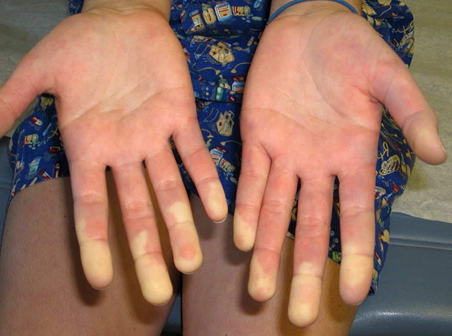
Figure 1. Raynaud’s disease fingers
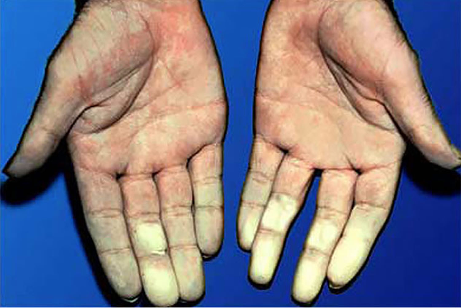
Figure 2. Scleroderma
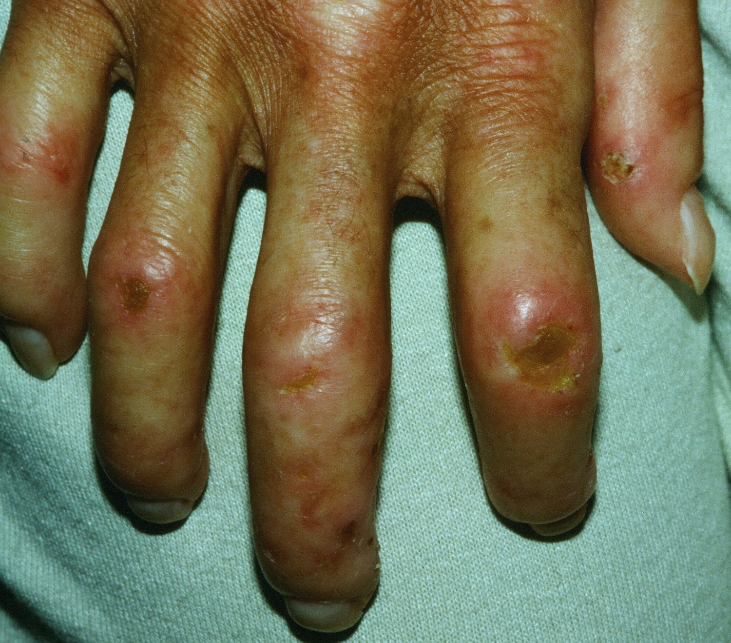
Figure 3. Polymyositis – mechanic’s hands

Mixed connective tissue disease causes
The precise cause of mixed connective tissue disease isn’t known but is believed to involve the interaction between your genes with your environment 1, 25. Mixed connective tissue disease (MCTD) is an autoimmune disorder. In autoimmune disorders, your immune system — responsible for defending your body against harmful substances like bacteria, viruses, fungi, and cancer cells — mistakenly attacks healthy cells.
Given the anti-U1-ribonucleoprotein (anti-RNP) antibody is the hallmark of mixed connective tissue disease (MCTD), it is believed that the anti-U1-ribonucleoprotein (anti-RNP) antibody and its antigen play a role in the pathogenesis of mixed connective tissue disease (MCTD) 26. The ribonucleoprotein (RNP) is a 70 kDa complex formed between RNA molecules and RNA-binding proteins 27. The ribonucleoprotein (RNP) molecules are usually in the nucleus of all human cells, where they help to manufacture messenger RNA (mRNA), and where the immune system cannot find them. However, in dead or dying cells, ribonucleoprotein (RNP) molecules can become exposed to the immune system. Since ribonucleoprotein (RNP) molecules are nearly identical in humans to their counterparts in single celled organisms without immune systems, the human immune system can be fooled into responding to ribonucleoprotein (RNP) as if it were from a dangerous invader. The U1-RNP complex is the main target of the anti-RNP antibody in mixed connective tissue disease (MCTD) 28. The anti-U1-ribonucleoprotein (anti-RNP) antibody immune response that helps define mixed connective tissue disease (MCTD) also appears to mediate some of the damage in mixed connective tissue disease (MCTD).
It is notable that the RNA component unique to the U1-small nuclear ribonucleoprotein, U1-RNA, is among the most prevalent RNAs present in cellular apoptotic debris, and that U1-RNA is an agonist for autoimmunity-associated endosomal Toll-lIke receptors, including TLR7 and TLR3 29, 30. These observations promote the hypothesis that immune recognition of apoptotic debris may play a key role in the cause of anti-RNP autoimmunity, as in mixed connective-tissue disease (MCTD) 25.
Two main mechanisms have been proposed for the pathogenic role of anti-U1-ribonucleoprotein (anti-RNP) antibodies. The first mechanism involves direct binding to endothelial cells through U1-RNP protein molecules or recognition of nucleosome RNP fragments in endothelial cell apoptotic blebs 31, 32. This mechanism may lead to the observable characteristics involving vascular diseases such as Raynaud phenomenon, skin sclerosis, and pulmonary hypertension. The second possible pathogenic mechanism is through immune complex formation, which can activate the complement cascade and lead to tissue inflammation and damage, such as myositis, arthritis, and interstitial lung disease.
Over time, some patients with mixed connective-tissue disease (MCTD) also develop anti-Smith autoantibodies—an expansion of the autoimmune response known as epitope spreading. “Epitope spreading” refers to a phenomenon in immunology where an immune response initially directed at one specific region of an antigen (an epitope) expands to include other epitopes on the same molecule or on different molecules, often as a consequence of tissue damage or inflammation or the development of new autoantibodies during the disease course, can lead to changes in clinical features 1. For example, a lower rate of skin sclerosis and a higher percentage of interstitial lung disease appeared in patients with “epitope spreading” compared to those who did not during a follow-up of patients with mixed connective-tissue disease (MCTD) 33. Escolà-Vergé 33 reported that epitope spreading occurred in 13 (43%) of 40 patients with mixed connective-tissue disease, mainly during the first 2 years after diagnosis. Compared with patients who did not have epitope spreading, patients with epitope spreading had a significantly lower prevalence of skin sclerosis (0% vs 44%) and a higher prevalence of interstitial lung disease (46% vs 15%).
Some people with mixed connective tissue disease (MCTD) have a family history of the condition. Several genes that control the immune system’s responsiveness to invaders and the ability to hide or destroy dead cell debris influence the risk of developing mixed connective tissue disease. Genetic typing of human leukocyte antigen B (HLA-B) gene and HLA-DRB1 gene showed that the risk variant forms of a gene (alleles) for mixed connective-tissue disease (MCTD) are HLA-B*08, HLA-DRB1*04:01, HLA-DRB1*15 and DRB1*09:01/DRB1*15; whereas protective alleles are DRB1*04:04, DRB1*13:01, and DRB1*13:02 34, 35. In a study of a nationwide mixed connective-tissue disease cohort in Norway, Flåm and colleagues 34 found that HLA-B*08 and DRB1*04:01 were risk alleles for mixed connective-tissue disease, while DRB1*04:04, DRB1*13:01 and DRB1*13:02 were protective. Risk alleles for SLE, systemic sclerosis, and polymyositis/dermatomyositis were distinct from those for mixed connective-tissue disease (MCTD) 34. Oka and colleagues 35 performed genotyping of HLA-DRB1 and -DQB1 in 116 Japanese mixed connective-tissue disease patients and 413 controls and analyzed the genotype frequencies. The analysis showed that HLA-DRB1*04:01 and DRB1*09:01 were risk alleles for Japanese mixed connective-tissue disease. DRB1*13:02 was also confirmed to be protective against mixed connective-tissue disease in Japanese patients 35.
Prior immune exposures to other things that look like RNP such as with prior viral infections or chemicals in the environment may also increase the risk. Additional effects of heredity and the environment on the risk for developing mixed connective tissue disease and on its manifestations and severity are likely. However, no clear environmental risk factor has been identified thus far 1, 25. Immune activation due to environmental factors in people with genetic predisposition is believed to play a role. Some environmental factors, such as infections, drugs, toxins, ultraviolet (UV) radiation, and chemicals, including vinyl chloride and silica, have some correlation with the development of mixed connective-tissue disease (MCTD) 36.
Molecular mimicry is the leading theory triggering the onset of disease 1. Amino acid sequences from non-self proteins in the environment may mimic host epitopes and induce autoantibody responses. For example, almost 91% of DNA from the serum of patients with mixed connective-tissue disease (MCTD) had a human immunodeficiency virus type 1 (HIV-1) conserved Pol sequence 37. Seventy-five percent of patients with mixed connective-tissue disease (MCTD) patients had antibodies to HIV GAG proteins p35 and p24 38.
Risk factors for mixed connective tissue disease
Mixed connective tissue disease can occur in people of any age. However, it appears to be most common in women under the age of 30.
Mixed connective tissue disease symptoms
Individuals with mixed connective tissue disease have symptoms that overlap with those of two or more connective tissue diseases. These diseases include systemic lupus erythematosus (SLE), polymyositis, dermatomyositis, scleroderma, and rheumatoid arthritis. The key features of early mixed connective-tissue disease (MCTD) are Raynaud phenomenon, inflammatory arthritis, swollen hands, and myalgia (muscle pain) or myositis (muscle inflammation) 39.
Early indications of mixed connective tissue disease can include:
- General feeling of being unwell. This malaise may be accompanied by increased fatigue and a mild fever.
- Cold and numb fingers or toes (Raynaud’s phenomenon). In response to cold or stress, your fingers or toes might turn white and then purplish blue. After warming, the fingers or toes turn red.
- Swollen fingers or hands. Some people experience swelling to the point where the fingers resemble sausages.
- Muscle pain (myalgia or myositis)
- Joint pain (arthralgia or arthritis). Joints may become deformed, similar to what occurs with rheumatoid arthritis.
- Rash. Red or reddish brown patches may appear over the knuckles.
- Esophageal hypomotility
- Pulmonary dysfunction
- Myositis
- Leukopenia
- Sclerodactyly (a condition where the skin of the fingers and toes thickens and tightens, often leading to a claw-like appearance and limited mobility)
- Pleuritis or pericarditis
- Pulmonary dysfunction
- Pulmonary hypertension
A condition known as Raynaud’s phenomenon may precede the development of additional symptoms of mixed connective tissue disease. Raynaud’s phenomenon, which is seen also in scleroderma, is characterized by painfully cold fingers and toes with blue and/or white color changes caused by spasm of blood vessels in the hands and feet in response to cold or stress. It occurs in approximately 90 percent of individuals with mixed connective tissue disease.
Pain in multiple joints (polyarthralgia) or inflammation of joints (arthritis) also occurs in the majority of affected individuals. Lupus-like skin inflammation in sun-exposed areas and hair loss are common, as are skin scarring changes on the fingers and face like those seen in scleroderma. Muscle weakness due to inflammation (myositis) of proximal muscle groups can also occur. Additional frequent symptoms include hand swelling and fatigue.
Dysfunction of the esophagus occurs in at least half of individuals with mixed connective tissue disease. The esophagus is the tube that carries food from the mouth to the stomach. Esophageal trouble most often manifests as heartburn (gastroesophageal reflux) and difficulty swallowing solid foods. Nearly half of individuals with mixed connective tissue disease may develop clinically significant lung involvement, typically sometime after the condition first emerges. mixed connective tissue disease lung disease may lead to breathing (respiratory) difficulties caused either by high blood pressure in the lungs (pulmonary hypertension) or by causing lung inflammation and scarring in and around the air sacs (interstitial lung disease).
Heart (cardiac) involvement is less common in mixed connective tissue disease than lung problems, but can be serious when it occurs.
Kidney (renal) disease occurs much less often in mixed connective tissue disease than in lupus (10 percent of individuals with mixed connective tissue disease) and is often mild in mixed connective tissue disease.
Neurologic abnormalities are noted in approximately 10 percent of individuals with mixed connective tissue disease.
The low levels of circulating red blood cells (anemia) and a reduction in the white blood cell count (leukopenia) occur in 30 to 40 percent of cases. Disease of the lymph nodes (lymphadenopathy), enlargement of the spleen (splenomegaly), enlargement of the liver (hepatomegaly), and intestinal involvement may also occur in some cases.
Although medications may be required to help control mixed connective tissue disease, the condition has been reported to eventually enter sustained remission in as many as 40% of cases.
Among patients with mixed connective tissue disease, patterns of organ targeting have been reported that suggest disease subtypes. Some patients have more vascular manifestations, and have higher risk for pulmonary hypertension. Some patients have more myositis manifestations and have higher risk for interstitial lung disease. Some patients with more classic rheumatoid arthritis manifestations may have a lower risk of major internal organ damage.
The experience with a large single-center cohort of mixed connective tissue disease patients suggests that the following three clinical subclusters of mixed connective tissue disease manifestations may exist 40:
- Predominantly vascular manifestations, including Raynaud phenomenon, pulmonary hypertension, and antiphospholipid syndrome with thromboses (who are at the greatest risk of mortality)
- A polymyositislike picture, including interstitial lung disease, esophageal dysmotility, and myositis
- Erosive polyarthritis with anti–cyclic citrullinated peptide (anti-CCP) antibodies and sclerodactyly.
Skin signs and symptoms
The most common skin change is Raynaud phenomenon, which is also the most common presenting feature of mixed connective-tissue disease, occurring between 50.3% and 93.2% at presentation and 57.5% to 99% after follow-up 18. Puffy digits, characterized by diffuse soft tissue swelling of the fingers, are also characteristic, although not always present, affecting 53% to 72% of patients at presentation and 46% to 92% after follow-up 18. Nailfold capillary abnormalities may be present on nailfold capillaroscopy. A scleroderma-like pattern to nailfold capillary abnormalities is the most commonly observed, with early-type lesions being predominant 41. Other possible skin signs and symptoms include hand edema, acrosclerosis, calcinosis cutis, telangiectasia, erythema nodosum, hair loss, and vasculitis of digits. Discoid plaques and a malar rash similar to systemic lupus erythematosus can also occur. Case reports describe panniculitis involving the abdomen, legs, and breasts 42, 43.
Musculoskeletal signs and symptoms
Patients with mixed connective tissue disease may develop inflammatory arthritis similar to systemic lupus erythematosus (SLE) or rheumatoid arthritis (RA) and this type of arthritis is reported in 65.3% to 86% of patients with mixed connective tissue disease at the time of presentation and in 49.7% to 89.6% after follow-up 18.
Muscle signs and symptoms
Inflammatory myopathy (myositis), clinically and histologically similar to polymyositis, often manifests as painless proximal muscle weakness with elevated levels of creatinine kinase and aldolase in the serum. Inflammatory myopathy (myositis) is reported in 13.5% to 27.9% of patients at the time of presentation and in 19% to 32.5% at follow-up 18. Rarely, myositis can also be the initial presentation 44.
Heart and lung signs and symptoms
Lung (pulmonary) involvement occurs in almost 73% of patients, with shortness of breath (dyspnea) being the most common symptom 45. Other symptoms include dry cough, pleuritic chest pain, wheezing, and hemoptysis. Interstitial lung disease can occur in 27.8% to 47% of patients, with a nonspecific interstitial pneumonitis pattern being the most common 18. Pulmonary hypertension can occur in 6.9% to 17.8% of patients 18. Other pulmonary signs and symptoms may include pleural effusion, pulmonary vasculitis, thromboembolic disease, alveolar hemorrhage, infections, and obstructive airway disease 46. Pericarditis is the most common heart involvement in mixed connective tissue disease affecting up to 40% of patients with mixed connective tissue disease 47. Pericardial effusion, mitral valve prolapse, myocarditis, accelerated atherosclerosis, and conduction abnormalities can also occur in patients with mixed connective-tissue disease.
Gastrointestinal signs and symptoms
Esophageal dysmotility leading to dysphagia (difficulty swallowing), odynophagia (painful swallowing), gastrointestinal reflux, and regurgitation are all common features, occurring in 34.8% to 38.9% of patients with mixed connective tissue disease at presentation and in 45.3% to 49.6% during follow-up 18. Less common gastrointestinal signs and symptoms are pancreatitis, megacolon, duodenal bleeding, portal hypertension, and autoimmune hepatitis. Rarely, patients with mixed connective tissue disease can present with protein-losing enteropathy 48.
Less common signs and symptoms
Kidney: Kidney involvement is less common and may occur in 15% to 25% of mixed connective-tissue disease patients. Most patients are typically asymptomatic 49. Membranous nephropathy is the most common finding. Kidney involvement can sometimes be associated with significant morbidity, risk of hypertension, and progressive kidney disease 50.
Central nervous system (brain and spinal cord): The involvement of the central nervous system is increasingly recognized as a manifestation of mixed connective tissue disease. Nervous system involvement has been described in up to 25% of patients with mixed connective tissue disease 51. Trigeminal neuralgia is the most common central nervous system manifestation. Other possible symptoms include headaches, peripheral neuropathy, aseptic meningitis, cerebral venous sinus thrombosis, and sensorineural hearing loss 52.
Hematological involvement: Anemia, leukopenia (low level of white blood cells in the blood), and thrombocytopenia (low level of platelet cells in the blood) may occur. Idiopathic immune–mediated thrombocytopenia and hemolytic anemia are not common manifestations and should prompt further evaluation for systemic lupus erythematosus (SLE) and other causes.
Mixed connective tissue disease complications
Mixed connective tissue disease can lead to serious complications, including:
- High blood pressure in the lungs (pulmonary hypertension). This condition is the main cause of death in people with mixed connective tissue disease.
- Interstitial lung disease. This large group of disorders can cause scarring in your lungs, which affects your ability to breathe.
- Heart disease. Parts of the heart may become enlarged, or inflammation may occur around the heart. Heart failure can occur. Heart disease is the cause of death in about 20 percent of people with mixed connective tissue disease.
- Kidney damage. About one-fourth of people with mixed connective tissue disease develop kidney problems. Kidney involvement is usually mild, but can lead to kidney failure.
- Digestive tract damage. Commonly, mixed connective tissue disease affects the digestive tract. You might have abdominal pain and problems with swallowing and digesting food.
- Anemia. About 75 percent of people with mixed connective tissue disease have iron deficiency anemia.
- Tissue death (necrosis). People with severe Raynaud’s phenomenon (Raynaud’s disease) can develop gangrene in the fingers.
- Hearing loss. Often unrecognized, hearing loss may occur in as many as half the people with mixed connective tissue disease. More research is needed to understand this association.
- Nerve damage. Sjogren syndrome can affect the nerve that carries feeling from your face to your brain (trigeminal nerve). If you have trigeminal neuralgia, even mild stimulation of your face — such as from brushing your teeth or putting on makeup — can trigger a jolt of severe pain.
Treatment side effects
Corticosteroids are commonly used to manage the signs and symptoms of mixed connective tissue disease. These medications are effective, but they carry risks.
Corticosteroids that you take by mouth affect your entire body. For this reason, they are the most likely type of corticosteroid to cause side effects. Side effects depend on the dose of corticosteroid you receive and may include:
- A buildup of fluid, causing swelling in your lower legs.
- High blood pressure.
- Problems with mood swings, memory, behavior, and other psychological effects, such as confusion or delirium.
- Upset stomach.
- Weight gain in the belly, face and back of the neck.
When taking corticosteroids by mouth for a longer term, you may experience:
- Problems with the eyes, such as glaucoma or cataracts.
- A round face, which is sometimes called moon face.
- High blood sugar, which can trigger or worsen diabetes.
- Increased risk of infections, especially with common bacterial, viral and fungal microorganisms.
- Bone fractures and thinning bones, called osteoporosis.
- Fatigue, loss of appetite, nausea and muscle weakness.
- Thin skin, bruising and slower wound healing.
Your doctor will likely monitor you for adverse effects, such as osteoporosis, muscle weakness and infection. You may need to take calcium and vitamin D supplements to help ease these adverse effects.
Mixed connective tissue disease diagnosis
Diagnosing mixed connective tissue disease can be difficult because the symptoms of the three diseases usually occur one after another over a long period of time. Doctors may suspect a diagnosis of mixed connective tissue disease after asking questions about the your medical history and performing a physical examination.
Your doctor is likely do these tests:
- Physical exam to check for swollen hands and painful, swollen joints
- Blood tests that reveal abnormally high levels of antibodies to the U1 small nuclear ribonucleoprotein (anti-RNP). Anti-RNP antibodies are required for diagnosis of mixed connective-tissue disease. Titers are typically high. The presence of anti–U1-70 kd is characteristic of mixed connective-tissue disease but not specific. Anti-RNP autoantibodies typically become undetectable in patients in remission.
Laboratory studies used in the workup for mixed connective-tissue disorder (MCTD) are as follows 53:
- Complete blood cell count (CBC)
- Urinalysis
- Routine blood chemistry
- Indicators of acute phase response (erythrocyte sedimentation rate [ESR] or C-reactive protein [CRP])
- Muscle enzymes if myositis is suspected clinically
- Antinuclear antibodies
- Anti–U1-ribonucleoprotein (RNP) antibodies
- Amylase and lipase – To assess for pancreatitis if clinically indicated
- Serologic indicators of pulmonary hypertension (such as brain natriuretic peptide [BNP]) if clinically indicated.
Other immune studies 53:
- Antiphospholipid antibodies including anti-cardiolipin antibody (ACL) and lupus anticoagulant (LA) may be associated with pulmonary hypertension
- Rheumatoid factor (RF) is frequently detected
- Other lupus-specific antibodies (eg, anti–double-stranded DNA antibodies [anti-dsDNA]) are typically absent
- Scleroderma-specific antibodies, including anticentromere, anti–Scl-70 (topoisomerase), and anti–PM-1 (Pm-Scl), are typically absent
- C3 and C4 complement levels are more likely to be depleted in lupus than in mixed connective-tissue disease.
Imaging studies used in the workup of patients with mixed connective-tissue disease include the following 53:
- Chest radiography: Chest x-ray may serve as a screening test for lung disease, including pulmonary infiltrates, interstitial lung disease, pleural effusions, and cardiomegaly.
- Electrocardiogram: An echocardiogram is a useful tool for screening for right heart disease and pulmonary hypertension and assessing the pericardium for the presence of effusion or thickening, valvular abnormalities, and overall cardiac function (exercise echocardiography may have increased sensitivity for identifying pulmonary hypertension) 54.
- Computed tomography (CT) scanning: Used to evaluate abdominal pain indicated for evidence of serositis, pancreatitis, or visceral perforation related to vasculitis. High-resolution CT is very sensitive in diagnosing interstitial lung disease. Common findings include ground-glass opacities, linear opacities, subpleural micronodules, septal thickening, and traction bronchiectasis, typically with peripheral and lower lobe predominance. Honeycombing, airspace consolidation, emphysema, and centrilobular nodules are less common findings 55.
- MRI: Used to assess neuropsychiatric signs or symptoms.
Additional tests 53:
- Pulmonary function testing is used to screen for declining diffusing capacity of lung for carbon monoxide (DLCO), possibly indicating pulmonary hypertension or to assess lung volumes and forced vital capacity to screen for suspicion/progression of interstitial lung disease 56. Patients with interstitial lung disease may show reductions in the diffusion capacity for carbon monoxide, forced vital capacity, and forced expiratory volume.
- Cardiac enzyme assays are helpful to assess for myocardial ischemia and myocarditis.
- Infection, stroke, or neuropsychiatric manifestations may be monitored with cerebrospinal fluid (CFS) analysis.
- The six-minute walk can be helpful to assess for cardiopulmonary insufficiency, possibly indicating pulmonary hypertension 57.
- Right heart catheterization. Definitive diagnosis of pulmonary hypertension in mixed connective-tissue disease requires right heart catheterization demonstrating mean pulmonary arterial pressure at rest >20 mmHg.
Other tests for evaluating gastrointestinal tract symptoms may include an esophagram, upper endoscopy, or colonoscopy. For assessing potential inflammatory muscle or peripheral nerve disease, an electromyogram and nerve conduction study may be used.
Mixed connective-tissue disease Differential Diagnosis
Due to nonspecific symptoms and different organ involvement, mixed connective-tissue disease (MCTD) mimics several other conditions, especially other connective tissue diseases. Differential diagnoses are as follows 1:
- Systemic lupus erythematosus (SLE) is an autoimmune disease characterized by the presence of an anti-nuclear antibody and varied manifestations, which may include inflammatory arthritis, mucocutaneous disease, serositis, pleuritis, pericarditis, renal disease, fevers, neuropsychiatric symptoms, leukopenia, autoimmune hemolysis, thrombocytopenia, antiphospholipid antibodies, low complements, and the presence of an anti-double stranded DNA antibody or anti-Smith antibody.
- Rheumatoid arthritis (RA) is typically symmetric polyarthritis, predominantly affecting the small joints of the hands, wrists, ankles, and feet. Up to 70% of patients have an abnormal rheumatoid factor and anti-cyclic citrullinated peptide antibody.
- Polymyositis is an idiopathic inflammatory myositis that causes proximal over distal muscle weakness. This condition is associated with abnormal muscle biochemistries, signs of muscle edema or inflammation on magnetic resonance imaging, and the absence of autoantibodies.
- Dermatomyositis is an idiopathic inflammatory myositis similar to polymyositis but includes varying presentations involving the skin, lungs, gastrointestinal tract, and joints. This condition is typically associated with either myositis-specific antibodies or myositis-associated antibodies.
- Scleroderma is a chronic multisystem disease characterized by widespread vascular dysfunction and progressive fibrosis of the skin and internal organs, which is associated with autoimmunity.
Mixed connective tissue disease treatment
There’s no cure for mixed connective tissue disease. But medication can help manage the signs and symptoms. Whenever possible, a rheumatologist experienced in diagnosis and treatment of connective-tissue disease disease should co-manage all patients with mixed connective-tissue disease (MCTD). Consultation with other specialists or subspecialists may be indicated for the evaluation and/or treatment of specific aspects of disease, such as pulmonary hypertension, interstitial lung disease, gastroesophageal reflux, or acute ischemia due to Raynaud phenomenon.
Treatment for mixed connective tissue disease depends on which organs are involved and the severity of the disease. Some people need continuous treatment, while others need it only during periods of heightened disease activity, called flares or flare-ups. If you have a more serious form of the disease, you might need continuous medication.
The type of medication prescribed depends on the severity of your disease and your symptoms. Medications can include:
- Corticosteroids. Drugs, such as prednisone (Deltasone, Rayos), can help prevent your immune system from attacking healthy cells and suppressing inflammation. Side effects of corticosteroids can include mood swings, weight gain, high blood sugar, increased blood pressure, weakened bones and cataracts.
- Antimalarial drugs. Hydroxychloroquine (Plaquenil) can treat mild mixed connective tissue disease and might prevent flare-ups.
- Calcium channel blockers. Medications, such as nifedipine (Adalat CC, Procardia) and amlodipine (Norvasc), help relax the muscles in the walls of your blood vessels and may be used to treat Raynaud’s phenomenon.
- Other immunosuppressants. Your doctor might prescribe other medications based on your specific signs and symptoms. For example, if they’re similar to those of lupus, your doctor might recommend medications typically prescribed for people with lupus.
- Pulmonary hypertension medications. Bosentan (Tracleer) or sildenafil (Revatio, Viagra) may be prescribed. Pulmonary hypertension is typically less responsive to steroids, and the guidance of an expert in pulmonary hypertension should direct advanced management. Vasodilators such as prostaglandins, including epoprostenol; endothelin receptor antagonists, including ambrisentan; phosphodiesterase 5 inhibitors, including sildenafil; and immunosuppression with corticosteroids and cyclophosphamide may be appropriate therapeutic considerations.
Your doctor is likely to monitor you closely for signs of pulmonary hypertension.
The management of Raynaud phenomenon includes symptomatic strategies such as avoiding caffeine, smoking, cold temperatures, and injury to the digits. Oral calcium channel blockers, such as nifedipine, which decreases peripheral resistance, are an option. Prostaglandins, endothelin receptor antagonists, phosphodiesterase 5 inhibitors, and topical nitroglycerins are also effective.
Arthritis and arthralgia typically respond to nonsteroidal anti-inflammatory drugs (NSAIDs) and hydroxychloroquine (anti-malaria drug). For refractory synovitis, corticosteroids, methotrexate, and other disease-modifying anti-rheumatic drugs (DMARDs) can be used.
Pleuritis, pericarditis, myositis, and aseptic meningitis typically respond to steroids. Steroid-sparing agents, such as methotrexate, cyclosporine, azathioprine, and mycophenolate mofetil, are commonly used as second-line agents. Steroid-resistant myositis may respond to intravenous immunoglobulin.
Gastrointestinal reflux treatment involves proton pump inhibitors (PPI) or histamine blockers, lifestyle changes, and dietary modifications, such as elevating the head of the bed and avoiding dietary triggers. Prokinetics and gastric fundoplication are possible options for those who fail twice-daily proton pump inhibitor (PPI) therapy. Individuals with esophageal motility disorder may require prokinetics. Patients with malabsorption should be on a lactose-free diet, and medium-chain triglycerides should substitute for long-chain fatty acids.
Patients with autoimmune hemolytic anemia and thrombocytopenia are initially treated with steroids. Clinicians can consider rituximab in resistant cases.
Although, no controlled studies have been performed in mixed connective tissue disease, some patients with mixed connective tissue disease have been included in previous trials of lupus, scleroderma, myositis, and rheumatoid arthritis patients. In general, it appears that these mixed connective tissue disease subgroups respond similarly to treatments as have been reported in larger classical rheumatic disease-specific patient cohorts. These observations and accumulated clinical experience by mixed connective tissue disease experts supports the use of antimalarials for potential lupus-like disease modifying effects, the use of vasodilators to treat Raynaud’s phenomenon, the use of proton pump inhibitors for GERD (gastroesophageal reflux disease), and the use of additional disease-modifying anti-rheumatic drugs (DMARDs) for rheumatoid arthritis-like polyarthritis.
Cohort studies of mixed connective tissue disease patients with pulmonary hypertension or other lung disease have suggested that these patients may be more likely to respond well to a course of aggressive immunosuppression than is typical for patients with similar lung disease stemming from other causes.
Low-to-moderate doses of corticosteroids are often effective for rapid control of disease flares, and may be used as part of long-term therapy in some patients, despite their substantial long-term drug toxicities. Scleroderma renal crisis, a serious complication of scleroderma that is more likely after the use of high dose corticosteroids, has been infrequently reported in mixed connective tissue disease.
Nonsteroidal anti-inflammatory drugs (NSAIDs) may also be used to help control mild inflammatory symptoms, though their use must be balanced with their risk for gastrointestinal complications. NSAIDs rarely can cause aseptic meningitis in some individuals; this seems to occur slightly more often in patients with mixed connective tissue disease compared to other groups.
Home remedies
Other ways to control symptoms of mixed connective tissue disease include:
- Nonsteroidal anti-inflammatory drugs (NSAIDs). Medications, such as ibuprofen (Advil, Motrin IB, others) or naproxen sodium (Aleve, others), might help relieve the pain and inflammation if your condition is mild.
- Protecting your hands from cold. Wearing gloves and taking other measures to keep your hands warm can help prevent Raynaud’s phenomenon.
- Not smoking. Smoking causes blood vessels to narrow, which can worsen the effects of Raynaud’s phenomenon.
- Reducing stress. Raynaud’s phenomenon is often triggered by stress. Relaxation techniques — such as slowing and focusing on your breathing — can help reduce your stress levels.
- Physical Activity and exercise. Convincing data support the value of an active lifestyle and an exercise program tailored to the needs of patients with arthritis of various types 58, 59. This approach also appears to be appropriate in mixed connective tissue disease.
Mixed connective tissue disease diet
Patients with hypertension, esophageal reflux, malabsorption, or other sclerodermatous-type bowel involvement may need special consideration.
Because atherosclerotic heart disease remains a major risk in all patients, advocate a heart-healthy diet. However, no specific dietary manipulations have been demonstrated to be effective in treating mixed connective tissue disease.
High-dose omega-3 fatty acids (a type of essential polyunsaturated fat occurring chiefly in fish oils that play a crucial role in various bodily functions) have been reported to inhibit the production of pro-inflammatory prostaglandins, and clinical trials have reported benefits of dietary supplementation with omega-3 fatty acids in several inflammatory and autoimmune diseases 60. Fish oils contain the omega−3 fatty acids eicosapentaenoic acid (EPA) and docosahexaenoic acid (DHA) that are known to reduce inflammation in the body and improve hypertriglyceridemia. Some studies have found that fish oil supplements may ease rheumatoid arthritis pain and stiffness. Side effects can include nausea, belching and a fishy taste in the mouth. Fish oil can get in the way of medicines you take. So check with your rheumatologist before trying it. Omega-3 fatty acids may have similar benefits to the long-term use of nonsteroidal anti-inflammatory drugs (NSAIDs), with less risk of worsening of hypertension and less risk of gastrointestinal or kidney toxicity. However, omega-3 fatty acids at this dose have platelet inhibitory effects that can increase bleeding risk in susceptible patients.
Mixed connective tissue disease prognosis
Most patients with mixed connective tissue disease (MCTD) have a good prognosis, although it depends on which organ is affected, the degree of inflammation, and the rate of disease progression 18, 3. Pulmonary hypertension is the most common disease-associated cause of death. Careful monitoring and aggressive treatment may improve the outcome of pulmonary hypertension 61. The mortality rate (death rate) varies between 3.1% and 10% in the medical literature 62, 10.
In a study involving the Hungarian population 62, the survival rate for 5 and 10 years after mixed connective tissue disease (MCTD) diagnosis was 98% and 96%, respectively. In a French cohort study involving 330 patients with mixed connective-tissue disease (MCTD) followed up on average for 8 years, 45.2% of patients achieved remission, 7.6% developed pulmonary hypertension, and 27.9% developed interstitial lung disease 10.
Pulmonary hypertension is the most common cause of death. Interstitial lung disease, infections, cardiovascular complications, and malignancies are other causes 63. The presence of immunoglobulin G anticardiolipin antibodies may be associated with more severe disease 1.
The identification of the anti-SMN complex antibodies (antibodies for Survival of Motor Neuron protein) also has prognostic significance 1. Mixed connective-tissue disease patients with anti-SMN complex antibodies had poorer disease-related outcomes compared to patients who did not have this autoantibody, and complications were mainly related to pulmonary hypertension and interstitial lung disease 9. The anti-Ro52 autoantibody also known as anti-TRIM21 antibodies also signify a worse prognosis in those with mixed connective-tissue disease, specifically related to the severity of interstitial lung disease 14.
“Epitope spreading” refers to a phenomenon in immunology where an immune response initially directed at one specific region of an antigen (an epitope) expands to include other epitopes on the same molecule or on different molecules, often as a consequence of tissue damage or inflammation or the development of new autoantibodies during the disease course, can lead to changes in clinical features 1. For example, a lower rate of skin sclerosis and a higher percentage of interstitial lung disease appeared in patients with “epitope spreading” compared to those who did not during a follow-up of patients with mixed connective-tissue disease (MCTD) 33. In addition, patients with mixed connective-tissue disease can clinically progress to other connective-tissue diseases, such as systemic sclerosis (scleroderma), rheumatoid arthritis, Sjogren syndrome, and systemic lupus erythematosus (SLE) 1. In a retrospective review involving patients with mixed connective-tissue disease followed over a median time of 8 years from diagnosis, approximately 25.6% evolved to a definite connective tissue disease 62. On the other hand, patients with other connective tissue diseases or undifferentiated connective tissue diseases can eventually develop mixed connective-tissue disease (MCTD) 1.
A long-term observational nationwide cohort study from Norway found that interstitial lung disease (ILD) was present in 41% of mixed connective-tissue disease patients and progressed in 19% of patients across the observation period of a mean of 6.4 years 64. The following were the strongest predictors of interstitial lung disease (ILD) progression 61:
- Male sex
- Presence of anti-ro52 antibodies
- Elevated anti-RNP titer
- Absence of arthritis
Mixed connective tissue disease life expectancy
In general, the long-term outlook (prognosis) for people with mixed connective tissue disease (MCTD) is favorable, but it mostly depends on the signs and symptoms present in each person.
The overall 10-year survival rate of the disease is about 80%. Some people have symptom-free periods lasting for many years with no treatment. Despite treatment, the disease gets worse in about 13% of people and can cause potentially fatal complications in six to 12 years. The prognosis is worse for people with features of systemic sclerosis and/or polymyositis. Pulmonary hypertension is the most common mixed connective tissue disease-associated cause of death.
Long-term outcome studies have established pulmonary hypertension as the most common mixed connective tissue disease-associated cause of death 65. Immunoglobulin G (IgG) anticardiolipin antibodies are a marker for development of pulmonary hypertension. Infections are also a major cause of death.
Cardiac disease, most often pericarditis, is also common in mixed connective tissue disease patients, with prevalence estimates ranging from 13% to 65%. Other cardiac abnormalities include conduction abnormalities, pericardial effusion, mitral valve prolapse, diastolic dysfunction, and accelerated atherosclerosis. In three prospective studies with 13-15 years of follow-up, mixed connective tissue disease patients had an overall mortality rate of 10.4%, and 20% of these deaths were directly attributable to cardiac causes 66.
- Sapkota B, Al Khalili Y. Mixed Connective Tissue Disease. [Updated 2024 Jul 27]. In: StatPearls [Internet]. Treasure Island (FL): StatPearls Publishing; 2025 Jan-. Available from: https://www.ncbi.nlm.nih.gov/books/NBK542198[↩][↩][↩][↩][↩][↩][↩][↩][↩][↩][↩][↩][↩]
- Zandman-Goddard G, Solomon M, Rosman Z, Peeva E, Shoenfeld Y. Environment and lupus-related diseases. Lupus. 2012 Mar;21(3):241-50. doi: 10.1177/0961203311426568[↩]
- Sharp GC, Irvin WS, Tan EM, Gould RG, Holman HR. Mixed connective tissue disease–an apparently distinct rheumatic disease syndrome associated with a specific antibody to an extractable nuclear antigen (ENA). Am J Med. 1972 Feb;52(2):148-59. doi: 10.1016/0002-9343(72)90064-2[↩][↩][↩]
- Bennett RM, O’Connell DJ. Mixed connective tisssue disease: a clinicopathologic study of 20 cases. Semin Arthritis Rheum. 1980 Aug;10(1):25-51. doi: 10.1016/0049-0172(80)90013-x[↩]
- Amigues JM, Cantagrel A, Abbal M, Mazieres B. Comparative study of 4 diagnosis criteria sets for mixed connective tissue disease in patients with anti-RNP antibodies. Autoimmunity Group of the Hospitals of Toulouse. J Rheumatol. 1996 Dec;23(12):2055-62.[↩]
- Alarcón-Segovia D, Cardiel MH. Comparison between 3 diagnostic criteria for mixed connective tissue disease. Study of 593 patients. J Rheumatol. 1989 Mar;16(3):328-34.[↩][↩]
- Alarcon-Segovia D, Villareal M. Classification and diagnostic criteria for mixed connective tissue disease. Kasukawa R, Sharp GC, eds. Mixed Connective Tissue Disease and Anti-Nuclear Antibodies. Amsterdam: Excerpta Medica; 1987. 33-40.[↩]
- Tanaka Y, Kuwana M, Fujii T, Kameda H, et al. 2019 Diagnostic criteria for mixed connective tissue disease (MCTD): From the Japan research committee of the ministry of health, labor, and welfare for systemic autoimmune diseases. Mod Rheumatol. 2021 Jan;31(1):29-33. https://doi.org/10.1080/14397595.2019.1709944[↩][↩]
- Kubo S, Tanaka Y. Evolution of diagnostic criteria and new insights into clinical testing in mixed connective tissue disease; anti-survival motor neuron complex antibody as a novel marker of severity of the disease. Immunol Med. 2024 Jun;47(2):52-57. doi: 10.1080/25785826.2024.2338593[↩][↩]
- Gunnarsson R, Hetlevik SO, Lilleby V, Molberg Ø. Mixed connective tissue disease. Best Pract Res Clin Rheumatol. 2016 Feb;30(1):95-111. doi: 10.1016/j.berh.2016.03.002[↩][↩][↩]
- Cansu DÜ, Korkmaz C. Pulmonary hypertension in connective tissue diseases: epidemiology, pathogenesis, and treatment. Clin Rheumatol. 2023 Oct;42(10):2601-2610. doi: 10.1007/s10067-022-06446-y[↩]
- Cappelli S, Bellando Randone S, Martinović D, et al. “To be or not to be,” ten years after: evidence for mixed connective tissue disease as a distinct entity. Semin Arthritis Rheum. 2012 Feb;41(4):589-98. doi: 10.1016/j.semarthrit.2011.07.010[↩]
- Yoshida S. Pulmonary arterial hypertension in connective tissue diseases. Allergol Int. 2011 Dec;60(4):405-9. doi: 10.2332/allergolint.11-RAI-0360[↩]
- Gunnarsson R, El-Hage F, Aaløkken TM, Reiseter S, Lund MB, Garen T; Norwegian MCTD study group; Molberg Ø. Associations between anti-Ro52 antibodies and lung fibrosis in mixed connective tissue disease. Rheumatology (Oxford). 2016 Jan;55(1):103-8. doi: 10.1093/rheumatology/kev300[↩][↩]
- Mixed Connective-Tissue Disease (MCTD). https://emedicine.medscape.com/article/335815-overview[↩][↩]
- Martínez-Barrio J, Valor L, López-Longo FJ. Facts and controversies in mixed connective tissue disease. Med Clin (Barc). 2018 Jan 12;150(1):26-32. English, Spanish. doi: 10.1016/j.medcli.2017.06.066[↩]
- Ciang NC, Pereira N, Isenberg DA. Mixed connective tissue disease-enigma variations? Rheumatology (Oxford). 2017 Mar 1;56(3):326-333. doi: 10.1093/rheumatology/kew265[↩]
- Alves MR, Isenberg DA. “Mixed connective tissue disease”: a condition in search of an identity. Clin Exp Med. 2020 May;20(2):159-166. doi: 10.1007/s10238-020-00606-7[↩][↩][↩][↩][↩][↩][↩][↩][↩]
- Maldonado ME, Perez M, Pignac-Kobinger J, Marx ET, Tozman EM, Greidinger EL, Hoffman RW. Clinical and immunologic manifestations of mixed connective tissue disease in a Miami population compared to a Midwestern US Caucasian population. J Rheumatol. 2008 Mar;35(3):429-37. https://pmc.ncbi.nlm.nih.gov/articles/PMC2919224[↩]
- Ungprasert P, Crowson CS, Chowdhary VR, Ernste FC, Moder KG, Matteson EL. Epidemiology of Mixed Connective Tissue Disease, 1985-2014: A Population-Based Study. Arthritis Care Res (Hoboken). 2016 Dec;68(12):1843-1848. doi: 10.1002/acr.22872[↩][↩]
- Nakae K, Furusawa F, Kasukawa R, et al. . A nationwide epidemiological survey on diffuse collagen diseases: Estimation of prevalence rate in Japan. Kasukawa R, Sharp G, eds. Mixed Connective Tissue Disease and Anti-nuclear Antibodies. Amsterdam: Excerpta Medica; 1987. 9.[↩][↩]
- Gunnarsson R, Molberg O, Gilboe IM, Gran JT; PAHNOR1 Study Group. The prevalence and incidence of mixed connective tissue disease: a national multicentre survey of Norwegian patients. Ann Rheum Dis. 2011 Jun;70(6):1047-51. doi: 10.1136/ard.2010.143792[↩][↩]
- Mixed Connective-Tissue Disease (MCTD). https://emedicine.medscape.com/article/335815-overview#a6[↩]
- Ferucci ED, Johnston JM, Gordon C, Helmick CG, Lim SS. Prevalence of Mixed Connective Tissue Disease in a Population-Based Registry of American Indian/Alaska Native People in 2007. Arthritis Care Res (Hoboken). 2017 Aug;69(8):1271-1275. doi: 10.1002/acr.23135[↩]
- Mixed Connective-Tissue Disease (MCTD). https://emedicine.medscape.com/article/335815-overview#a5[↩][↩][↩]
- Venables PJ. Mixed connective tissue disease. Lupus. 2006;15(3):132-7. doi: 10.1191/0961203306lu2283rr[↩]
- Ribonucleoprotein. https://www.sciencedirect.com/topics/neuroscience/ribonucleoprotein[↩]
- Greidinger EL, Zang YJ, Jaimes K, Martinez L, Nassiri M, Hoffman RW. CD4+ T cells target epitopes residing within the RNA-binding domain of the U1-70-kDa small nuclear ribonucleoprotein autoantigen and have restricted TCR diversity in an HLA-DR4-transgenic murine model of mixed connective tissue disease. J Immunol. 2008 Jun 15;180(12):8444-54. doi: 10.4049/jimmunol.180.12.8444[↩]
- Greidinger EL, Zang Y, Martinez L, Jaimes K, Nassiri M, Bejarano P, Barber GN, Hoffman RW. Differential tissue targeting of autoimmunity manifestations by autoantigen-associated Y RNAs. Arthritis Rheum. 2007 May;56(5):1589-97. doi: 10.1002/art.22601[↩]
- Hardy MP, Audemard É, Migneault F, Feghaly A, Brochu S, Gendron P, Boilard É, Major F, Dieudé M, Hébert MJ, Perreault C. Apoptotic endothelial cells release small extracellular vesicles loaded with immunostimulatory viral-like RNAs. Sci Rep. 2019 May 10;9(1):7203. doi: 10.1038/s41598-019-43591-[↩]
- Okawa-Takatsuji M, Aotsuka S, Uwatoko S, Takaono M, Iwasaki K, Kinoshita M, Sumiya M. Endothelial cell-binding activity of anti-U1-ribonucleoprotein antibodies in patients with connective tissue diseases. Clin Exp Immunol. 2001 Nov;126(2):345-54. doi: 10.1046/j.1365-2249.2001.01669.x[↩]
- Biggiogera M, Bottone MG, Martin TE, Uchiumi T, Pellicciari. Still immunodetectable nuclear RNPs are extruded from the cytoplasm of spontaneously apoptotic thymocytes. Exp Cell Res. 1997 Aug 1;234(2):512-20. doi: 10.1006/excr.1997.3657[↩]
- Escolà-Vergé L, Pinal-Fernandez I, Fernandez-Codina A, Callejas-Moraga EL, Espinosa J, Marin A, Labrador-Horrillo M, Selva-O’Callaghan A. Mixed Connective Tissue Disease and Epitope Spreading: An Historical Cohort Study. J Clin Rheumatol. 2017 Apr;23(3):155-159. doi: 10.1097/RHU.0000000000000500[↩][↩][↩]
- Flåm ST, Gunnarsson R, Garen T; Norwegian MCTD Study Group; Lie BA, Molberg Ø. The HLA profiles of mixed connective tissue disease differ distinctly from the profiles of clinically related connective tissue diseases. Rheumatology (Oxford). 2015 Mar;54(3):528-35. doi: 10.1093/rheumatology/keu310[↩][↩][↩]
- Oka S, Higuchi T, Furukawa H, Shimada K, Hashimoto A, Komiya A, Matsui T, Fukui N, Suematsu E, Ohno S, Kono H, Katayama M, Nagaoka S, Migita K, Tohma S. Predisposition of HLA-DRB1*04:01/*15 heterozygous genotypes to Japanese mixed connective tissue disease. Sci Rep. 2022 Jun 15;12(1):9916. doi: 10.1038/s41598-022-14116-x[↩][↩][↩]
- Khanna A, Suri JC, Ray A, Sharma RK. Silica associated mixed connective tissue disorder in a stone crusher. Indian J Occup Environ Med. 2013 May;17(2):76-7. doi: 10.4103/0019-5278.123172[↩]
- Prokop J, Jagodzinski PP. Identification of retroviral conserved pol sequences in serum of mixed connective tissue disease and systemic sclerosis patients. Biomed Pharmacother. 2004 Jan;58(1):61-4. doi: 10.1016/j.biopha.2003.10.001[↩]
- Ranki A, Kurki P, Riepponen S, Stephansson E. Antibodies to retroviral proteins in autoimmune connective tissue disease. Relation to clinical manifestations and ribonucleoprotein autoantibodies. Arthritis Rheum. 1992 Dec;35(12):1483-91. doi: 10.1002/art.1780351212[↩]
- Ferrara CA, La Rocca G, Ielo G, Libra A, Sambataro G. Towards Early Diagnosis of Mixed Connective Tissue Disease: Updated Perspectives. Immunotargets Ther. 2023 Jul 26;12:79-89. doi: 10.2147/ITT.S390023[↩]
- Szodoray P, Hajas A, Kardos L, Dezso B, Soos G, Zold E, Vegh J, Csipo I, Nakken B, Zeher M, Szegedi G, Bodolay E. Distinct phenotypes in mixed connective tissue disease: subgroups and survival. Lupus. 2012 Nov;21(13):1412-22. doi: 10.1177/0961203312456751[↩]
- Ornowska S, Wudarski M, Dziewięcka E, Olesińska M. Naifold capillaroscopy in mixed connective tissue disease patients. Clin Rheumatol. 2024 May;43(5):1703-1709. doi: 10.1007/s10067-024-06879-7[↩]
- Varma R, Szilagyi S, Harshan M. Breast involvement in mixed connective tissue disease. Radiol Case Rep. 2019 Jan 17;14(4):430-435. doi: 10.1016/j.radcr.2019.01.001[↩]
- Nakashima M, Suzuki K, Okada M, Kataharada K, Takada K, Nakanishi T, Kobayashi M, Ohsuzu F. Panniculitis in a patient with mixed connective tissue disease. Mod Rheumatol. 2004;14(3):250-3. doi: 10.1007/s10165-004-0301-x[↩]
- S L, Tony K, Raghupathy, V S, Malepati B. A Rare Case of Mixed Connective Tissue Disease (MCTD) with Intricate Features of Lupus, Polymyositis and Rheumatoid Arthritis Presenting with Severe Myositis. J Clin Diagn Res. 2015 Mar;9(3):OD05-7. doi: 10.7860/JCDR/2015/11575.5695[↩]
- Sullivan WD, Hurst DJ, Harmon CE, Esther JH, Agia GA, Maltby JD, Lillard SB, Held CN, Wolfe JF, Sunderrajan EV, et al. A prospective evaluation emphasizing pulmonary involvement in patients with mixed connective tissue disease. Medicine (Baltimore). 1984 Mar;63(2):92-107. doi: 10.1097/00005792-198403000-00003[↩]
- Prakash UB. Lungs in mixed connective tissue disease. J Thorac Imaging. 1992 Mar;7(2):55-61. doi: 10.1097/00005382-199203000-00007[↩]
- Ungprasert P, Wannarong T, Panichsillapakit T, Cheungpasitporn W, Thongprayoon C, Ahmed S, Raddatz DA. Cardiac involvement in mixed connective tissue disease: a systematic review. Int J Cardiol. 2014 Feb 15;171(3):326-30. doi: 10.1016/j.ijcard.2013.12.079[↩]
- Furuya T, Suzuki T, Onoda N, Tamura K, Sato K, Demura H, Kashiwazaki S. Mixed connective tissue disease associated with protein losing enteropathy: successful treatment with intravenous cyclophosphamide therapy. Intern Med. 1992 Dec;31(12):1359-62. doi: 10.2169/internalmedicine.31.1359[↩]
- Murakami T, Endo S, Moriki T, Doi T, Matsumoto Y. Mixed connective tissue disease developing into MPO-ANCA-positive polyangiitis. Intern Med. 2011;50(6):591-5. doi: 10.2169/internalmedicine.50.3958[↩]
- Kitridou RC, Akmal M, Turkel SB, Ehresmann GR, Quismorio FP Jr, Massry SG. Renal involvement in mixed connective tissue disease: a longitudinal clinicopathologic study. Semin Arthritis Rheum. 1986 Nov;16(2):135-45. doi: 10.1016/0049-0172(86)90047-8[↩]
- Bennett RM, Bong DM, Spargo BH. Neuropsychiatric problems in mixed connective tissue disease. Am J Med. 1978 Dec;65(6):955-62. doi: 10.1016/0002-9343(78)90747-7[↩]
- Agrawal A, Kohat AK, Sahu C, Kansal C, Fatima A. Headache and cerebral venous sinus thrombosis as a first presentation of mixed connective tissue disease: a rare case and literature review. Acta Neurol Belg. 2024 Oct;124(5):1741-1743. doi: 10.1007/s13760-024-02548-3[↩]
- Mixed Connective-Tissue Disease (MCTD) Workup. https://emedicine.medscape.com/article/335815-workup[↩][↩][↩][↩]
- Niklas K, Niklas A, Mularek-Kubzdela T, Puszczewicz M. Prevalence of pulmonary hypertension in patients with systemic sclerosis and mixed connective tissue disease. Medicine (Baltimore). 2018 Jul;97(28):e11437. doi: 10.1097/MD.0000000000011437[↩]
- Kozuka T, Johkoh T, Honda O, Mihara N, Koyama M, Tomiyama N, Hamada S, Nakamura H, Ichikado K. Pulmonary involvement in mixed connective tissue disease: high-resolution CT findings in 41 patients. J Thorac Imaging. 2001 Apr;16(2):94-8. doi: 10.1097/00005382-200104000-00005[↩]
- Khanna D, Gladue H, Channick R, Chung L, Distler O, Furst DE, Hachulla E, Humbert M, Langleben D, Mathai SC, Saggar R, Visovatti S, Altorok N, Townsend W, FitzGerald J, McLaughlin VV; Scleroderma Foundation and Pulmonary Hypertension Association. Recommendations for screening and detection of connective tissue disease-associated pulmonary arterial hypertension. Arthritis Rheum. 2013 Dec;65(12):3194-201. doi: 10.1002/art.38172[↩]
- Kusunose K, Yamada H, Hotchi J, Bando M, Nishio S, Hirata Y, Ise T, Yamaguchi K, Yagi S, Soeki T, Wakatsuki T, Kishi J, Sata M. Prediction of Future Overt Pulmonary Hypertension by 6-Min Walk Stress Echocardiography in Patients With Connective Tissue Disease. J Am Coll Cardiol. 2015 Jul 28;66(4):376-84. doi: 10.1016/j.jacc.2015.05.032[↩]
- About Physical Activity and Arthritis. https://www.cdc.gov/arthritis/prevention/index.html[↩]
- 8 Ways Exercise Helps Your Joints. https://www.arthritis.org/health-wellness/healthy-living/physical-activity/getting-started/8-ways-exercise-helps-joints[↩]
- Simopoulos AP. Omega-3 fatty acids in inflammation and autoimmune diseases. J Am Coll Nutr. 2002 Dec;21(6):495-505. doi: 10.1080/07315724.2002.10719248[↩]
- Mixed Connective-Tissue Disease (MCTD). https://emedicine.medscape.com/article/335815-overview#a7[↩][↩]
- Chevalier K, Thoreau B, Michel M, Godeau B, et al. Clinical presentation, course, and prognosis of patients with mixed connective tissue disease: A multicenter retrospective cohort. J Intern Med. 2024 Apr;295(4):532-543. doi: 10.1111/joim.13752[↩][↩][↩]
- Hajas A, Szodoray P, Nakken B, Gaal J, Zöld E, Laczik R, Demeter N, Nagy G, Szekanecz Z, Zeher M, Szegedi G, Bodolay E. Clinical course, prognosis, and causes of death in mixed connective tissue disease. J Rheumatol. 2013 Jul;40(7):1134-42. doi: 10.3899/jrheum.121272[↩]
- Reiseter S, Gunnarsson R, Mogens Aaløkken T, Lund MB, Mynarek G, Corander J, Haydon J, Molberg Ø. Progression and mortality of interstitial lung disease in mixed connective tissue disease: a long-term observational nationwide cohort study. Rheumatology (Oxford). 2018 Feb 1;57(2):255-262. doi: 10.1093/rheumatology/kex077[↩]
- Burdt MA, Hoffman RW, Deutscher SL, et al. Long-term outcome in mixed connective tissue disease: longitudinal clinical and serologic findings. Arthritis Rheum. 1999 May. 42(5):899-909.[↩]
- Ungprasert P, Wannarong T, Panichsillapakit T, Cheungpasitporn W, Thongprayoon C, Ahmed S, et al. Cardiac involvement in mixed connective tissue disease: a systematic review. Int J Cardiol. 2014 Feb 15. 171(3):326-30.[↩]


